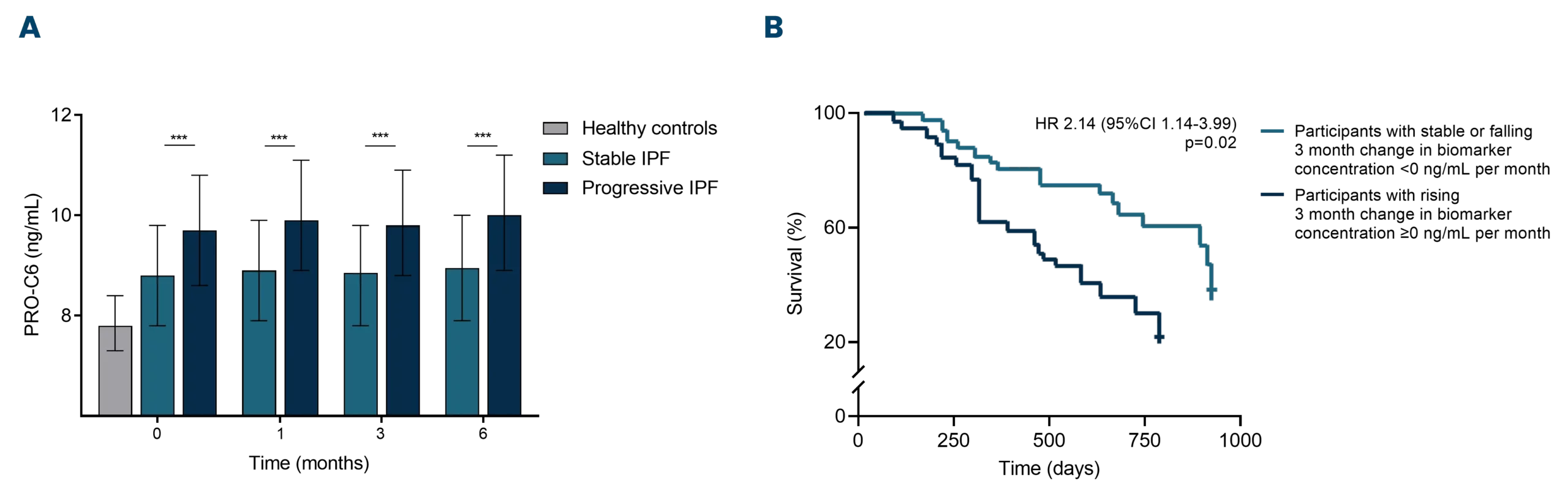
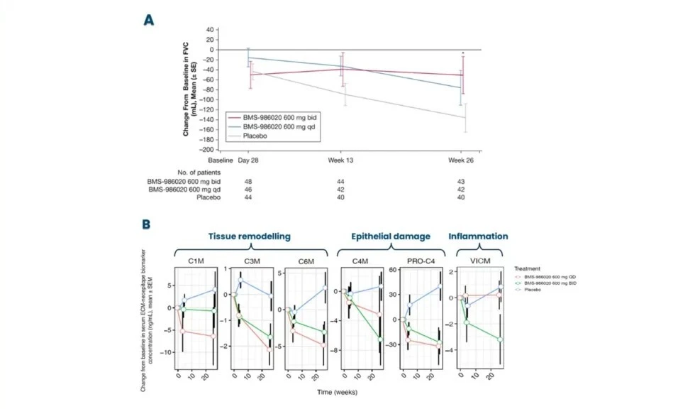
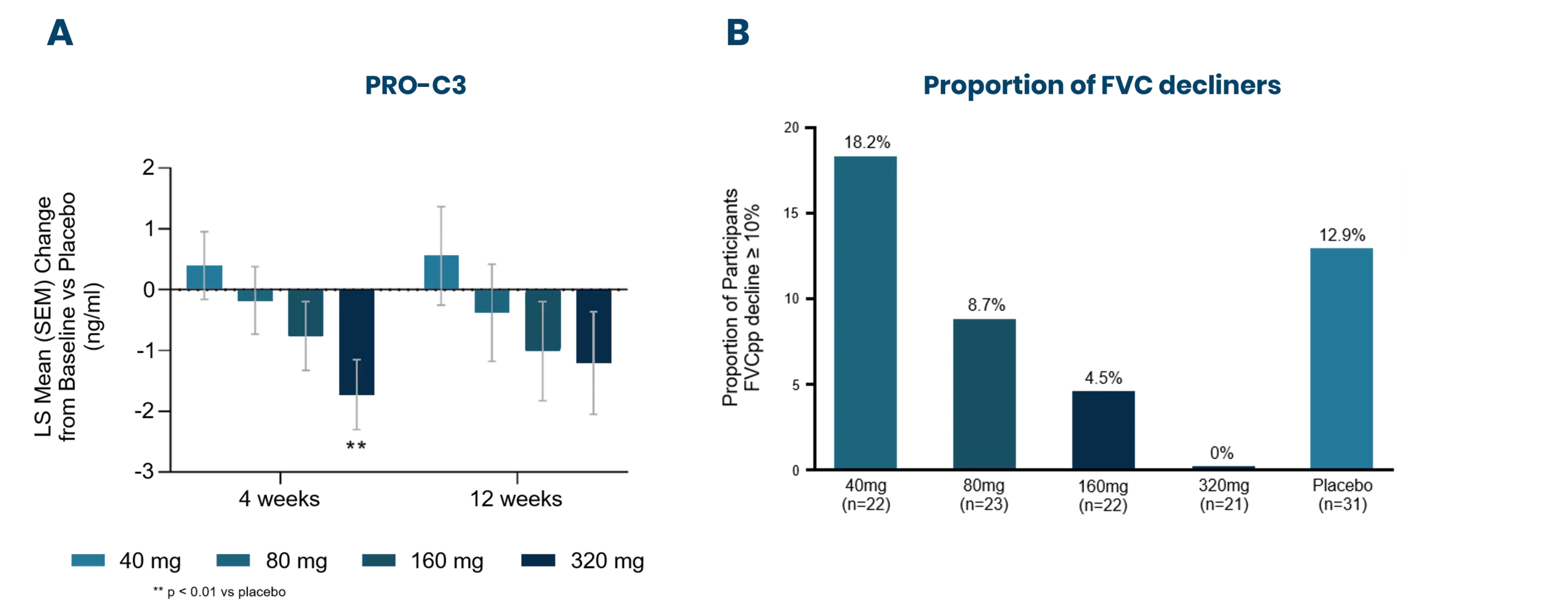
Progressive pulmonary fibrosis, including idiopathic pulmonary fibrosis (IPF), is associated with abnormal lung tissue remodeling and excessive extracellular matrix deposition. The Nordic ProteinFingerPrint TechnologyTM offers extracellular matrix (ECM) biomarkers that quantify the synthesis and degradation of key proteins in the fibrotic lung.

These biomarkers can be used early on for patient stratification, aiding in the inclusion of fast-progressing patients in clinical trials. The most common primary endpoint in clinical trials for IPF is to slow the decline in lung function (forced vital capacity [FVC]). Nordic Bioscience’s biomarkers have the potential to reflect treatment effects faster and more precisely than spirometry, enabling shorter and more effective trials. These biomarkers can also be used for precision medicine by monitoring and continuous evaluation of anti-fibrotic effects and potentially aid in therapy selection for the individual patient. Additionally, the biomarkers are translational and can be used across preclinical model systems to demonstrate anti-fibrotic effects throughout the drug development pipeline.

Nordic Bioscience develops biomarkers for pulmonary fibrosis that identify patients at high risk of progression and/or death. In the prospective, multicenter, observational cohort study PROFILE, our pulmonary team showed that extracellular matrix (ECM) biomarkers were significantly elevated in fast-progressing IPF patients (FVC decline ≥ 10% or death at 12 months) compared with slow progressors (figure 1A)[1]. Moreover, IPF patients with high baseline biomarker levels were at increased risk of mortality. Additionally, it has been shown that IPF subjects with increasing biomarker levels over three months were at greater risk of mortality compared to those with stable biomarker levels (figure 1B)[2].

These results demonstrate how the Nordic ProteinFingerPrint TechnologyTM can be used for patient stratification and for monitoring disease progression over time.

Figure 1: ECM biomarkers were measured in patients with IPF as part of the prospective, multicenter, observational cohort study, PROFILE. A) PRO-C6 was significantly elevated in fast-progressing IPF patients (FVC decline ≥ 10% or death at 12 months) when compared to slow progressors. B) IPF patients with increasing levels of C1M were at greater risk of mortality compared to those with stable or declining levels. HR: hazard ratio.

Browse our pulmonary fibrosis biomarker panel

Nordic Bioscience’s biomarkers are pharmacodynamic and can serve as indicators of treatment effect. In particular, the LPA1 antagonist BMS-986020 significantly improved FVC decline in a phase 2b clinical trial of IPF (figure 2A)[3] and reduced the levels of biomarkers reflecting tissue remodeling, epithelial damage, and inflammation (figure 2B)[4].

Importantly, a significant decrease in some of these biomarkers was detected at week 4, prior to effects on FVC (figure 2B and [5]). Similar effects were observed with the dual integrin inhibitor Bexotegrast (PLN-74809) in the INTEGRIS-IPF phase 2a clinical trial, which reduced both type III collagen synthesis and FVC decline at 4 weeks (figure 3)[6].

These findings highlight the value of the Nordic ProteinFingerPrint TechnologyTM in clinical trials for tracking pharmacodynamic effects over time. As Nordic Bioscience’s biomarkers have the potential to detect treatment effects earlier than traditional spirometry, they can contribute to the design of more efficient and shorter clinical trials.


Figure 2: In a phase 2b clinical trial with an LPA1 antagonist, 143 IPF patients were randomized to receive placebo, 600 mg BMS-986020 once daily (QD) or twice daily (BD) for 26 weeks. A) Change from baseline in forced vital capacity (FVC) for the placebo and active treatment arms at day 28, weeks 13 and 26. B) Change from baseline in Nordic Bioscience’s biomarkers reflecting tissue remodelling (C1M, C3M, C6M), epithelial damage (C4M, PRO-C4), and inflammation (VICM). Biomarkers were assessed in serum samples at baseline, day 28 and week 26. SE: Standard Error, SEM: Standard Error of the Mean.

Figure 3: The INTEGRIS-IPF phase 2a clinical trial evaluated the effects of a dual integrin inhibitor in 119 IPF patients. Patients received placebo, 40 mg, 80 mg, 160 mg, or 320 mg of Bexotegrast (PLN-74809). The study met its primary endpoint, and the figure shows the 12-week interim data. A) Change from baseline in serum PRO-C3 levels vs Placebo at 4 and 12 weeks. B) Proportion of participants having a decline in FVC in percent predicted (FVCpp) ≥ 10% at week 12. LS: Least Squares, SEM: Standard Error of the Mean

At Nordic Bioscience we believe that translational science may improve drug development. Therefore, we evaluated the ability of our biomarkers to translate and reverse translate between pulmonary fibrosis patients and in vitro, in vivo, and ex vivo model systems. Our team has shown that ECM biomarkers, such as PRO-C6, can be dose-dependently modulated by anti-fibrotic therapies when quantified in blood from IPF patients and in supernatants/blood from models of pulmonary fibrosis (figure 4)[7,8,9]. We have shown effects on our biomarkers in the primary fibroblast model Scar-in-a-Jar (SiaJ), in the precision-cut lung slice (PCLS) model, and in the mouse and rat bleomycin model.

These results demonstrate how the Nordic ProteinFingerPrint TechnologyTM may be used pharmacodynamically and translationally, demonstrating anti-fibrotic effects throughout different drug development stages.

Figure 4. Nordic Bioscience’s biomarker PRO-C6 was dose-dependently modulated by anti-fibrotic treatment across the model systems A) Scar-in-a-Jar (SiaJ) and B) precision-cut lung slices (PCLS), as well as C) in patients with idiopathic pulmonary fibrosis (IPF). BL: Baseline. 


Figure 5. Illustration of the changes in the ECM of the small airways and alveoli in a healthy lung and a diseased lung. BM: basement membrane, IM: interstitial matrix.

Pulmonary fibrosis is characterized by a dysregulated ECM remodeling. In particular, IPF is believed to be initiated by injury to the alveolar epithelial layer, disrupting the underlying basement membrane. Ineffective attempts to repair the epithelium and basement membrane activates fibroblasts which start to secrete ECM proteins which are then deposited in the interstitial matrix.

The normally thin layer of tissue in the distal lungs is thickened, hindering gas transfer in the alveoli. The fibrillar type I and III collagens are the main ECM proteins deposited in the fibrotic tissue. The production of type VI collagen is also affected and causes the release of the pro-fibrotic peptide, endotrophin, which is believed to play a crucial role in fibrosis development. Inflammatory cells recruited to the site of injury release proteases which degrade ECM proteins in an attempt to repair the damaged lung and remove fibrotic tissue, resulting in an enhanced lung tissue degradation. The balance between deposition and degradation of ECM proteins is essential to maintain tissue homeostasis (figure 5)[10].

Nordic Bioscience’s biomarkers are available to evaluate these different disease processes, including tissue remodeling, epithelial damage, wound healing, and inflammation, in our CAP-accredited laboratory.

Pulmonary fibrosis is a chronic interstitial lung disease in which progressive scarring of the lungs leads to fibrosis and loss of lung function. People with pulmonary fibrosis suffer from dry, persistent cough and progressive fatigue. Patients with IPF have a poor prognosis with an average survival of 3-5 years after diagnosis without effective treatment.

How many people have pulmonary fibrosis?
Despite being considered a rare disease, millions of people suffer from pulmonary fibrosis, and about 50,000 new cases of IPF are diagnosed each year. Risk factors include age, gender, tobacco use, and a family history of pulmonary fibrosis, as some genes are associated with IPF.

How is pulmonary fibrosis diagnosed?
IPF is diagnosed by lung function tests, a chest X-ray and a high-resolution CT scan to identify the fibrotic pattern associated with pulmonary fibrosis. A lung biopsy may also be needed to make a diagnosis.

How is IPF treated?
Currently, two antifibrotic agents are approved by the FDA and EMA for the treatment of IPF, namely pirfenidone and nintedanib. However, neither of these can stop the disease but only slow its progression. In addition, both agents have been associated with patient tolerability issues and have limited effect in improving a patient's quality of life.

Biomarkers for other respiratory indications

Nordic Bioscience's assays and services are research use only products and services and do not qualify for medical or diagnostic purposes.

Please don't hesitate to contact us if you have any questions or other inquiries.

Are you interested in learning more about Nordic Bioscience?
Enter your information in the form and a representative will contact you shortly.

By submitting this form you agree to our terms and conditions.