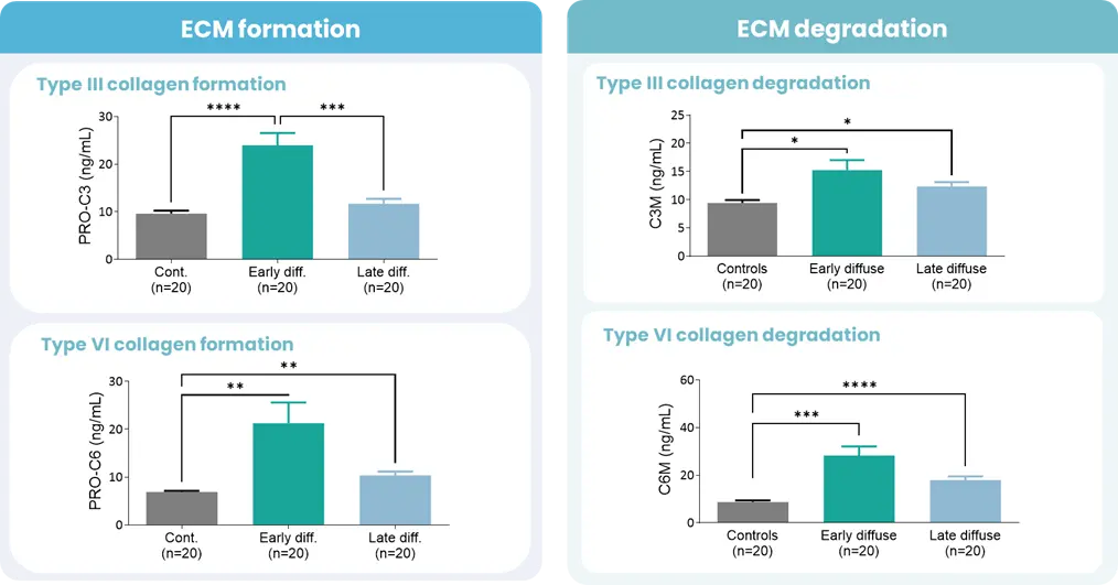
Autoimmune diseases, such as scleroderma—systemic sclerosis (SSc)—affect several tissues, leading to fibrotic tissue buildup of the skin and internal organs. Fibrosis is caused by an increased synthesis of extracellular matrix (ECM), which plays a key role in SSc pathogenesis.
Extracellular matrix-specific biomarkers that help us quantify and monitor fibrosis of the skin internal organs are key when we want to understand the disease mechanisms and prediction of scleroderma/SSc progression.
Utilizing biomarkers can help address several aspects of unmet needs in SSc. A well-designed biomarker program enables precise diagnosis, prediction of clinical course and organ involvement, evaluation of therapeutic responsiveness, and identification of novel therapeutic targets.
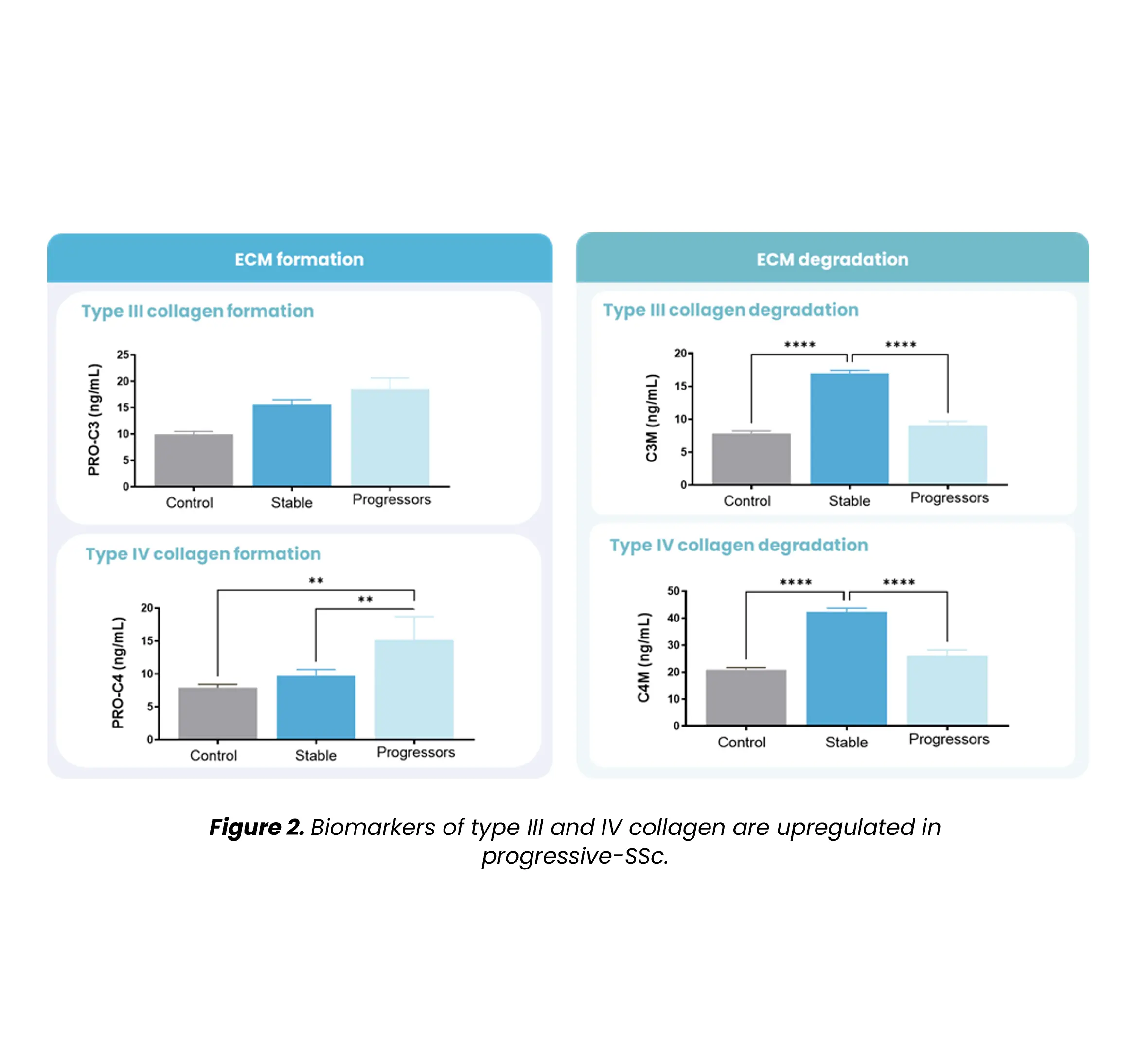
Figure 2. Biomarkers of SSc progression | A balanced extracellular matrix (ECM) is necessary to preserve organ integrity and tissue homeostasis. In SSc, as shown in Figure 2, this balance is altered, and the fibrosis biomarkers (PRO-C3 and PRO-C6) are associated with disease progression (10% decline in FVC%, or increased mRSS >25% during a one-year clinical follow-up). At the same time, tissue degradation biomarkers were associated with stable disease. This has been shown in a study published in The Lancet, in 2021. |
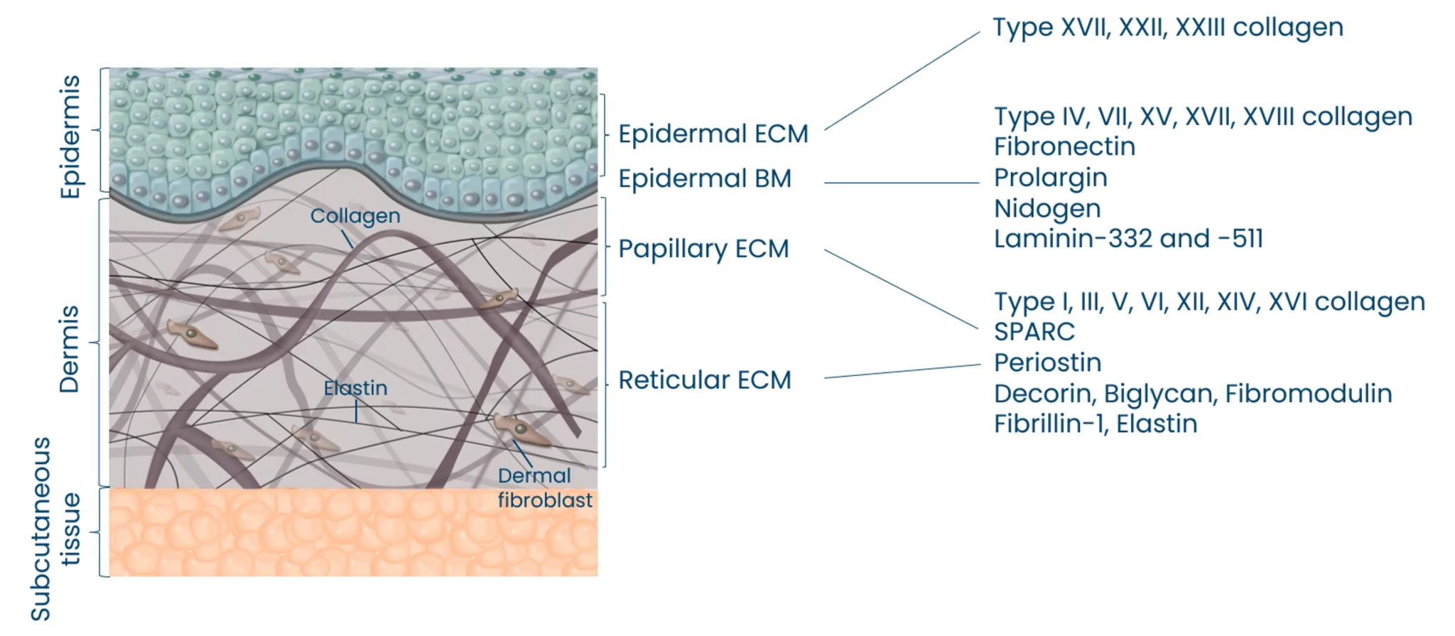
The skin is the largest organ of the human body, and consists of three different layers; epidermis, dermis and subcutis. These three layers have distinct tissue architectures and functions. Skin functions are dependent on a complex composition of extracellular matrix (ECM) proteins. The ECM of the skin can be divided into the epidermal ECM, epidermal basement membrane and the papillary/reticular ECM. The major ECM components of the skin are collagens, which are located in the different tissue compartments to maintain tissue architecture. The Nordic ProteinFingerPrint Technology™ allows for the quantification of the different layers of the skin, and also the quantification of immune cell activity involved in different pathologies (Mast Cells, Neutrophils, and macrophages). |
Systemic sclerosis (SSc) is a chronic rheumatic disease with one of the highest mortalities within rheumatology. It is an autoimmune, fibro-inflammatory skin disease characterized by a distinctive triad of microvascular damage (vasculopathy), immune activation with autoimmunity, and generalized fibrosis of multiple organs, including the skin and lungs. The great heterogeneity of disease cause and presentation creates considerable challenges in developing treatment and treating patients.
The prevalence of SSc varies considerably around the world. Northern Europe and parts of Asia have a prevalence of >150 per million, whereas southern Europe, North America, and Australia have a higher prevalence of up to 443 per million. As with other autoimmune diseases, women are about six times more likely to get SSc than men.
Skin fibrosis is assessed by the modified Rodnan Skin Score (mRSS) and the presence of vascular damage, including Raynaud’s Phenomenon, is included in the diagnostic assessment. Furthermore, the presence of auto-antibodies and pulmonary involvement (either presented as interstitial lung disease or pulmonary arterial hypertension) is included in the diagnosis.
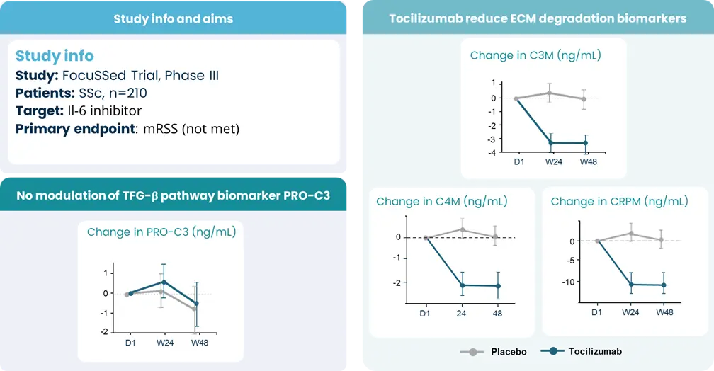
There is no cure or disease-modifying drug for SSc, but symptoms and inflammation can be treated using NSAIDs, steroids, or biologics. The type of treatment depends on the symptoms and organs involved. Several novel drugs are in development targeting either the inflammatory and/or the fibrotic axis.
Systemic Sclerosis is characterized by microvascular damage (vasculopathy), immune activation with autoimmunity, and generalized fibrosis of multiple organs, including the skin and lungs. The different organs that may be affected by SSc have tissues consisting mainly of collagens. The collagens are remodeled as part of normal homeostasis of the organ– that is, collagens are broken down and rebuilt as part of normal repair and maintenance of the tissue. In SSc, the balance of tissue remodeling, i.e. formation and degradation of ECM proteins, is interrupted resulting in fibrosis of the organs. Similarly, vascular remodeling leads to increased or disrupted vascularization, which may be detected by increased turnover of the ECM proteins of the vasculature. Nordic Bioscience's biomarkers can directly quantify this fibrosis and microvascular damage in a serum sample.
Search and find publications that we have published.
Please don't hesitate to contact us if you have any questions or other inquiries.