The currently applied detection tools for atherosclerotic plaques rely on imaging techniques or blood-based biomarkers. Evaluating plaque presence by imaging can be difficult due to the spatial distribution of the plaque throughout the circulatory system.

The current biomarkers and diagnostic tools in myocardial infarction (MI) are based on molecules released from the stressed heart and imaging- or ECG-based methods. The most established blood-based biomarker for MI is high-sensitive troponin, which is an exceptional tool in the acute phase but does not inform on the short- and long-term prognosis of patients experiencing MI.

Imaging-based techniques do not provide insight into plaque composition, a critical component for plaque stability. Blood-based biomarkers measuring lipids and cholesterol are also flawed because atherosclerosis is not only caused by lipid accumulation. The currently applied biomarkers are therefore imprecise and only portray part of the underlying factors of the condition.

Extracellular matrix (ECM) proteins constitute a significant proportion of the atherosclerotic plaque. During normal tissue homeostasis, the balance between formation and degradation is finely balanced. Disruption of this balance can lead to an unstable plaque. Accurately quantifying ECM changes in atherosclerotic plaques could therefore add incremental value to the panel of established biomarkers and assessment techniques, potentially improving the prognostic power.

Moreso, MI leads to ECM remodeling in the heart, which results in the further release of ECM formation and degradation epitopes. These epitopes could provide specific information on patient outcomes following MI.

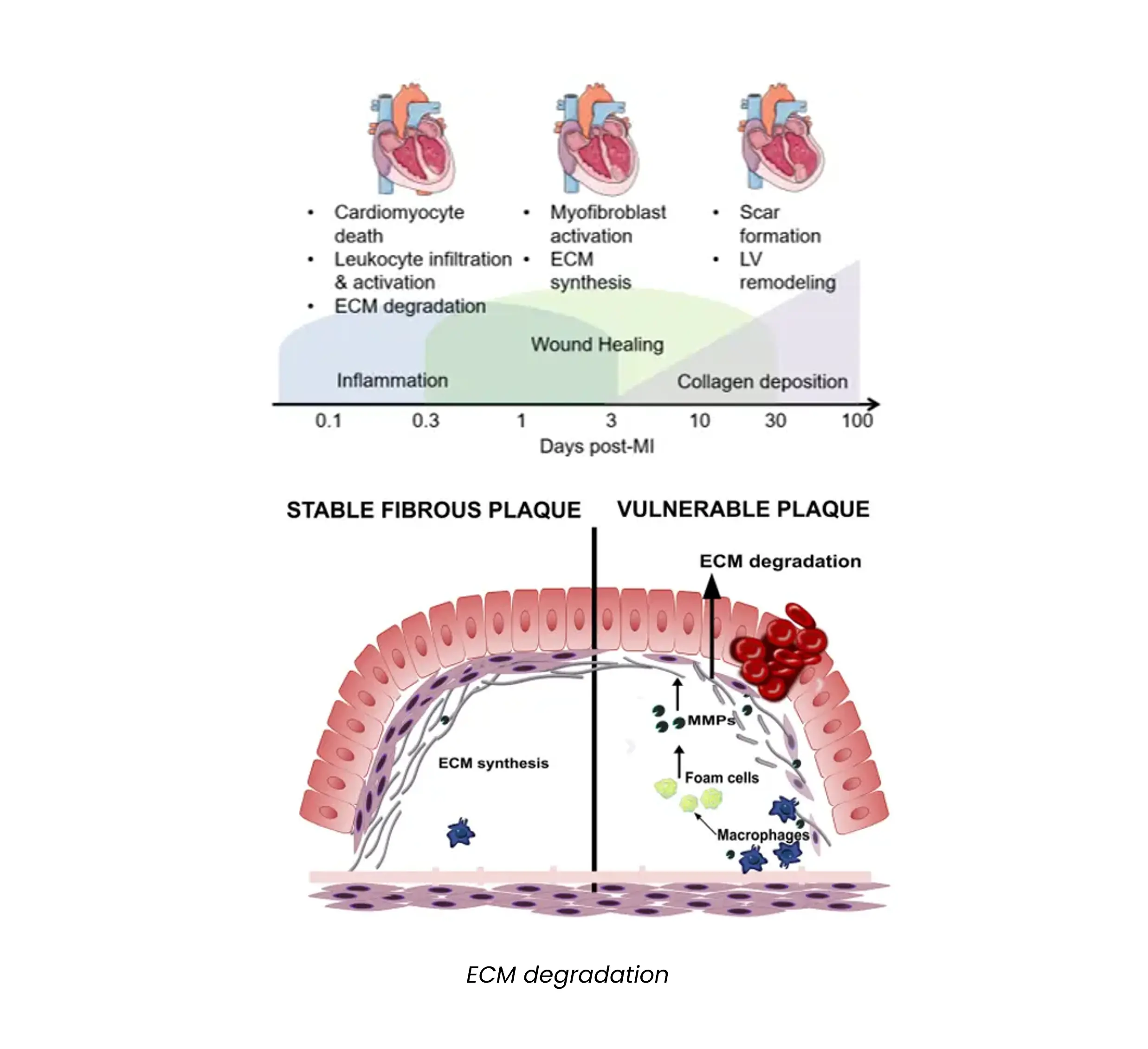
The main component determining whether a plaque is stable or vulnerable is the presence of a stable fibrous cap, which maintains the plaque in place. In the presence of inflammation, the fibrous plaque is damaged, causing plaque instability, which increases the risk of thrombosis.

During this process, proteins of the ECM constituting the fibrous cap are degraded and released in circulation, where they can be measured by Nordic Bioscience's biomarker technology.

Over the course of MI, the remodeling of the ECM is involved in the initial phases, characterized by tissue destruction mediated by inflammatory cells, and in later phases, characterized by ECM formation to substitute the necrotic tissue. An excessive ECM remodeling can cause the formation of scar tissue that causes a long-term impairment of heart contractility.

Nordic Bioscience biomarkers can be used in every phase of heart remodeling, informing on the balance between ECM formation and degradation leading to modifications in the cardiac tissue.

Browse our list of atherosclerotic plaque instability biomarkers

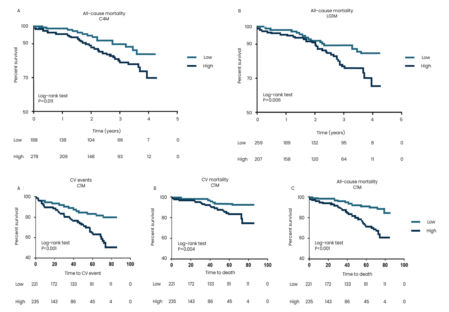
During atherosclerosis, inflammatory cells produce proteases such as matrix metalloproteinases (MMPs), which degrade proteins of the ECM, such as the basement membrane proteins type IV collagen and laminin and the interstitial type I collagen, ultimately destabilizing the plaque.

Nordic Bioscience's biomarkers of laminin (LG1M), type IV (C4M) and type I (C1M) collagen degradation have shown to be associated with adverse events in atherosclerotic patients, potentially linked to the presence of vulnerable plaques (Figure 1.)

Figure 1. Tissue profiling in atherosclerosis and myocardial infarction (MI)

Cardiomyocyte necrosis is a common feature subsequent to MI as a result of ischemia. Due to the low regenerative capacity of the heart, the lost cardiomyocytes are replaced by ECM proteins, a process referred to as replacement fibrosis.

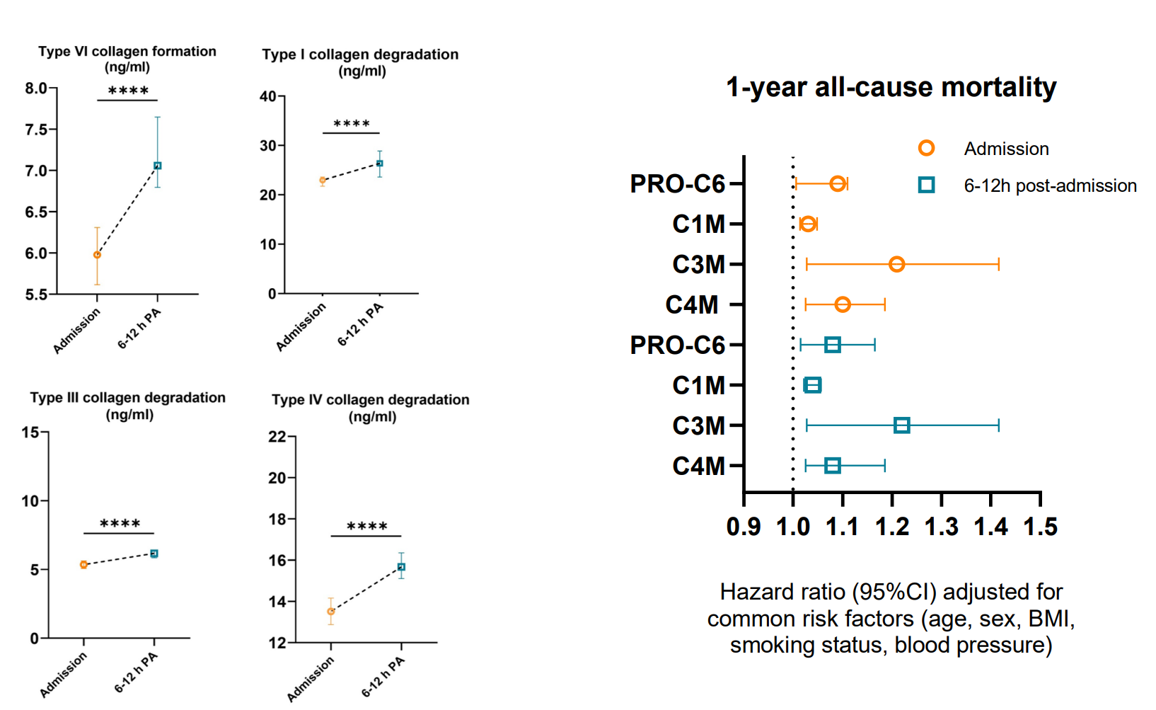
This leads to differential regulation of the collagens found in the heart, including type I, III, IV, and VI collagens. In a study of 221 patients with STEMI, degradation of type I collagen (C1M), type III collagen (C3M), type IV collagen (C4M) and formation of type VI collagen (nordicPRO-C6™) significantly increased in circulation during the first hours after admission to the hospital, and levels at both admission and 6-12h post-admission were associated with an increased risk of mortality at 1 year after adjustment for common risk factors (Figure 2).

Figure 2. Cardiac biomarkers significantly increased in circulation during the first hours after admission to the hospital


Atherosclerosis is a systemic disease affecting the cardiovascular system. It is a leading cause of cardiovascular complications worldwide, often causing acute events leading to mortality or a significant decrease in quality of life.

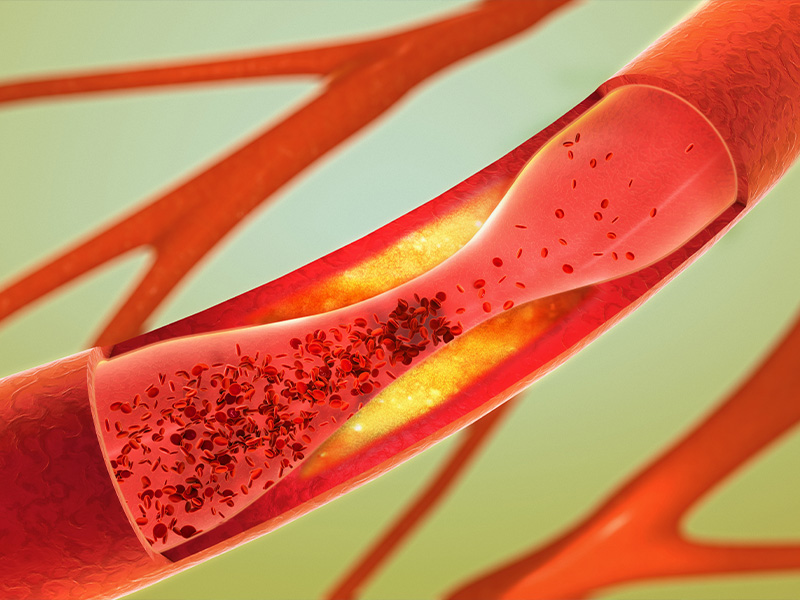
Atherosclerosis is characterized by the formation of plaques in the arterial wall containing retained lipids, inflammatory cells, apoptotic cells, and increased extracellular matrix (ECM) protein formation. Over time, the plaque hardens which narrows the arteries and restricts blood flow. The biological composition of the plaque determines the risk of plaque rupture. Acute plaque rupture can cause local thrombosis which leads to partial- or total occlusion of the affected artery. If this happens in one of the two major arteries supplying blood to the heart, the result is often a myocardial infarction (MI), also known as a heart attack. MI is a major cause of mortality and cardiovascular-related admission to hospitals, and it often leads to heart failure (HF).

How many suffer from atherosclerosis and MI?

Atherosclerosis is a very common and asymptomatic condition: it is therefore difficult to accurately determine incidence. In the Western world, more than 370.000 people die every year from coronary heart disease (CHD), a condition caused by atherosclerosis in the coronary arteries, and approximately 800.000 people suffer from stroke annually, resulting in more than 140.000 deaths. MI is a frequent event in cardiovascular patients suffering from CHD, with more than 10 million events registered annually worldwide and 1.5 million cases in the United States.

How are atherosclerosis and MI diagnosed?

Atherosclerosis is commonly assessed by computed tomography angiography. Cardiac magnetic resonance imaging is also frequently being implemented in diagnosis and risk assessment of plaque rupture. Atherosclerosis can also be assessed invasively by angiography but is not recommended as a screening tool. Blood-based biomarkers are also implemented in diagnosis, such as C-reactive protein (CRP), lipids and lipoproteins.

Patients experiencing MI often complain about chest pain and discomfort in the chest, shoulders, and arms. Patients can also experience shortness of breath, nausea, and feeling faint. However, these symptoms are not unique to cardiovascular diseases, and further diagnostic measures are applied.

Because MI leads to tissue damage, cardiac-specific biomarkers are released such as troponin, and can be measured in the blood of patients. Electrocardiogram (ECG) is the most common non-invasive tool used for diagnosis of MI and it is often used repeatedly over minutes to hours to monitor changes in the cardiac rhythm after admission to hospital. ECG is employed to separate a MI with ST-segment elevation myocardial infarction (STEMI) and a MI with non-ST-segment elevation myocardial infarction (NSTEMI). Imaging techniques, such as angiography, are used to assess the blockage of the coronary arteries.

How are atherosclerosis and myocardial infarction treated?

The best treatment for atherosclerosis is to treat risk factors, such as elevated LDL, blood pressure, and diabetes. Exercise and healthy dietary habits are also important to reduce risk. Statins are the main treatment options for lowering LDL levels. To control blood pressure multiple drug classes are implemented, most commonly including angiotensin-converting enzyme (ACEs) inhibitors and angiotensin II receptor blockers (ARBs). Clinical treatment of atherosclerosis includes bypass surgery and insertion of stents.

When a MI is caused by a total coronary blockage, early treatment is crucial for patient outcome. In general, treatment strategies are aimed at unblocking the occluded artery to restore blood supply to the infarcted area(s) of the heart. Agents that reduce blood coagulation and clotting, can reduce mortality upwards of 50% in acute MI patients. Both STEMI and NSTEMI patients are treated with percutaneous coronary intervention (PCI) in combination with stents to increase blood flow through the affected artery(ies). Following an MI, lifestyle modifications and medications are recommended to reduce the likelihood of a recurring MI.

Biomarkers for other cardiovascular indications

Nordic Bioscience's assays and services are research use only products and services and do not qualify for medical or diagnostic purposes.

Please don't hesitate to contact us if you have any questions or other inquiries.

Are you interested in learning more about Nordic Bioscience?
Enter your information in the form and a representative will contact you shortly.

By submitting this form you agree to our terms and conditions.