The field of immuno-oncology is based on decades of research into how tumors have acquired mechanisms to circumvent and evade elimination by the immune system. By intervening with the ability of tumors to escape the immune system and by re-activating and re-directing immune cells in the tumor microenvironment, immuno-oncology therapies enable the body's own defense system to recognize and eliminate the tumor.

While chemo- and targeted therapies have prevailed in the treatment of solid tumors, immuno-oncology therapies such as checkpoint inhibitors have revolutionized the field by inducing durable long-term responses in cancer patients. However, despite the substantially extended clinical benefit experienced by some patients, a large percentage of patients included in these clinical trials do not respond to treatment and are left with few, if any, other treatment options.

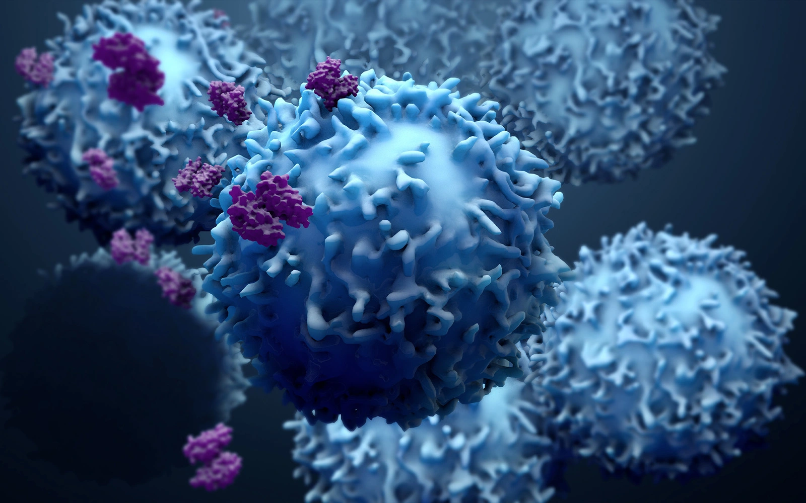
One essential characteristic of patients benefitting from immuno-oncology therapies such as checkpoint inhibitors is that their T-cells are present and/or recruited to the tumor. That is, if T-cells are not homing to the tumor, treatment will have a poor effect. Therefore, it is important to identify patients with a T-cell permissive tumor microenvironment.

Analysis of the tumor microenvironment in patients with a variety of solid tumors has revealed that patients can be divided into phenotypes based on T-cell infiltration: tumors with T-cell infiltration ("hot tumors"/"inflamed tumors") and tumors without T-cell infiltration ("cold tumors"/"excluded tumors").

Interestingly, it has been shown that:

  1. T-cells are actively degrading the basement membrane extracellular matrix when transmigrating from the blood and into the tumor, and
  2. The architecture of the extracellular matrix defines the localization and migration of T-cells within the tumor with T-cells being entrapped in the dense interstitial extracellular matrix and therefore cannot reach and kill the tumor cells.

By combining the serological assessment of a biomarker related to tumor fibrosis (PRO-C3) with the serological assessment of a biomarker related to active T-cell infiltration in the tumor microenvironment (C4G), it is possible to identify in our CAP-accredited lab the fraction of patients with benefit from checkpoint inhibitor therapy from the periphery.

C4G - Granzyme B degraded type IV collagen (T-cell infiltration)

Activated T-cells release the protease granzyme B (GzB), which can cleave type IV collagen during tumor infiltration. In other words, GzB promotes T-cell transmigration via basement membrane remodeling. Nordic Bioscience has identified GzB-specific cleavage sites on type IV collagen by mass spectrometry and developed an assay (C4G) with the ability to predict response and outcome in cancer patients treated with checkpoint inhibitors. C4G has been validated in several indications, with other immuno-oncology modalities and in other disease areas.

PRO-C3 - Pro-peptide of type III collagen (Tumor fibrosis)

Cancer-associated fibroblasts (CAFs) are the main drivers of the increased extracellular matrix and collagen deposition leading to tumor fibrosis (desmoplasia). Consequent to the impact of desmoplasia on efficient infiltration and activity of T-cells in the tumor, tools to measure and quantify the desmoplastic reaction have great applicability in the immuno-oncology setting. PRO-C3 (the N-terminal pro-peptide on type III collagen) an assay developed by Nordic Bioscience for measuring true formation of fibroblast-derived collagen has shown to predict response and outcome in cancer patients treated with checkpoint inhibitors. PRO-C3 is also validated in several indications, with other modalities and in other disease areas.

Visit our cancer biomarker portfolio and choose a panel that fits your clinical research or drug development targets!


C4G biomarkerPRO-C3 biomarker
Immune inflamedImmune excluded
Peripheral measure of T-cell infiltrationPeripheral measure of tumor fibrosis

T-cells use Granzyme B to degrade type IV collagen when infiltrating the tumor that resulting in the release of C4G into circulation

T cells may be retained in a high-density collagen barrier associated with the release of collagen type III fragments (PRO-C3) into circulation


It is well established that immuno-oncology therapies can have a great impact on the composition and quality of the cells of the tumor microenvironment and therefore also on the composition and quality of the extracellular matrix and the Nordic ProteinFingerPrint Technology™ biomarkers. This potentially allow pharmacodynamic profiling of novel treatments by measuring these biomarkers directly in serum/plasma samples over the course of an immuno-oncology clinical trial. Likewise, these biomarkers may enlighten on the mode-of-action of a given immuno-oncology compound alone or in combinations.

VICM - Matrix metalloprotease degraded, citrullinated vimentin (Macrophage activity)

Tumor-associated macrophages (TAMs) are the dominant leukocyte population found in the tumor microenvironment where they are key mediators in maintaining the inflammatory processes in the tumor. Monitoring how macrophage activity is affected by a given immuno-oncology treatment may shed light on the MoA. The protein fragment VICM (matrix metalloprotease degraded and citrullinated vimentin) has been shown to be released from activated macrophages and dose-dependently decreased by anti-GM-CSFRα therapy. Moreover, changes in the VICM biomarker are linked to response to checkpoint blockade supporting a link between immuno-oncology therapy and TAM activity.


Visit our cancer biomarker portfolio and choose a panel that fits your clinical research or drug development targets!


Immuno-oncology treatment response relies on complex interactions between tumor cells, the stromal cells (immune cells, fibroblasts etc.), soluble factors (TGF-beta) and the extracellular matrix. This entails a need for biomarkers reflecting such tumor microenvironment alterations. Many immuno-oncology-related biomarkers currently being evaluated for predicting and monitoring response are based on fresh tissue biopsies which, for one, are often not obtainable, but also suffer from heterogeneity due to "sampling error" and lack of reflection on the dynamic component of such tumor microenvironment alterations. Therefore, peripheral biomarkers (liquid biopsies) in the setting of IO therapies are urgently needed.

Proteins are constantly being produced, degraded and modified as part of maintaining healthy tissue. However, these processes become uncontrolled in the tumor microenvironment. Ultimately, all these activity changes in the tumor microenvironment manifest in alterations in the quality and composition of the extracellular matrix. Compared to genomics and IHC, looking at specific neo-epitopes on extracellular matrix proteins associated with given pathophysiological processes will provide a higher correlation with actual disease activity and immuno-oncological processes.

At Nordic Bioscience we focus our biomarker efforts on targeting and quantifying post-translational modifications, cleavage sites, and neo-epitopes on proteins or peptide fragments in circulation. We call these protein fingerprints. This approach is in fundamental contrast to most precision medicine approaches in the immuno-oncology space where the primary focus is on genomics and staining of total protein content by immunohistochemistry. Quantifying unique neo-epitopes on circulating peptides originating from the same protein may provide the complete opposite prognostic value.

Same protein - Different neo-epitope - Opposite prognostic value

Overall survival in metastatic melanoma patients  treated with Ipilimumab (anti-CTLA4)


Nordic Bioscience's assays and services are research use only products and services and do not qualify for medical or diagnostic purposes.

Please don't hesitate to contact us if you have any questions or other inquiries.

Are you interested in learning more about Nordic Bioscience?
Enter your information in the form and a representative will contact you shortly.

By submitting this form you agree to our terms and conditions.