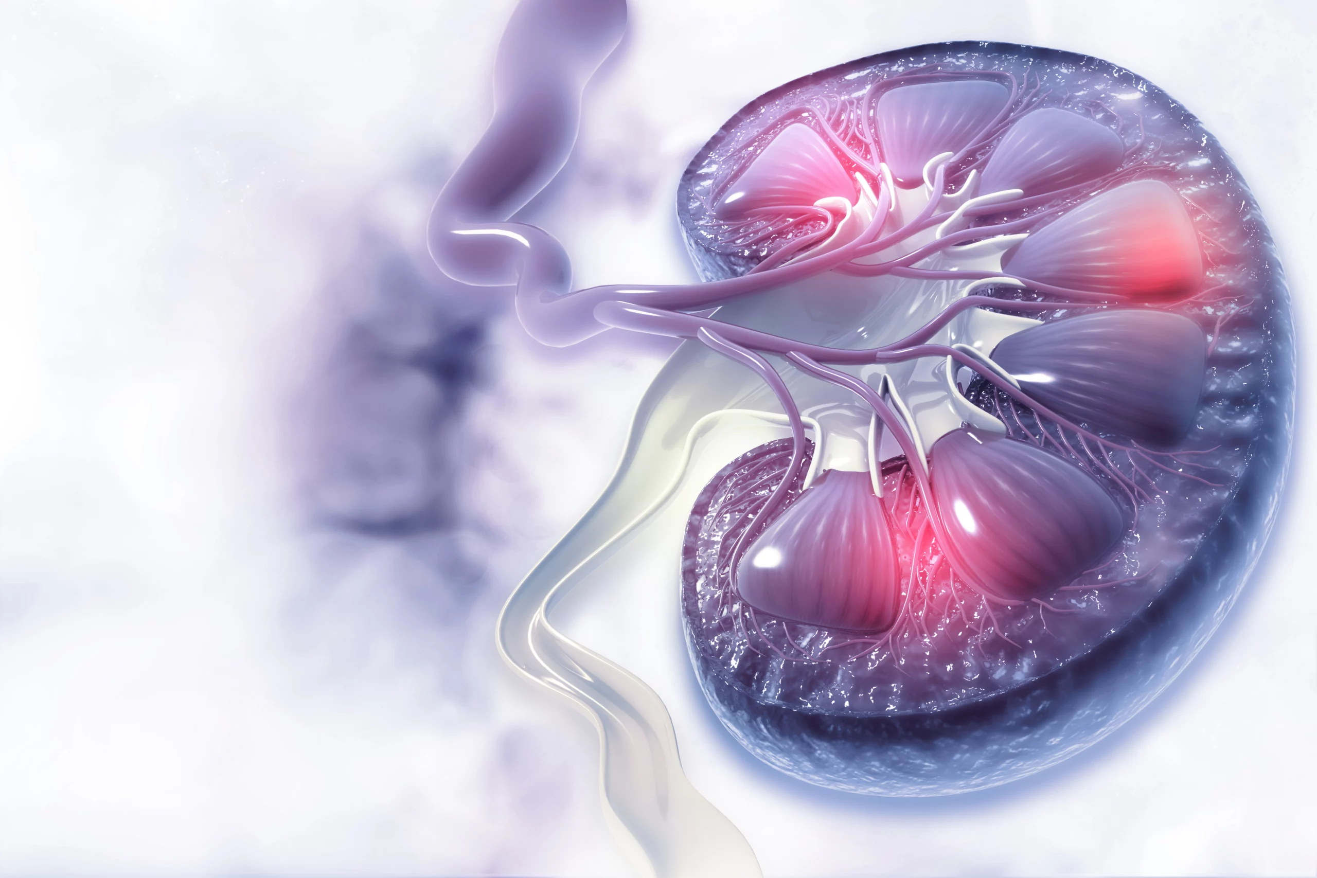
Chronic kidney disease (CKD) is caused by progressive deterioration of kidney function due to loss of functional units in the kidneys. The functional units, called nephrons, consist of the glomerulus and tubules. The main site of filtration is called the glomerulus. It consists of an arteriole that winds around itself to form a spherical structure. The glomerulus is held together by the mesangial matrix, while the surface of the endothelial cells of the arteriole is covered by the foot processes of podocytes and the specialized glomerular basement membrane (GBM).

The GBM and the slit membranes of the interlocking foot processes of podocytes form the filtration barrier. After filtration, the filtrate enters the tubules (proximal tubules, loop of Henle, and distal tubules), where various molecules are reabsorbed or excreted into the urine; after this process, the urine enters the collecting ducts. During life, nephrons are lost due to injury and aging. Loss of nephrons is irreversible, but humans are born with a large reservoir of nephrons and a large proportion can be lost before symptoms appear. Because the kidney is the primary site for excretion of waste products, impaired kidney function has far-reaching effects throughout the body. For example, patients with CKD have a significantly increased risk of cardiovascular events.

How many people have CKD?
Large-scale, nationally representative screening programs conducted in Australia, Norway, and the United States in the 2000s have shown that more than 10% of the adult population has CKD. In the Western world, diabetes is the most common cause of CKD, followed by hypertension. With the increase in lifestyle diseases such as diabetes and hypertension and increased life expectancy, CKD is expected to become an increasing problem worldwide. In 2017, there were 697.5 million cases of CKD worldwide and about 1.2 million people died from CKD.

How is CKD treated?
If detected early, it is possible to halt or slow the progression of CKD pharmacologically. However, because of the reservoir of nephrons, CKD is usually not diagnosed until it is advanced. Patients who reach end-stage renal disease require dialysis or kidney transplantation. The main classes of treatment for CKD are medications to regulate blood pressure or treat the underlying causes of the disease (e.g. GLP-1 agonists and SGLT2 inhibitors for diabetes, which have a positive effect on kidney health).

How is CKD diagnosed?
Patients are diagnosed with CKD when the concentration of creatinine in the blood and/or protein or albumin in the urine is above a certain threshold. Because serum creatinine increases only after the loss of many nephrons and not all patients have proteinuria or albuminuria, better diagnostic tools are needed to identify patients at risk of developing CKD.

 As mentioned earlier, current diagnostic markers for CKD are not very sensitive. Therefore, novel diagnostic biomarkers can be used as complementary tools to identify patients at high risk for developing CKD or with early disease who can be successfully treated to halt disease progression. There is also an unmet need for prognostic biomarkers capable of identifying patients at higher risk for progression to end-stage renal disease. Patients with active disease can be selected to enrich clinical trials.

A predictive biomarker would identify patients most likely to respond to therapy. This would have a major impact on drug development by reducing the duration, size, and cost of trials. Biomarkers can also aid drug development by monitoring therapeutic efficacy when concentrations in biological fluids are affected by treatment. A number of diagnostic biomarkers and biomarkers of disease activity are currently being studied in CKD, but their ability to stratify risk and predict response to treatment is limited.

Visit our kidney biomarker portfolio and choose a panel that fits your clinical research or drug development targets!

In the normal extracellular matrix (ECM) of the kidney, ECM proteins are remodeled as part of normal homeostasis, i.e., degraded and replaced by newly synthesized proteins. Collagens are the most abundant ECM proteins in the renal stroma. As a result of repeated or prolonged impairment, the balance between ECM degradation and formation is disturbed, leading to net collagen formation.

Abnormal collagen formation leads to renal fibrosis, which is the most important predictor of end-stage renal disease progression. The Nordic ProteinFingerPrint Technology™ allows quantification of protein fragments generated by ECM turnover directly in serum and/or urine, allowing monitoring of the processes of fibrogenesis and fibrolysis.

In the kidneys, the extracellular matrix is distributed among three main compartments: the tubulointerstitial matrix, the glomerular basement membrane (GBM) supporting endothelial cells, and the tubular basement membrane supporting epithelial cells, and the mesangial matrix.

Biomarkers describing the altered turnover of these compartments would allow characterization and quantification of structural changes in the tissue. The ECM in these different compartments is characterized by the presence of different proteins or protein isoforms. Consequently, measurement of specific formation or degradation products would provide information on which compartments are most affected by the disease or treatment.

Collagen biomarkers and tissue characterization:

Tubulointerstitial matrix

  • Degradation: reC1M, C3M, C5M, C6M, C6Ma3, C7M
  • Formation: PRO-C1, PRO-C3, PRO-C5, PRO-C6

 Mesangial matrix :

  • Degradation: C4M, C5M
  • Formation: PRO-C4, PRO-C5, PRO-C18

 Glomerular basement membrane:

  • Degradation: C4Ma3, TUM
  • Formation: PRO-C4

 Tubular basement membrane:

  • Degradation: C4M
  • Formation: PRO-C4

Nordic ProteinFingerPrint Technology™ biomarkers measured in serum and urine are either elevated or decreased in CKD patients compared with healthy individuals and correlate with biomarkers of renal function (creatinine, cystatin C, albuminuria) and inflammation (e.g., CRP, IL-6).


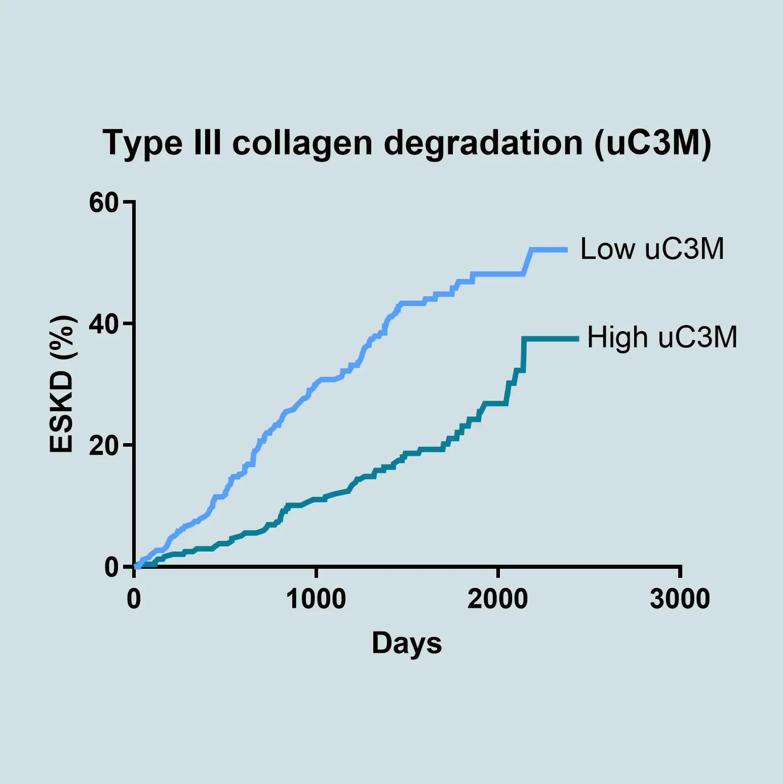
Tubulointerstitial matrix degradation quantified by MMP-9 degraded type III collagen (C3M) in urine

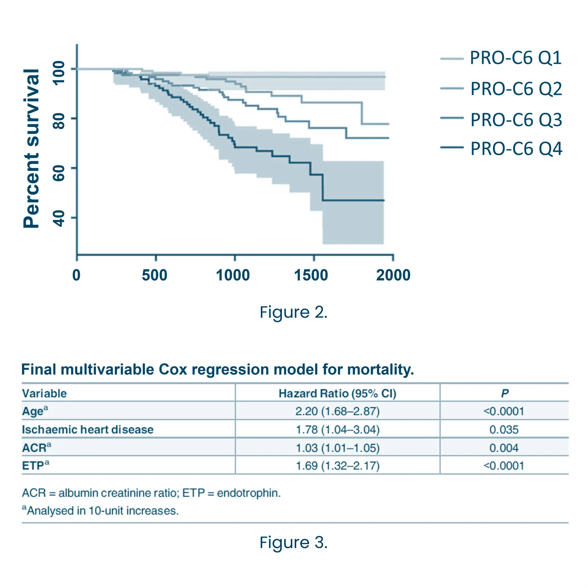
Tubulointerstitial matrix formation quantified  by released pro-peptide of type VI collagen containing endotrophin, a known matrikine with potential deleterious functions (PRO-C6) in serum

Sparding et al Nephrology Dialysis Transplantation, 2018 (FP275)

Sparding et al Nephrology Dialysis Transplantation, 2018 (FP274)

Nordic ProteinFingerPrint Technology™ biomarkers measured in serum and urine are associated with the severity of CKD and correlate with the extent of fibrosis in histopathological specimens.


Urinary fragments reflecting type III collagen degradation (C3M) and formation (PRO-C3) shift with increasingdiseaseseverity.


Nordic ProteinFingerPrint Technology™ biomarkers measured at baseline are prognostic of adverse outcomes including CKD progression, cardiovascular events, and mortality.


Prognostic ability of PRO -C6 in patients with type 2 diabetes with microalbuminuria without symptoms of coronary artery disease (BNPcure study). *Danger ratios (HR) with 95% CI are per doubling. PRO -C6 was adjusted for conventional cardiovascular risk factors (age, sex, systolic blood pressure, LDL cholesterol, smoking, HbA1c, plasma creatinine, and UAER). Cardiovascular events were defined as a composite of cardiovascular mortality, stroke, ischemic cardiovascular disease, and heart failure. Disease progression was defined as a decrease in eGFR of more than 30%.

Nordic ProteinFingerPrint Technology™ biomarkers enable pharmacodynamic profiling of novel treatments by measuring protein fragments that directly reflect degradation and formation in a sample.

Early modulation of Nordic ProteinFingerPrint Technology™ biomarkers after treatment of patients with type 2 diabetes with a GLP1 agonist. Pharmacodynamic biomarkers could aid in dose finding and provide information on target/mode of action.


Nordic ProteinFingerPrint Technology™ biomarkers at baseline can predict response to treatment by PPARγ agonists (reduction in HbA1c).


The applications presented here are for research use only.

Please don't hesitate to contact us if you have any questions or other inquiries.

Are you interested in learning more about Nordic Bioscience?
Enter your information in the form and a representative will contact you shortly.

By submitting this form you agree to our terms and conditions.