Cardiomyopathy management has been based solely on the morphological and functional changes of the heart for a long time. The last decades of imaging evolution and genetic testing have contributed immensely to disease diagnosis and management. Heart failure biomarkers including natriuretic peptides and troponin isoforms have also been used to assess cardiac stress and cardiomyocyte cell death. However, heart failure is often the endpoint of cardiomyopathies hence there is an unmet need for circulating biomarkers for early diagnosis and/or prognostic ability.
Cardiomyopathies are characterized by structural changes in the cardiac muscle due to severe extracellular matrix (ECM) remodeling taking place under pathological conditions. Several studies have shown cardiac fibrosis being present in all different types of cardiomyopathies and being associated with a worse prognosis.
Cardiomyopathy patients range from asymptomatic to symptomatic fast progressors. Being able to quantify changes in the ECM turnover early on can be very informative. With the use of highly specific, non-invasive biomarkers targeting extracellular matrix components circulating in the blood, it would be possible to get a great insight into disease progression and influence decision-making.
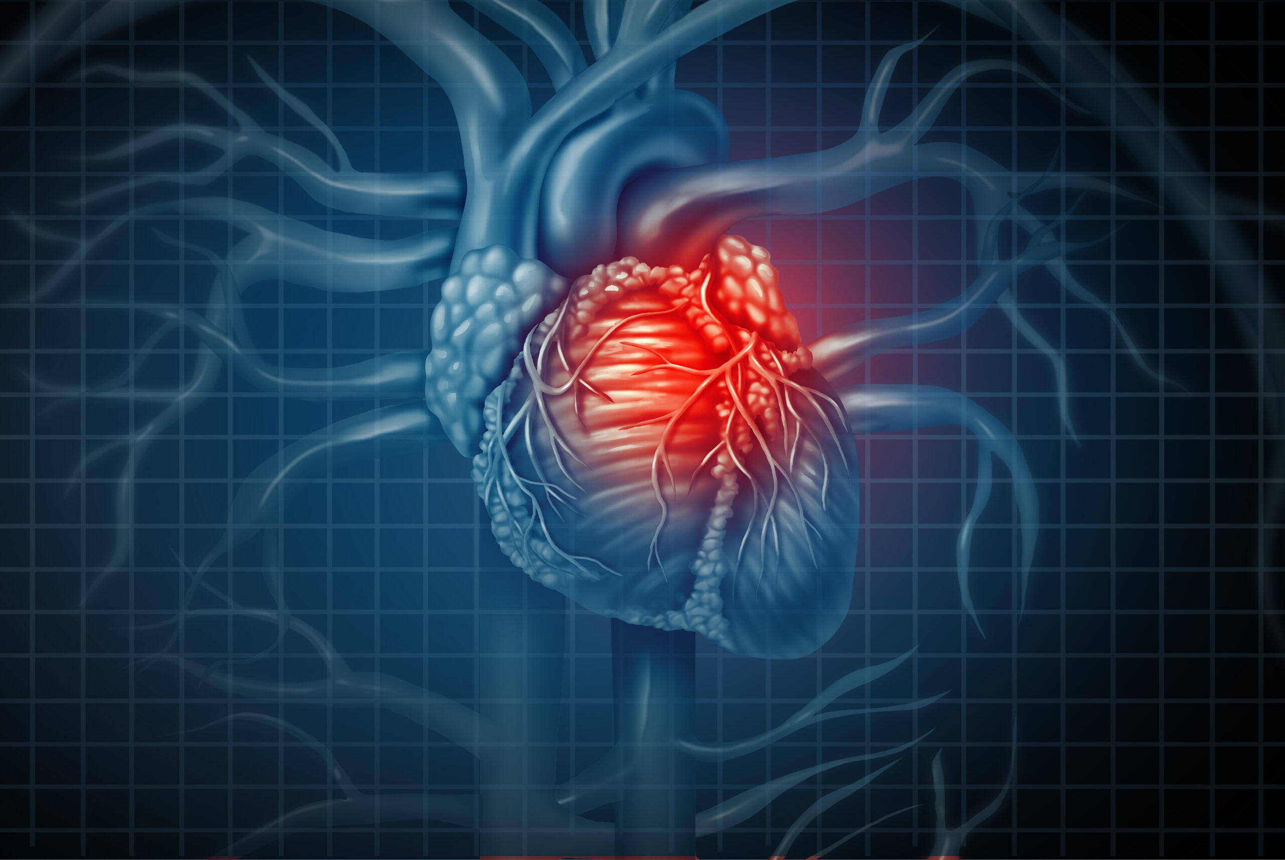
Cardiomyopathy is a term describing a disease that affects the cardiac muscle, also known as the myocardium. All cardiomyopathies are characterized by morphological and/or functional changes in the myocardium. There are several types of cardiomyopathies including:
How many have cardiomyopathies?
DCM and HCM are the most common cardiomyopathies with a prevalence of about 1 in 250 for DCM and 1 in 500 for HCM. ARC has a prevalence of about 1 in 5000, whereas RCM and LVNC are classified as rare. According to the GBD 2020 study from the University of Washington, 6.11 million (95% UI, 5.02–7.22 million) prevalent cases of cardiomyopathy were reported in 2020 on a global scale.
How are cardiomyopathies treated?
Cardiomyopathies are considered in principle inherited disorders, although some phenotypes are seen in both genetic and non-genetic contexts. That means that treatment focuses more on delaying disease progression and managing other associated complications rather than treating the genetic cause. Treatment depends on the severity of the patient and can vary from lifestyle changes, medical therapy, surgical myectomy, implantable defibrillators, heart transplantation, and others. Cardiomyopathies with no genetic background are often milder and easier to manage.
How are cardiomyopathies diagnosed?
Cardiomyopathies are clinically diagnosed and managed with the use of genetic testing, assessment of clinical symptoms, electrocardiogram (ECG) measurements, and cardiac imaging to detect functional, structural, and morphological alterations.
In hypertrophic cardiomyopathy, diagnosis is performed with ECG as a first screening, followed by echocardiography or magnetic resonance imaging (MRI). The main diagnostic characteristic is ECG abnormalities caused by a characteristic left ventricular hypertrophy ≥1.5 cm (LVH). In dilated cardiomyopathy, diagnosis is performed based on echocardiographic, computer tomography (CT), or MRI evidence of LV enlargement and impaired systolic function. In arrhythmogenic cardiomyopathy, diagnosis is performed by assessing the size of the right ventricle normalized to body surface area and less frequently with right ventricular endomyocardial biopsy. Other criteria include the presence of epsilon waves, left bundle morphology ventricular tachycardia, and premature ventricular contractions >500 in 24 h. In restrictive cardiomyopathy, diagnosis is performed with hemodynamic or echocardiographic Doppler, and the main diagnostic characteristic is restrictive ventricular filling, with normal systolic ventricular function, normal or reduced end-diastolic ventricular dimensions, and absence of LVH. Although detailed imaging is helpful endomyocardial biopsy is necessary to create causality in RCM, especially for non-genetic forms. In left ventricular non-compaction cardiomyopathy, diagnosis is performed with higher-resolution echocardiography, CT, and MRI, and the main diagnostic characteristic is hyper trabeculation of the LV apex. According to the latest guidelines, LVNC is considered a common cardiac trait that sometimes occurs with various forms of cardiomyopathy, and which also occurs in people without cardiomyopathy.
Search and find publications that we have published.
Please don't hesitate to contact us if you have any questions or other inquiries.