Most biomarker candidates die in the valley of death. What made PRO-C3 different?

Biomarker discovery has accelerated dramatically, but clinically viable products rarely follow. Thousands of protein signals are associated with disease every year, yet, relatively few become reliable tools for clinical development or patient management. Many stall in the biomarker valley of death: the long and resource-intensive space between initial discovery and validated clinical utility.

High-throughput omics and proteomics can identify thousands of disease-associated signals. But an association alone does not make a measurement an actionable biomarker. Biological specificity, analytical performance, clinical evidence and a clearly defined Context of Use must follow.

A new expert opinion in the Journal of Hepatology (IF 40 as of August 2026) proposes a higher standard. While the paper takes the liver field as an example, it’s findings are applicable to most diseases. It introduces the concept of the unicorn biomarker: a rare biomarker with diagnostic, prognostic and pharmacodynamic utility, supported by rigorous technical, clinical and regulatory development. PRO-C3, invented by Nordic Bioscience, is highlighted as one of the potential unicorn biomarkers approaching this standard. 

Most candidates die in the biomarker valley of death
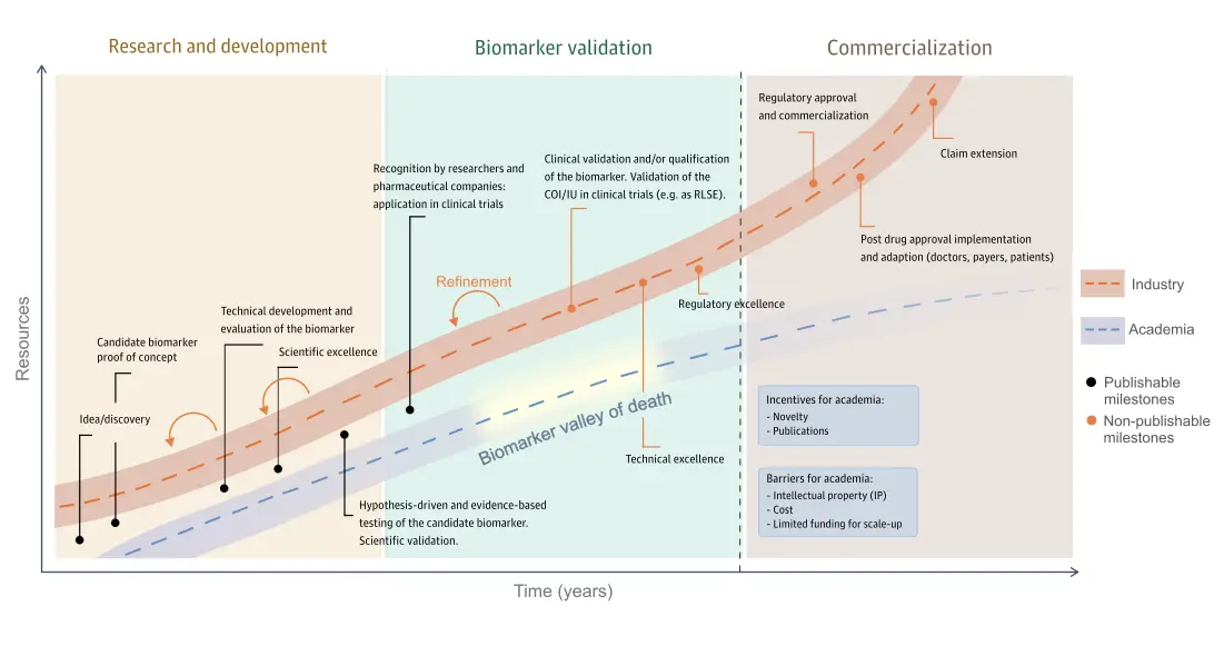
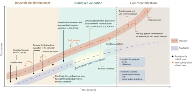
Fig.1 The path to becoming a unicorn biomarker consisting of three stages: Research and development, biomarker validation, and commercialization.

Discovering a promising protein signal is only the beginning. To progress toward clinical utility, a candidate must undergo technical development, scientific and clinical validation, regulatory alignment and, ultimately, implementation on a reliable and scalable platform. Many candidates never progress beyond discovery or early validation, while others stall when the cost and technical requirements increase.

The journey begins with research and development, where a biomarker is discovered through scientific hypothesis testing. Most biomarkers remain in research and development, as advancing further requires investment in technical excellence and quality control. The second stage is the biomarker validation stage where biomarkers are validated in clinical trials and technical performance on the final assay design is rigorously tested. This phase is often called the “biomarker valley of death” because the work is costly, technically demanding, and rarely publishable, causing many candidates to stall.

The final stage is commercialization, which focuses on achieving regulatory approval for specific contexts of use (CoUs) or intended uses (IUs) and implementing marketing strategies to generate a return on the investment. Here, academia and industry collaborate to validate, adopt, market, and implement the biomarker for clinical applications.

What does it take to become Unicorn biomarker?
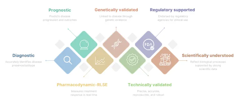
Fig. 2 An outline of the requirements a unicorn biomarker has to fulfill.

Unicorn biomarkers are rare and valuable, as they must fulfill various requirements (Fig. 3). Unlike biomarkers that serve a single purpose, they provide value across multiple stages of research and clinical development. They combine diagnostic utility, prognostic utility and pharmacodynamic utility, which is a combination of requirements that can be challenging because the criteria often is conflicting. While diagnostic and prognostic biomarkers must reliably reflect stable disease characteristics, pharmacodynamic biomarkers need to be sufficiently sensitive and dynamic to detect treatment-induced changes.

Beyond clinical utility, a unicorn biomarker must also be scientifically understood, technically validated to ensure reliable and reproducible results, and regulatory supported to enable its use in clinical development and, ultimately, patient care. Only when these scientific, technical, clinical, and regulatory criteria come together can a biomarker be considered a true unicorn biomarker.

Why the exact protein measurement matters

Proteins exist in different fragments, modified forms, or structural regions, each reflecting different biological processes. As a result, two measurements of the same protein may not necessarily provide the same clinical or biological insight if they target different parts of the molecule.

This is why the exact molecular target matters in biomarker development. The value of a biomarker does not only depend on identifying the right protein, but on measuring the right part of that protein in the right biological and clinical context.
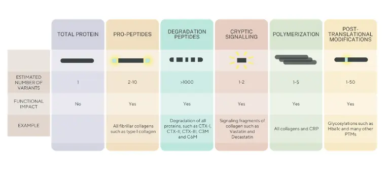
Fig. 3 The structural complexity of proteins and implications for quantification.

This is the principle behind our Nordic ProteinFingerPrint Technology™. By targeting specific protein fragments generated during extracellular matrix remodeling, the technology is designed to measure defined biological processes rather than total protein abundance alone. This enables a more precise understanding of whether tissue is being formed, degraded, or modified by disease-related enzyme activity, helping researchers connect biomarker measurements to disease mechanisms, progression, and treatment response.

PRO-C3: Journey toward Unicorn Biomarker Status

One example approaching this standard is nordicPRO-C3™. PRO-C3 detects a defined neo-epitope generated during type III collagen formation, linking the measurement to active fibrogenesis rather than total collagen abundance.

Its development has extended well beyond initial discovery. PRO-C3 has demonstrated diagnostic utility as part of the ADAPT score, prognostic value in fibrotic disease and pharmacodynamic utility for measuring treatment-related changes in fibrosis activity. It has also been used extensively in pharmaceutical clinical development.

The launch of Elecsys PRO-C3 by Roche Diagnostics transferred the measurement onto the globally established cobas platform as an IVD-labelled assay. This progression from a biologically defined neo-epitope to a clinically studied and commercially deployed assay is why the paper identifies PRO-C3 as a potential unicorn bomarker.

Why this matters for drug developers

Unicorn biomarkers are valuable to drug developers because they link biological mechanisms with measurable treatment effects. When associated to a clear Context of Use, they can support patient enrichment by identifying individuals with relevant disease activity, disease stage, or risk of progression. This can reduce heterogeneity in trials and improve the ability to detect a meaningful treatment effect.

Their pharmacodynamic utility is especially relevant to drug developers, as they can provide earlier evidence that a therapy is affecting the intended biological pathway before traditional clinical outcomes become visible. This can support proof-of-mechanism, dose selection, treatment monitoring, and go/no-go decisions, helping developers make more informed choices before moving into larger and more costly studies. By providing earlier and more mechanistic grounded information, they can reduce uncertainty, improve trial design, and help lower the risk of late-stage failure.

By combining diagnostic, prognostic, and pharmacodynamic utility with strong scientific, technical, and clinical validation, unicorn biomarkers represent an aspirational standard for biomarker development.

PRO-C3 is not an isolated discovery. It represents Nordic Biosciennce’s approach to biomarker development: begin with biologically meaningful protein fragments and continue through the technical, clinical and regulatory work required to make the measurement useful. We build biomarkers to cross the biomarker valley of death.

Myths and misconceptions about biomarkers

  • While some biomarkers may be subject to biological variability, many others such as NT-proBNP for heart failure or prostate-specific antigen (PSA) for prostate cancer have well-characterized reference ranges and robust assay reproducibility. In fact, standardization and quality control measures have significantly improved the reliability of many biomarker assays.

  • Although oncology has led the way, biomarkers are now well-established in many fields, including cardiology (e.g., troponin for myocardial infarction), hepatology (e.g., ELF or FIB-4 for liver fibrosis) [67], diabetes (HbA1c) and rheumatology (e.g., anti-CCP for rheumatoid arthritis). Their utility spans diagnosis, prognosis, and treatment monitoring across multiple disease domains.

  • While regulatory-grade validation is ideal for clinical implementation (particularly for RLSE or VSE CoUs), exploratory biomarkers can and should be incorporated into early-phase trials to guide hypothesis generation and decision-making and generating data that will enable eventual clinical validation for regulatory use later. Waiting for full validation can delay insights into their potential link to drug MoA or clinical efficacy and hinder development [81]. Further, the use of a biomarker during pre-clinical phases of drug development increases the likelihood of final regulatory approval from 8% to 25%.

  • Although biomarker assays introduce additional costs, these are often small compared with the overall expense of clinical trials. Biomarkers can support clinical development by providing mechanistic insights into treatment response and disease biology, thereby informing critical decisions related to efficacy, safety, and proof of mechanism. As a result, biomarkers may contribute to more efficient and informative clinical trials.
  • When biomarkers have a defined CoU, including their role, target population, and setting, they streamline rather than complicate regulatory pathways. In fact, the FDA’s BEST Resource provides a structured framework for qualifying biomarkers.

Discuss your biomarker strategy

Explore how biologically specific biomarkers could support patient enrichment, proof of mechanism, treatment monitoring or development decisions in your clinical program.

    Early-onset CRC is rising fast. The fibrotic barrier may explain why treatments fail

    A recent analysis published in JAMA highlights a major epidemiological shift: Colorectal cancer (CRC) is now the leading cause of cancer-related death among adults under 50 in the United States.

    CRC has long been perceived as a disease of older populations. The rapid rise in early-onset cases challenges that assumption and reinforces the need to rethink both biology and treatment strategies.

    Why standard therapies still fall short: the fibrotic stroma as a resistance mechanism

    Therapeutically, the backbone remains surgery in localized disease, combination chemotherapy regimens such as FOLFOX and FOLFIRI, anti-VEGF or anti-EGFR targeted therapies in selected patients, and immunotherapy in MSI-high/dMMR tumors. While these approaches have extended survival, resistance and relapse remain frequent, particularly in advanced disease.

    One key and often underestimated driver of progression and resistance is fibrosis. CRC tumors are embedded in a collagen-rich, fibrostenotic microenvironment shaped by cancer-associated fibroblasts (CAFs). This dense extracellular matrix (ECM) does not merely provide structure. It represents a desmoplastic barrier that limits drug penetration, reduces treatment efficacy, and supports tumor progression.

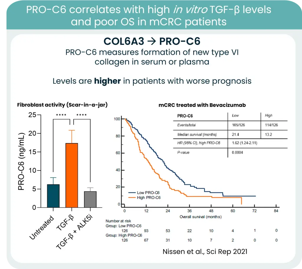
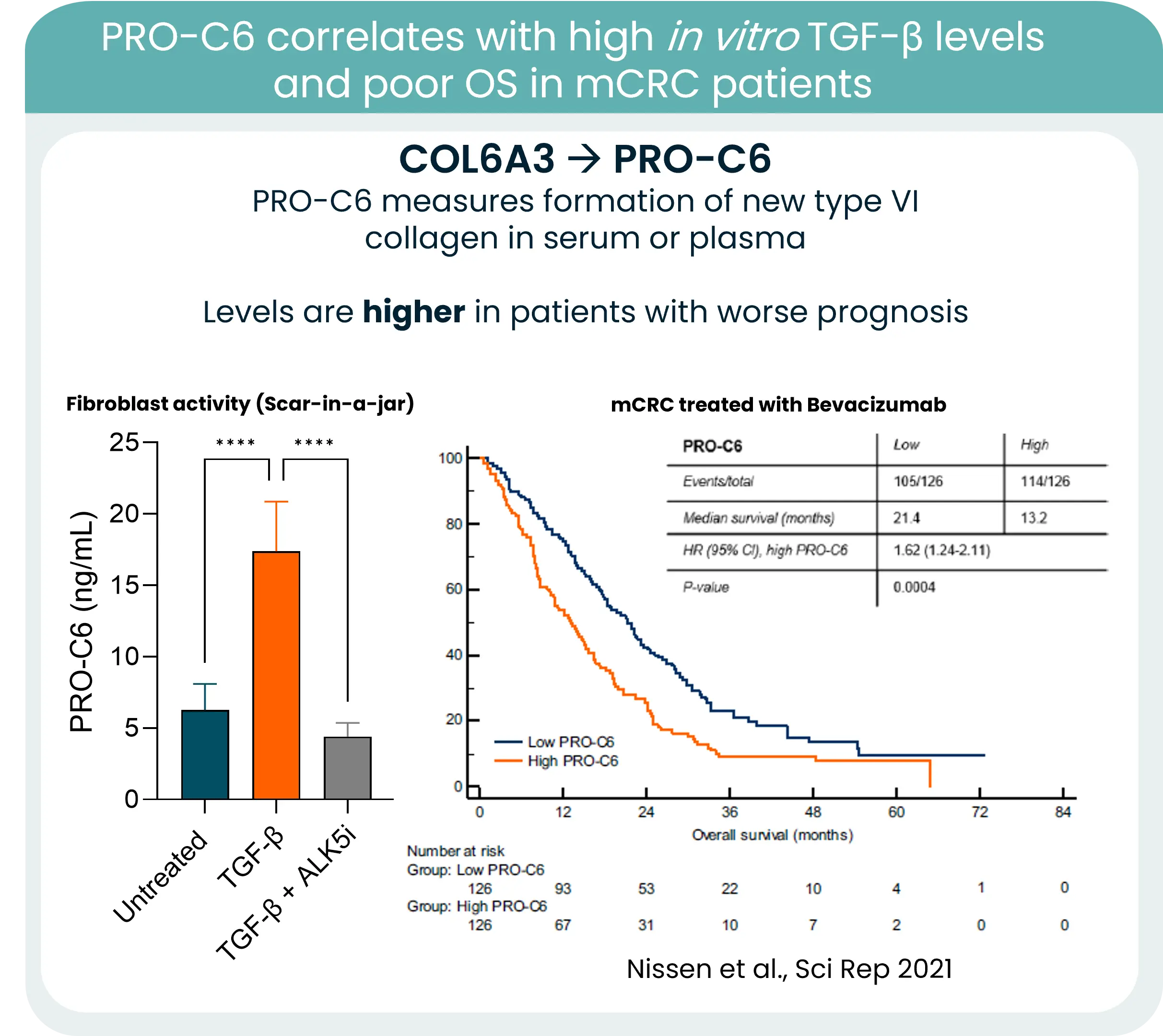
    PRO-C6 correlates with TGF-β levels and overall survive in mCRC patients

    From biology to measurement: collagen remodeling biomarkers

    Collagens are central components of this barrier, and increased collagen deposition and remodeling consistently associate with poorer outcomes. One notable example is type VI collagen. Its gene expression is strongly associated with KRAS mutations and high TGF-β levels in CRC. New type VI collagen formation releases protein fragments to the bloodstream, which can be quantified in serum using our PRO-C6 biomarker and correlate to high TGF-β levels and poor OS.

    In this context, measuring collagen formation and degradation becomes highly relevant. Quantifying ECM remodeling can provide insight into tumor aggressiveness, stromal activation, and potentially treatment response.

    As early-onset CRC continues to rise, integrating stromal blood-based biomarkers into clinical strategy may refine risk stratification and open new opportunities for stroma-targeted interventions.

    The future of CRC management will not rely solely on targeting tumor cells. It will require understanding and measuring the fibrotic environment that sustains them.

    α-synuclein—a critical protein in Parkinson’s disease

    Understanding Parkinson’s Disease Progression Through Protein Biomarkers

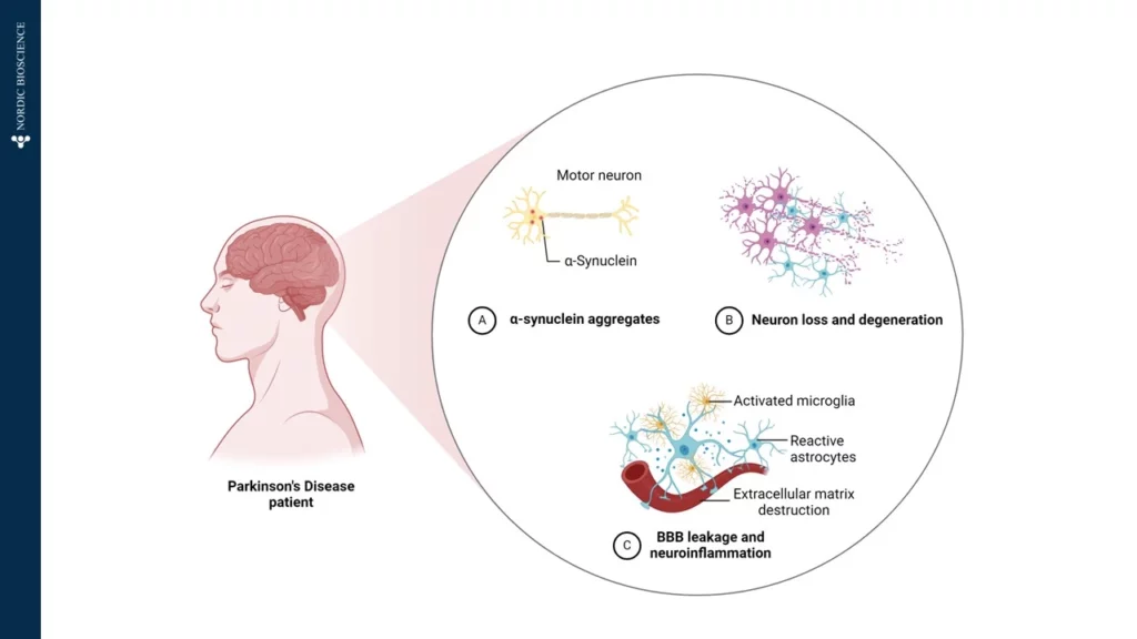
    Parkinson’s Disease (PD) is a complex neurodegenerative disorder, primarily affecting the brain’s control over movement, thought, memory, and emotion. Early symptoms often manifest as tremors due to impaired motor skills. Underlying these visible symptoms is a cascade of molecular changes, beginning with alterations in specific proteins—one of which, α-synuclein, plays a critical role in PD pathology. In Figure 1, we have illustrated how α-Synuclein aggregates, which impairs the motor neuron.

    Figure 1. Patients diagnosed with Parkinson’s Disease is affected by A) α-Synuclein aggregates affecting the motor neurons, B) Neuron loss and degeneration, and C) Blood-Brain-Barrier (BBB) leakage and neuroinflammation, by activated microglia, reactive astrocytes and extracellular matrix destruction.

    In a healthy brain, α-synuclein supports neuronal communication. However, in Parkinson’s, this protein undergoes abnormal processing, driven partly by the enzyme Calpain-1, which cleaves α-synuclein into smaller, altered fragments. This early cleavage disrupts cellular function and promotes the formation of toxic aggregates, which accumulate, kill neurons, and drive disease progression. Intriguingly, these fragmented proteins can cross the blood-brain barrier and enter the bloodstream, providing a potential “window into the brain” for tracking disease activity from a simple blood sample.

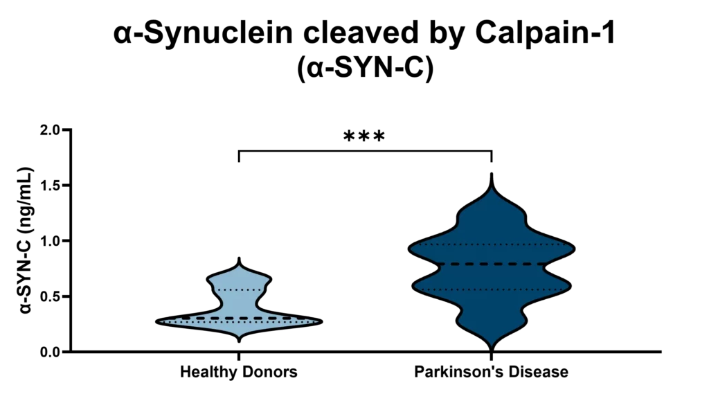
    At Nordic Bioscience, we have developed an innovative approach to harness this biomarker potential. Using our ProteinFingerPrint Biomarker Technology™, we can detect Calpain-1-cleaved α-synuclein fragments in blood serum with high precision. Our specific assay, α-SYN-C, captures the unique “fragment fingerprint” of PD by quantifying these cleaved fragments, which are significantly elevated in the blood of PD patients compared to healthy individuals, as illustrated in Figure 2.

    Figure 2. Patients with Parkinson’s Disease has significant higher levels of α-SYN-C in serum, compared to healthy donors. The α-SYN-C biomarker detects levels of α-Synuclein cleaved by Calpain-1 in serum. The assay is technically validated for measurements in human blood samples.

    This biomarker offers a non-invasive, accessible tool for monitoring Parkinson’s Disease progression and evaluating therapeutic responses. By examining α-SYN-C levels in blood samples, our technology not only provides insights into PD mechanisms but also opens doors for developing targeted therapies that address the disease’s underlying pathology. Through this work, we aim to support more accurate PD diagnostics and more effective, individualized treatments in the fight against neurodegeneration.

    Get in touch

    Are you interested in exploring collaboration possibilities?
Enter your information in the form and a representative will contact you shortly.

      NordicPro-C3™ predictor of liver disease and alcohol-related liver disease

      Liver fibrosis is a progressive condition characterized by the excessive accumulation of scar tissue in the liver, often resulting from chronic liver diseases. Fibroblast activity plays a critical role in the progression of fibrosis, leading to organ function loss and liver-related complications. Liver fibrosis accounts for approximately 2 million deaths worldwide, while alcohol-related liver disease (ArLD) causes over 330,000 cirrhosis deaths globally every year.

      In two recent studies, we investigated the fibrogenesis biomarker nordicPRO-C3™ in the context of advanced liver disease and full-spectrum fibrosis in alcohol-related liver disease. Our goal was to assess the nordicPRO-C3™ biomarker’s potential prognostic value and clinical utility as a predictor of outcome, and our results are very promising.

      The nordicPRO-C3™ biomarker is capable of identifying advanced liver fibrosis and alcohol-related liver disease, providing a new clinical utility tool

      In the first paper, published in JHEP Reports, we conducted investigations using two distinct cohorts of patients with compensated cirrhosis of mixed etiologies. In both cohorts, a 2-fold increase in nordicPRO-C3™ at baseline was associated with a significant hazard increase for liver-related events. Notably, nordicPRO-C3™ exhibited prognostic significance.

      The identification of nordicPRO-C3™ as an independent prognostic factor for liver-related clinical outcomes has important implications for both drug development and clinical practice. By understanding the dynamic range of nordicPRO-C3™, researchers can improve its utility as a predictive marker in drug trials, enabling the development of targeted therapies to mitigate fibrosis progression and improve patient outcomes.

      In a clinical setting, the integration of nordicPRO-C3™ measurement alongside established markers such as fibrosis-4 index (FIB-4) or transient elastography (TE) may enhance risk stratification and inform treatment decisions. Early identification of patients at higher risk for liver-related events can facilitate timely interventions, such as disease monitoring, lifestyle modifications, and appropriate therapeutic interventions.

      In the second paper, published in Liver International, we assessed nordicPRO-C3™ models to predict liver-related events in patients with a history of excessive alcohol use but without a confirmed diagnosis of chronic liver disease. Our findings our promising for improving risk stratification and clinical outcomes in alcohol-related liver disease.

      A prospective cohort study involving patients with alcohol-related Liver Disease (ArLD), was conducted and divided into a derivation cohort of secondary care patients and a validation cohort of primary care patients. Baseline variables, including nordicPRO-C3™, were utilized to develop a prediction model known as the ALPACA score. The prognostic accuracy of the ALPACA score was compared to existing tools such as the enhanced liver fibrosis (ELF) test, FIB-4, TE, and the ADAPT score (developed for fatty liver disease).

      The ALPACA score demonstrated excellent discriminative accuracy in both the derivation and validation cohorts, comparable to TE and the ELF test, and superior to FIB-4, nordicPRO-C3™ alone, and the ADAPT score. Importantly, the ALPACA score provided reliable prognostic performance independent of the baseline fibrosis stage. This breakthrough has the potential to revolutionize risk stratification and patient management in primary and secondary care settings.

      NordicPRO-C3™ has recently become available on the Roche-COBAS high-precision platform, paving the way for its commercial availability worldwide in the near future. This accessibility will enable widespread adoption of nordicPRO-C3™-based scores, leading to improved risk prediction and better outcomes for individuals.

      Get in touch

      Are you interested in exploring collaboration possibilities?
Enter your information in the form and a representative will contact you shortly.

        Serological biomarkers are different

        Biomarkers are more than just omics categories

        Biomarkers aren’t just confined to different omics categories; various classes are referring to the types of serological measurements.

        Consider cytokines, growth factors, receptors, kinases, transcription factors, intracellular proteins, extracellular proteins, and a subset within that—Extracellular Matrix (ECM) proteins. The common denominator in most pathologies is a loss of balance between different ECM proteins, especially collagens.

        Biomarkers are more than just omics categories

        This excessive destruction and deposition of proteins significantly propel the progression of end-stage diseases, culminating in organ dysfunction, failure, and, ultimately, death.

        Diverse cytokines and growth factors orchestrating via specific receptors and kinases lead to ECM destruction and deposition, making ECM the converging pathway for multiple stimuli.

        By the end of the day, what matters is to reverse ECM deterioration, or even undo, organ damage and function decline. Reversing organ damage can be achieved by stopping ECM deterioration. To truly reverse organ damage and restore organ functionality in patients, we need to repair the ECM to its normal balance.

        We at Nordic Bioscience believe that the answer lies in effecting change at the tissue level—the key to reversing organ damage and, in turn, revitalizing organ function.

        Have you considered if your treatment or pathway is affecting tissue formation or degradation? If so, feel free to browse our unique ECM biomarker portfolio.

        Get in touch

        Are you interested in exploring collaboration possibilities?
Enter your information in the form and a representative will contact you shortly.

          The CPa9-HNE (NordicCPa9™) biomarker in Crohn’s and Colitis

          CPa9-HNE ELISA has emerged as a novel serum calprotectin biomarker

          Status quo: Crohn’s and Colitis biomarkers

          Conventional serum calprotectin biomarkers are often not as clinically useful as the fecal versions, which is related to the half-life of calprotectin in blood (only 5-6 hours), leading to dissociation of calprotectin protein. The short half-life of calprotectin in blood reduces the window in which the current serum calprotectin ELISA assay can detect calprotectin dimer protein, which is composed of monomers S100A8 and S100A9.

          By applying the Protein Fingerprint technology, we have identified a neo-epitope of the S100A9 monomer of calprotectin derived from proteolytic cleavage by human neutrophil elastase (HNE). This ELISA assay is referred to as the CPa9-HNE (nordicCPa9™) assay and is a new and innovative method for quantification of calprotectin in serum developed by Nordic Bioscience.

          Inflammatory Bowel Disease (IBD) flares in the intestines.

          A new tool to measure… calprotectin

          Patients with Crohn’s disease and ulcerative colitis have been found to show significantly higher levels of CPa9-HNE (nordicCPa9™) in their serum than healthy subjects, with the AUC reaching 0.98 (CI: 0.97-1.00, p<0.0001) for CD and 0.96 (CI: 0.92-1.00, p<0.0001) for UC, proving the assay’s efficacy in distinguishing between disease states.

          In comparison, the MRP8/14 serum calprotectin assay from Bühlmann Lab, while also used to identify IBD, showed poorer separation between patients and healthy subjects, with AUCs of 0.72 (CI: 0.59-0.86, p=0.0025) for UC and 0.70 (CI: 0.56-0.84, p=0.0046) for CD, respectively.

          The nordicCPa9™ biomarker showed a better correlation with the endoscopic score for Crohn’s disease (SES-CD) and ulcerative colitis (MES) than the FCP. At the same time, the MRP8/14 assay and neutrophil count showed no significant correlation with endoscopic scores for IBD patients.

          To investigate the accuracy of the nordicCPa9™, we performed a received operator characteristic curve, demonstrating nordicCPa9™ with acceptable accuracy to identify patients with IBD in remission vs. active disease.

          Finally, we demonstrated that the nordicCPa9™ biomarker could detect the calprotectin neo-epitope only in the supernatant of in vitro activated primary human neutrophils, but not in inactive primary human neutrophils. This was in contrast to the MRP8/14 serum CP assay of the Bühlmann lab, as both inactive and activated primary human neutrophils secreted detectable levels of calprotectin.

          NordicCPa9™ ELISA proved to be a novel serum calprotectin biomarker with significant clinical potential as a biomarker for patients with IBD to monitor disease activity and neutrophil activity.

          However, circulating calprotectin metabolites released from locally inflamed mucosa are not be dissociated further. They can be readily quantified using an ELISA-based technique called Protein Fingerprint technology. This refined ELISA technique quantifies protein metabolites or neo-epitopes derived from proteolysis that reflect local tissue inflammation and remodeling.

          Read the full article here.

          Get in touch

          Are you interested in exploring collaboration possibilities?
Enter your information in the form and a representative will contact you shortly.

            NEJM Evidence study shows PRO-C6 to Segregate HFpEF

            PRO-C6 Findings In NEJM Evidence to Help Patient Segregation in HFpEF

            A persistent problem remains a challenge in HFpEF – patient heterogeneity.

            We need the right patients for the right treatment, but how do we get it to them? We believe that one approach to solving this problem is to improve patient segregation and endotyping.


            In collaboration with Bristol Myers Squibb and University of Pennsylvania, we recently identified a subset of patients at a very high risk of adverse outcomes, all characterized by increased fibroblast activity. Using our PRO-C6 biomarker assay we can accurately quantify this risk profile and differentiate patients based on how likely they are to be re-hospitalized or have all-cause mortality due to heart failure.

            These findings were recently published in NEJM Evidence.

            So how do we deal with patient heterogeneity?

            We believe it is time to give physicians and clinicians a better tool than what is available now.

            This is endotrophin endotyping, heart failure risk stratification redefined.

            Get in touch

            Are you interested in exploring collaboration possibilities?
Enter your information in the form and a representative will contact you shortly.

              Biomarkers in Drug Development

              The Protein Fingerprint Technology

              Through our Protein Fingerprint technology, we identify fragments of the Extracellular Matrix (ECM) that are released from affected tissues and develop blood-based biomarker assays to quantify disease activity with the aim of precision medicine.

              By quantifying tissue remodeling, our biomarkers provide true prognostic and predictive value, as we can select the patients who are most likely to respond to a given treatment and thereby function as surrogate biomarkers for clinical trials. Our biomarker assays not only enable faster drug development but also improve disease outcomes for chronically ill patients by predicting treatment response.

              Watch more videos about Nordic Bioscience’s biomarkers and the ECM on our YouTube channel!

              Get in touch

              Are you interested in exploring collaboration possibilities?
Enter your information in the form and a representative will contact you shortly.

                Use of Translational Biomarkers in Pulmonary Fibrosis

                PRO-C6 as a pharmacodynamic biomarker

                During the development of pulmonary fibrosis, there are changes in the extracellular matrix. We see the destruction of the basement membrane and the formation of various collagens in the interstitial matrix.

                We can monitor these changes in the ECM in a blood sample using Protein Fingerprint biomarker technology.

                Watch more videos about the extracellular matrix and the Protein Fingerprint on our YouTube channel!

                Get in touch

                Are you interested in exploring collaboration possibilities?
Enter your information in the form and a representative will contact you shortly.

                  Endotrophin as a Novel Biomarker in HFpEF – Identification of a High-risk Endotype

                  Identification of a High-risk Endotype

                  Heart failure with preserved ejection fraction (HFpEF) is a highly complex and heterogeneous syndrome that causes substantial morbidity and mortality, positioning this disease as one of the biggest unmet medical needs today.

                  The past years have seen great scientific and clinical efforts on elucidating the driving mechanisms, but much remains unknown and successful therapies at still largely absent.

                  One of the biggest challenges to treating HFpEF is the heterogeneous nature of the syndrome. Risk assessment by conventional biomarkers has not led to any significant therapies, but selection of a more homogenous patient population might be the way forward.

                  Fibrosis is a hallmark feature of HFpEF pathology, acting as a driver of outcome. Understanding the intricate pathways that lead to fibrogenesis, and how they ultimately impact pathogenesis, is important for understanding how to treat HFpEF. We have developed a biomarker of fibroblast activity, PRO-C6, which independently and with high precision can identify a subset of HFpEF patients with a very elevated risk of adverse outcome.

                  Watch more videos about Nordic Bioscience’s biomarkers and technology on our YouTube channel!

                  Get in touch

                  Are you interested in exploring collaboration possibilities?
Enter your information in the form and a representative will contact you shortly.