Biomarkers of Extracellular Matrix Remodeling Reflect Pharmacodynamic Effects on IMU-856, an Oral Epigenetic Modulator of Barrier Regeneration
Introduction
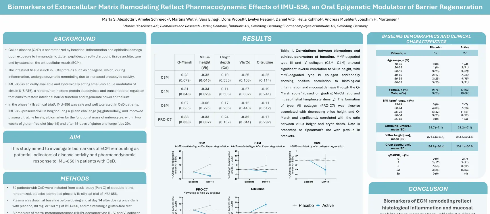
Celiac disease (CeD) is characterized by intestinal inflammation and epithelial damage upon exposure to immunogenic gluten peptide, directly disrupting tissue architecture and by extension the extracellular matrix (ECM). The intestinal tissue is rich in ECM proteins such as collagens, which, during inflammation, undergo enzymatic remodeling due to increased proteolytic activity.
IMU-856 is an orally available and systematically acting small-molecule modulator of sirtuin 6 (SIRT6), a histone/non-histone protein deacetylase and transcriptional regulator that aims to restore intestinal barrier function and regenerate bowel epithelium.
This study aimed to investigate biomarkers of ECM remodeling as potential indicators of disease activity and pharmacodynamic response to IMU-856 in patients with CeD.
Biomarkers of ECM remodeling reflect histological inflammation and mucosal architecture parameters, offering a direct insight into intestinal barrier integrity. These biomarkers potentially reflect treatment-induced improvement in intestinal tissue remodeling upon treatment with IMU-856.
Get in touch
Are you interested in exploring collaboration possibilities? Enter your information in the form and a representative will contact you shortly.
How can a simple blood sample track the progression of Parkinson’s disease?
Parkinson’s disease is a neurological condition that affects the brain and other parts of the nervous system. This animation explores the complex neurodegenerative processes occurring within the brain’s substantia nigra and the groundbreaking role of alpha-synuclein fragments as biomarkers.
Watch this video to learn about the fragmentation of alpha-synuclein, the breakdown of the blood-brain barrier, and how blood-based biomarkers can help identify patients at risk of progression and track treatment response.
Get in touch
Are you interested in exploring collaboration possibilities? Enter your information in the form and a representative will contact you shortly.
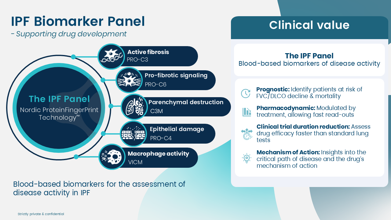
Extracellular matrix (ECM) remodeling is a central driver of disease progression in idiopathic pulmonary fibrosis (IPF) and other ILDs. At the cellular level, these remodeling processes ultimately lead to the decline in lung function seen in patients.
For drug developers, this means that modifying ECM remodeling is a key therapeutic objective.
But to evaluate whether novel compounds are working, we need reliable tools that measure the biological processes driving the disease.
We have developed a panel of ECM remodeling biomarkers based on our ProteinFingerprint Technology™ and 30 years of biomarker expertise.
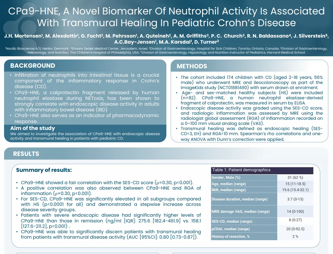
NordicCPa9™, a biomarker of neutrophil activity is associated with transmural healing in pediatric Crohn’s disease
Introduction
CPa9-HNE (nordicCPa9™), a calprotectin fragment released by human neutrophil estate during NETosis, has been shown to strongly correlate with endoscopic disease activity in adults with inflammatory bowel disease (IBD). NordicCPa9™ also serves as an indicator of pharmacodynamic response. This study aimed to investigate the association of nordicCPa9™ with endoscopic disease activity and transmural healing in patients with pediatric CD.
This study showed that nordicCPa9™, a biomarker of neutrophil activity, is associated with mucosal and radiologic mucosal damage in pediatric CD. These findings highlight the potential use of CPa9-HNE as a complementary tool for the endoscopic or radiographic assessment of intestinal mucosal damage/healing and provide an opportunity for further studies in children with CD.
Get in touch
Are you interested in exploring collaboration possibilities? Enter your information in the form and a representative will contact you shortly.
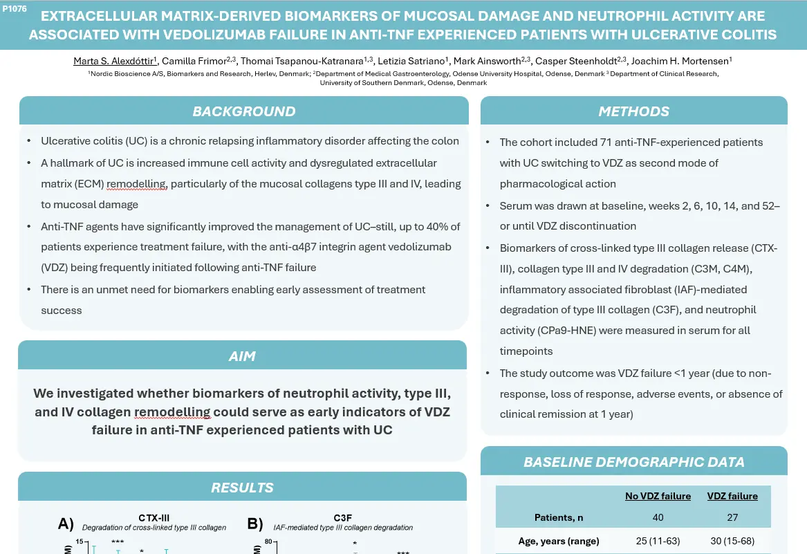
ECM-derived biomarkers of mucosal damage and neutrophil activity are associated with the failure of VZD
Introduction
Ulcerative colitis (UC) is a chronic relapsing inflammatory disorder affecting the colon. A hallmark of UC is increased immune cell activity and dysregulated extracellular matrix remodeling, particularly of the mucosal collagens type III and IV, leading to mucosal damage. Anti-TNF agents have significantly improved the management of UC-still, up to 40% of patients experience treatment failure, with the anti-α4β7 integrin agent vedolizumab (VDZ) being frequently initiated following anti-TNF failure. This presents the unmet need for biomarkers enabling early assessment of treatment success.
This study investigates whether biomarkers of neutrophil activity, type III, and IV collagen remodeling could serve as early indicators of VDZ failure in anti-TNF experienced patients with UC.
The early increase in CTX-III, reflecting fibrosis resolution, was associated with the absence of VDZ failure, whereas increased neutrophil activity (CPa9-HNE), IAF-mediated collagen type III degradation (C3F) and mucosal damage (C3M, C4M) were associated with the failure of VZD. These findings suggest that biomarkers reflecting neutrophil activity and inflammatory events in the mucosa may serve as promising tools for the early and dynamic assessment of later VDZ treatment outcomes in anti-TNF experienced patients with UC.
Get in touch
Are you interested in exploring collaboration possibilities? Enter your information in the form and a representative will contact you shortly.
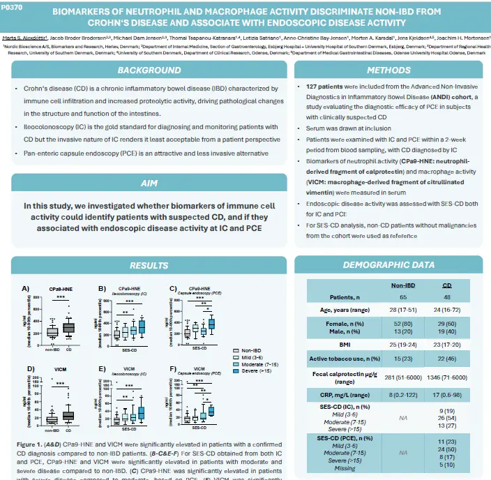
Biomarkers of neutrophil and macrophage activity discriminate non-IBD from CD and associate with endoscopic disease activity
Introduction
Crohn’s disease (CD) is a chronic inflammatory bowel diseases (IBD) characterized by immune cell infiltration and increased proteolytic activity, driving pathological changes in the structure and function of the intestines. Ileocolonoscopy (IC) is the gold standard for diagnosing and monitoring patients with CD but the invasive nature of IC renders it as least acceptable from a patient perspective. Pan-enteric endoscopy (PCE) is an attractive and less invasive method.
This study aimed to investigate whether biomarkers of immune cell activity could identify patients with suspected CD, and if they associated with endoscopic disease activity at IC and PCE.
CPa9-HNE [neutrophil activity] and VICM [macrophage activity] could identify patients with confirmed CD and were associated with endoscopic disease activity. Both biomarkers provided stronger discriminative performance when assessing disease activity by utilizing PCE. Biomarkers of neutrophil and macrophage activity may therefore provide additonal information to endoscopic assessment in the diagnosis and monitoring of CD.
Get in touch
Are you interested in exploring collaboration possibilities? Enter your information in the form and a representative will contact you shortly.
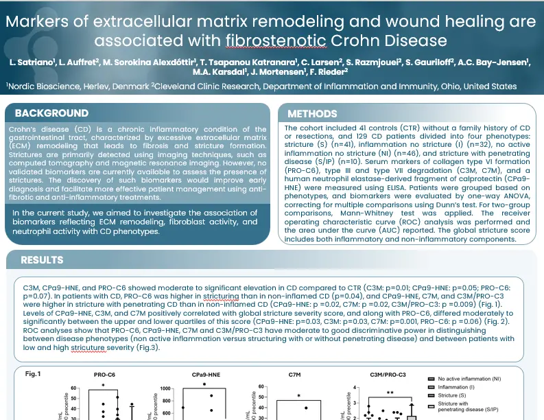
Markers of extracellular matrix remodeling and wound healing are associated with fibrostenotic Crohn’s disease
Introduction
Crohn’s disease (CD) is a chronic inflammatory condition of the gastrointestinal tract, characterized by excessive extracellular matrix (ECM) remodeling that leads to fibrosis and stricture formation. Strictures are primarily detected using imaging techniques, such as computed tomography and magnetic resonance imaging. However, no validated biomarkers are currently available to assess the presence of strictures. The discovery of such biomarkers would improve early diagnosis and facilitate more effective patient management using anti-fibrotic and anti-inflammatory treatments.
This study aimed to investigate the association of biomarkers reflecting ECM remodeling, fibroblast activity, and neutrophil activity with CD phenotypes.
The fibrosis marker (PRO-C6) was associated with stenosis and was elevated in CD patients with high levels of global stricture scores. Markers of mucosal damage (C7M, C3M/PROC3) and inflammation (CPa9-HNE) were associated with patients with stenosis and penetrating disease and showed a positive association with the global stricture score. These data suggest that markers of ECM remodeling could be valuable tools for assessing fibrostenosis in patients with CD.
Get in touch
Are you interested in exploring collaboration possibilities? Enter your information in the form and a representative will contact you shortly.
Fibroblast Activity and ECM Turnover: Biomarkers for Outcomes in Fibrosis and Cancer
Watch the replay of this webinar to learn more about why the extracellular matrix (ECM) sits at the center of chronic disease and why fibroblast activity is emerging as a practical, quantifiable lever for prognosis and drug development across fibrosis and oncology.
We highlight key takeaways from over 700 publications, with a focus on the most decision-relevant insights. We also showcase registry data from Denmark documenting that:
4/10 individuals live with ECM changes
1/4 live with fibrosis
55% of deaths are linked to diseases involving ECM remodeling
ECM remodeling is not background biology, but rather a measurable disease process that cuts across organ systems and indications. A substantial share of the population lives with ECM changes, and fibrosis remains a dominant contributor to mortality. This webinar focuses on the practical question: how do we quantify fibroblast-driven matrix turnover in a way that predicts outcomes and can guide therapy development?
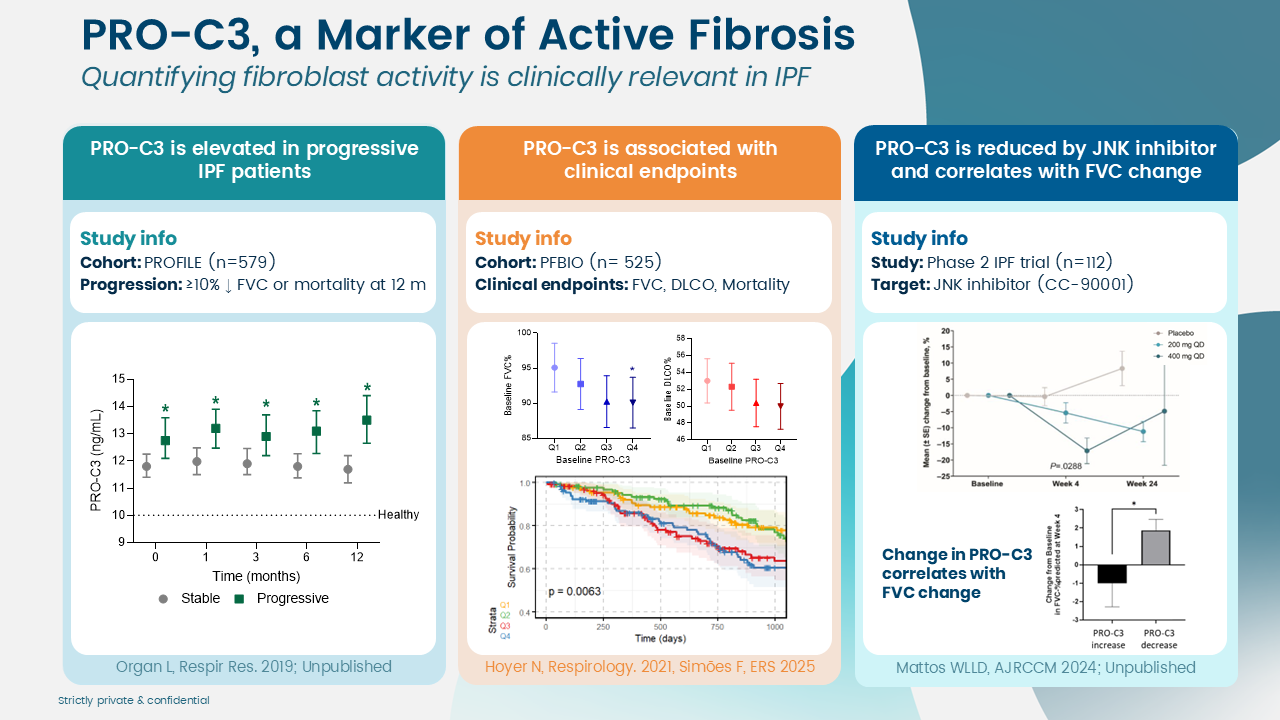
Across liver, lung, skin, intestinal, cardiovascular, kidney disease, and across solid tumors, fibroblast activity is repeatedly associated with prognosis. Regulatory momentum also reflects this direction: fibroblast activity biomarkers such as PRO-C3 and PRO-C6 (type III and type VI collagen formation) are supported for prognostic use.
We will connect mechanistic framing (what fibroblasts are doing) with translational readouts (what biomarkers capture in blood), then move into two applied deep dives:
Chronic liver disease: prognostic performance of ECM biomarkers, with emphasis on PRO-C3
Solid tumors: CAF-linked matrix signatures, including collagen type XI, and how baseline fibroblast activity stratifies survival
Dr. Morten Karsdal joined Nordic Bioscience in 2001 and became CEO in June 2010, leading the company to significant advancements in biomarker development and disease biology.
Dr. Karsdal is a KOL in extracellular matrix research, with more than 700 publication, 44,684 citations, and an impressive H-index of 106.
Dr. Karsdal is an honorary professor of inflammation research at the University of Southern Denmark, where he continues to supervise PhD students, fostering the next generation of researchers.
Dr. Karsdal chairs the Extracellular Matrix Pharmacology Congress, an important forum for advancing drug development by focusing on the extracellular matrix (ECM) as a key factor in most chronic diseases. He is renowned for his deep expertise in fibrosis, rheumatology (including rheumatoid arthritis and osteoarthritis), diabetes, and other chronic conditions, particularly in relation to ECM and biomarker research.
Dr. Karsdal has led the development of FDA-approved and supported molecular diagnostics, as well as more than 100 commercialized biomarker assays, including ELISA assays and high precision automated platforms.
He has extensive experience in clinical trial design and the clinical application of biochemical markers, often serving as a consultant to major pharmaceutical companies for the use of serological biomarkers in clinical trials.
In 2016, he and his research team authored the first edition of “Biochemistry of Collagens, Laminins and Elastin,” published by Elsevier Science. The book, now in its 3rd edition as of 2023, is a key resource on collagens and structural proteins, with a focus on their applications in chronic diseases.
Dr. Diana Julie Leeming
Dr. Diana Julie Leeming is the Senior Director of Fibrosis, Hepatic, and Pulmonary Research at Nordic Bioscience.
She joined Nordic Bioscience in 2004 and assumed the role of Director of Fibrosis in 2010, later being promoted to Senior Director in 2024.
Dr. Leeming focuses on developing serologically assessed markers to evaluate extracellular matrix remodeling in patients with pulmonary or hepatic fibrosis, aiding in diagnosis and pharmacodynamic evaluation.
She is a principal inventor of the PRO-C3 assay, a fibrogenesis marker utilized in multiple clinical trial studies.
Dr. Leeming has authored over 280 peer-reviewed publications, demonstrating her extensive contributions to the field.
Her H-index is 69, her I10-index is 195, and her research has garnered over 15,736 citations as of February 2026.
Dr. Nicholas Willumsen
Dr. Nicholas Willumsen is Director of Oncology at Nordic Bioscience, a position he has held since 2022.
He joined Nordic Bioscience in 2012 and became Head of the Oncology Department in 2016.
Dr. Willumsen leads a research group focused on the development of blood-based biomarkers to quantify tumor matrix components in serum from cancer patients.
His work aims to elucidate pharmacodynamic effects, treatment efficacy, and resistance mechanisms across cancer therapies.
The group’s research spans the full translational pipeline, from biomarker discovery to preclinical studies and clinical validation.
A central focus is understanding tumor–extracellular matrix interactions and their role in disease progression and treatment response.
Dr. Willumsen’s research supports the use of non-invasive biomarkers to guide oncology drug development and clinical decision-making.
He has authored peer-reviewed publications with an H-index of 31 and an i10-index of 53.
His work has received 3,246 citations as of February 2026.
This webinar is hosted co-hosted together with the International Society of Extracellular Matrix Pharmacology.
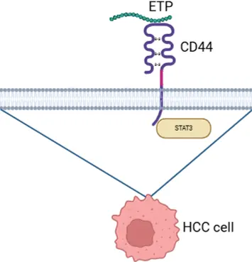
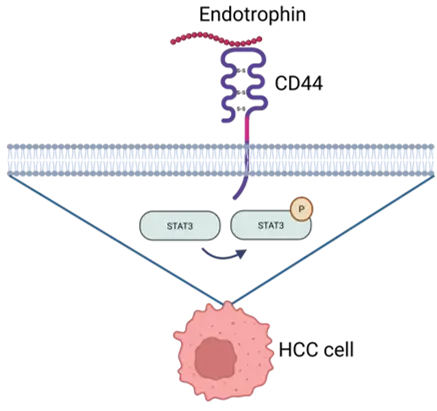
For years, Endotrophin has been discussed as a circulating signal linked to fibroinflammatory disease severity and adverse outcomes. But there has been a persistent gap between association and a clearly defined, targetable mechanism. Now, a new report addresses that gap by identifying CD44 as a receptor for Endotrophin and connecting Endotrophin–CD44 binding to STAT3 signaling in hepatocellular carcinoma (HCC).
This publication anchors that association mechanistically by showing that Endotrophin is produced in fibrotic liver by COL6A3‑rich hepatic stellate cells (HSCs) and is not merely a degradation product, but a bioactive hormone-like peptide that signals into neighboring cells.
In the study’s experimental system, Endotrophin binding to CD44 activated STAT3 signaling and was linked to phenotypes relevant to tumor progression, including epithelial–mesenchymal transition (EMT), proliferation, and sorafenib resistance.
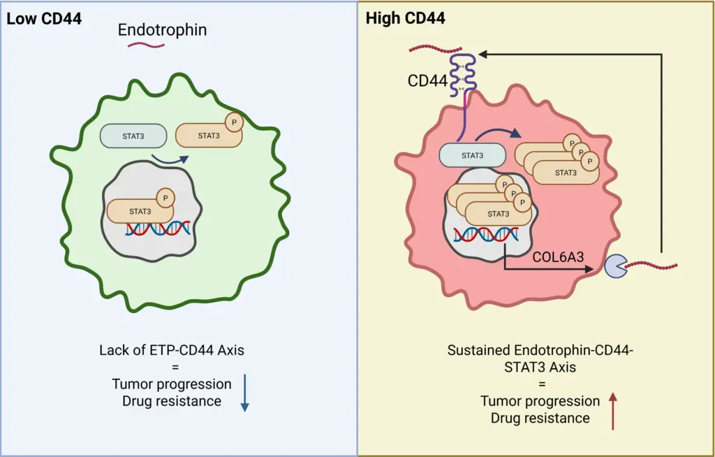
Fibroblast-derived Endotrophin signals into CD44+ cells and sustains itself
The paper further describes that hepatic stellate cell–derived Endotrophin targets pericentral CD44+ tumor cells, induces COL6A3 expression, and sustains Endotrophin production via a STAT3-dependent feedback loop.
Schematic of Endotrophin binding to CD44 and downstream STAT3 signaling. Created in BioRender. Gongora, F. (2026) https://BioRender.com/juay2se
When the Endotrophin-CD44 axis is activated, Endotrophin/PRO-C6 can serve as target engagement or stratification tools. Created in BioRender. Laursen, C. (2026) https://BioRender.com/2kd3vyi
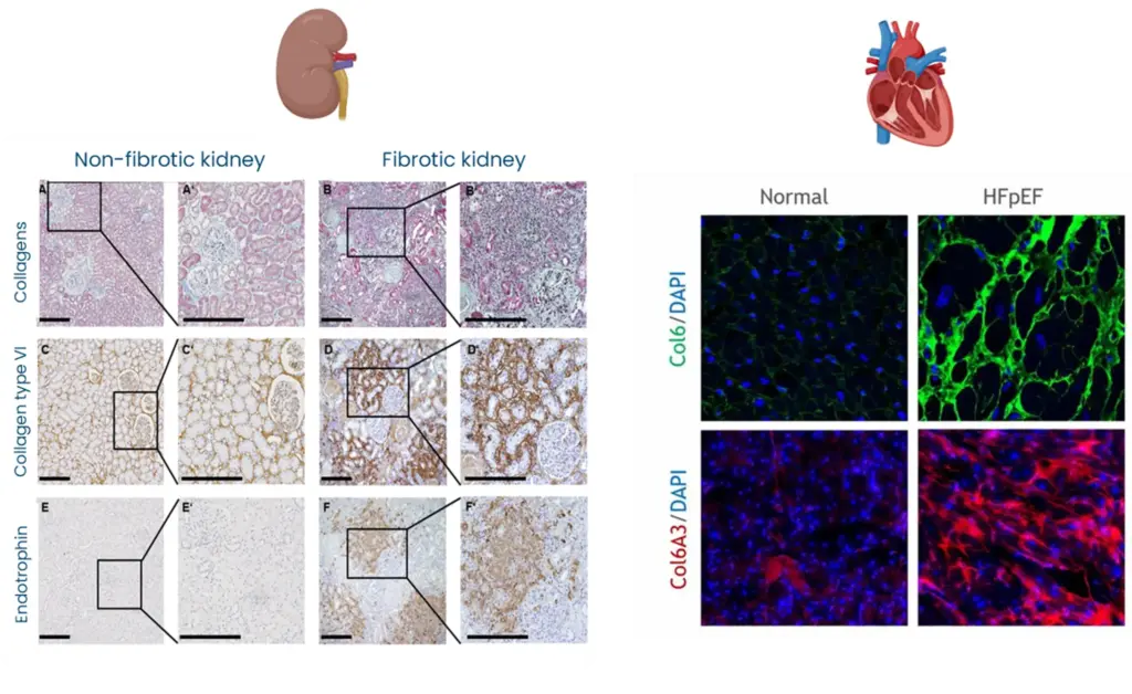
By defining CD44–STAT3 as the principal Endotrophin signaling route in HCC, the paper suggests a shared signaling logic by which Endotrophin‑rich fibrotic niches (kidney, heart, adipose tissue) can drive organ damage and risk of outcome through similar downstream pathways.
Interested in how Endotrophin relate to fibroblast activity?
Circulating Endotrophin likely reflects an activated Endotrophin-CD44–STAT3 loop in fibrotic organs. High circulating Endotrophin is not only a marker of fibroblast activity but a surrogate for an ongoing pathogenic signaling circuit. Inhibiting the Endotrophin–CD44–STAT3 axis (genetic Col6a3/Cd44 deletion, STAT3 inhibition, or CD44‑binding‑defective Endotrophin mutants) attenuates fibrosis, EMT, steatosis and chemoresistance, positioning this axis as a therapeutic target in metabolic dysfunction-associated HCC.
Endotrophin‑rich fibrotic niches can drive organ damage and risk of outcome
Endotrophin does not only stage and prognosticate fibrotic disease but can also identify patients in whom Endotrophin‑driven fibro-inflammatory signaling is active and may be actionable by Endotrophin‑neutralizing antibodies or STAT3‑targeted interventions.
Why measure outcomes and risk signals across chronic disease
Endotrophin in the blood reflects fibro-inflammatory remodeling associates with cardiac and kidney fibrosis, mortality, and progression of MASH, cirrhosis, and HCC, with levels rising across the spectrum of metabolic diseases.
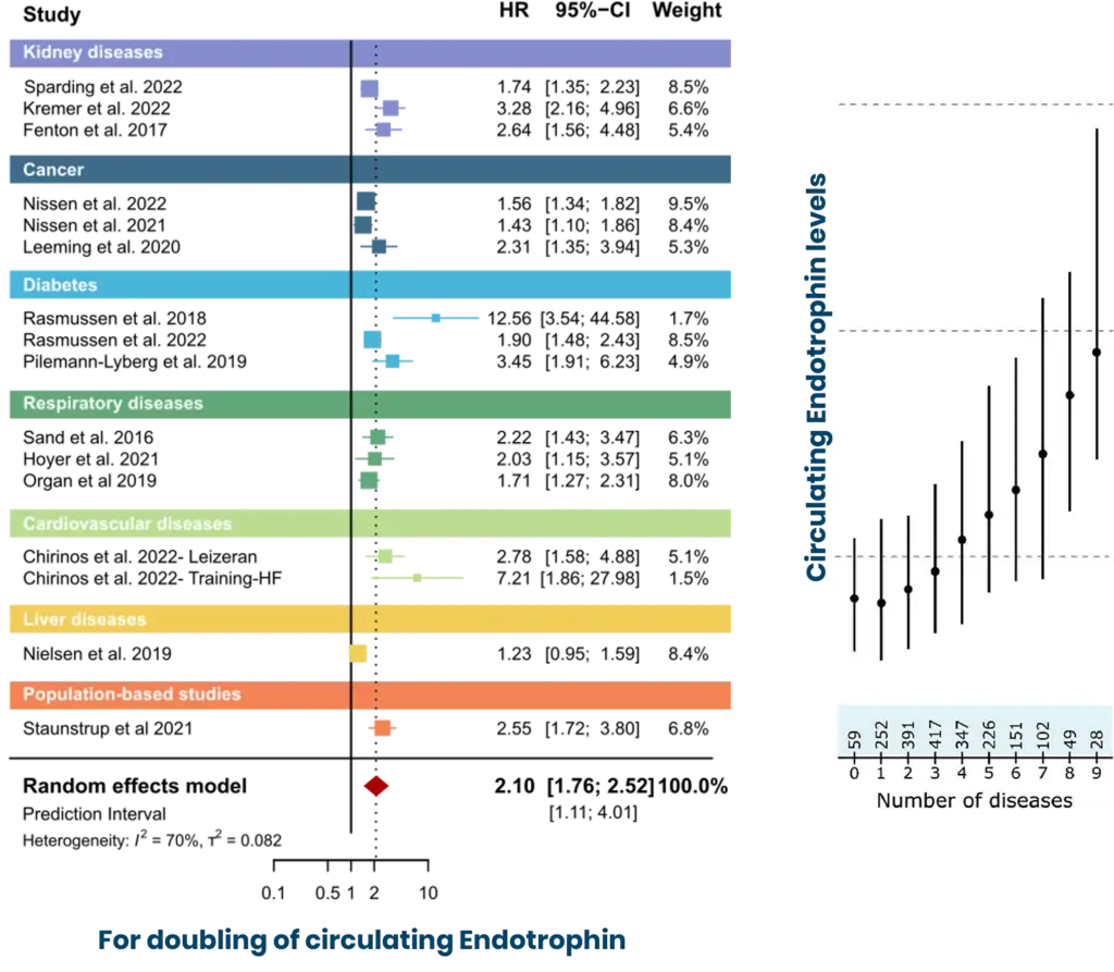
Endotrophin is not just “another fibrosis marker,” but a circulating readout of active fibro-inflammatory biology. In a systematic review and individual participant data meta-analysis, higher circulating Endotrophin independently associated with increased mortality risk across a broad range of chronic diseases: a core clinical reason to measure Endotrophin and nordicPRO-C6™ in programs where baseline biology and outcome risk need to be quantified.
Mechanistically, Endotrophin upregulates and releases into circulation as a consequence of recurring pathophysiological mechanisms across diseases. The functional implication of circulating Endotrophin supports PRO-C6 as an operational biomarker in clinical programs, where a blood-based measurement aligned with fibroblast-driven remodeling biology can be used at scale and longitudinally.
How does CD44 connect to existing literature on Endotrophin?
The new receptor paper changes how to interpret associations by providing a defined signaling handle. CD44 was identified as an Endotrophin receptor, and Endotrophin–CD44 engagement activated STAT3 signaling with disease-relevant phenotypes. When a circulating molecule is both outcome-associated and connected to a receptor axis, measuring Endotrophin and PRO-C6 becomes a practical component of biology-aware stratification and monitoring in fibro-inflammatory clinical programs. Nordic’s measurement approach centers on assays that quantify Endotrophin-related biology in circulation, including PRO-C6-based measurement supported through clinical evidence.
If your program involves fibro-inflammatory biology, position Endotrophin measurement as an evidence-aligned readout to support:
Baseline characterization of fibroblast-associated signaling burden,
Risk stratification informed by published outcome associations across chronic disease,
Mechanistic hypothesis testing in settings where CD44/STAT3 biology is part of the rationale.
A recent analysis published in JAMA highlights a major epidemiological shift: Colorectal cancer (CRC) is now the leading cause of cancer-related death among adults under 50 in the United States.
CRC has long been perceived as a disease of older populations. The rapid rise in early-onset cases challenges that assumption and reinforces the need to rethink both biology and treatment strategies.
Why standard therapies still fall short: the fibrotic stroma as a resistance mechanism
Therapeutically, the backbone remains surgery in localized disease, combination chemotherapy regimens such as FOLFOX and FOLFIRI, anti-VEGF or anti-EGFR targeted therapies in selected patients, and immunotherapy in MSI-high/dMMR tumors. While these approaches have extended survival, resistance and relapse remain frequent, particularly in advanced disease.
One key and often underestimated driver of progression and resistance is fibrosis. CRC tumors are embedded in a collagen-rich, fibrostenotic microenvironment shaped by cancer-associated fibroblasts (CAFs). This dense extracellular matrix (ECM) does not merely provide structure. It represents a desmoplastic barrier that limits drug penetration, reduces treatment efficacy, and supports tumor progression.
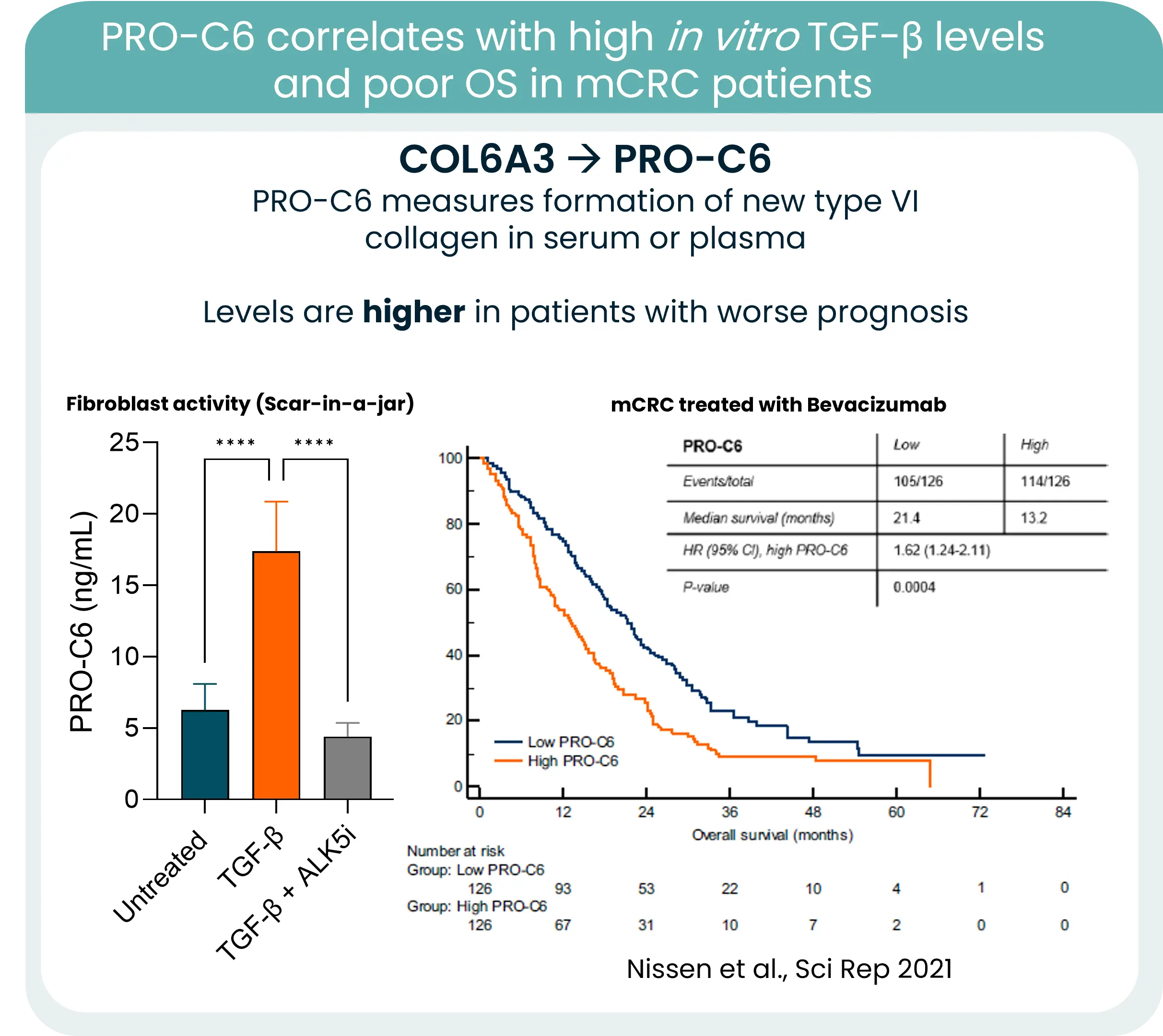
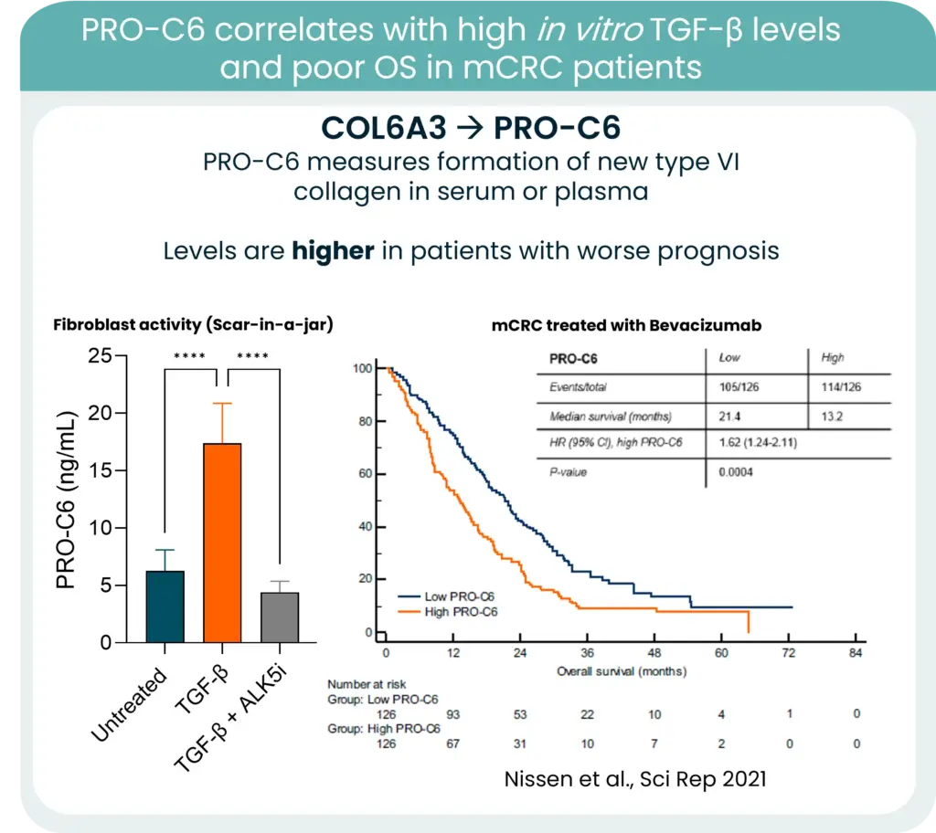
PRO-C6 correlates with TGF-β levels and overall survive in mCRC patients
From biology to measurement: collagen remodeling biomarkers
Collagens are central components of this barrier, and increased collagen deposition and remodeling consistently associate with poorer outcomes. One notable example is type VI collagen. Its gene expression is strongly associated with KRAS mutations and high TGF-β levels in CRC. New type VI collagen formation releases protein fragments to the bloodstream, which can be quantified in serum using our PRO-C6 biomarker and correlate to high TGF-β levels and poor OS.
In this context, measuring collagen formation and degradation becomes highly relevant. Quantifying ECM remodeling can provide insight into tumor aggressiveness, stromal activation, and potentially treatment response.
As early-onset CRC continues to rise, integrating stromal blood-based biomarkers into clinical strategy may refine risk stratification and open new opportunities for stroma-targeted interventions.
The future of CRC management will not rely solely on targeting tumor cells. It will require understanding and measuring the fibrotic environment that sustains them.