Most biomarker candidates die in the valley of death. What made PRO-C3 different?

Biomarker discovery has accelerated dramatically, but clinically viable products rarely follow. Thousands of protein signals are associated with disease every year, yet, relatively few become reliable tools for clinical development or patient management. Many stall in the biomarker valley of death: the long and resource-intensive space between initial discovery and validated clinical utility.

High-throughput omics and proteomics can identify thousands of disease-associated signals. But an association alone does not make a measurement an actionable biomarker. Biological specificity, analytical performance, clinical evidence and a clearly defined Context of Use must follow.

A new expert opinion in the Journal of Hepatology (IF 40 as of August 2026) proposes a higher standard. While the paper takes the liver field as an example, it’s findings are applicable to most diseases. It introduces the concept of the unicorn biomarker: a rare biomarker with diagnostic, prognostic and pharmacodynamic utility, supported by rigorous technical, clinical and regulatory development. PRO-C3, invented by Nordic Bioscience, is highlighted as one of the potential unicorn biomarkers approaching this standard. 

Most candidates die in the biomarker valley of death
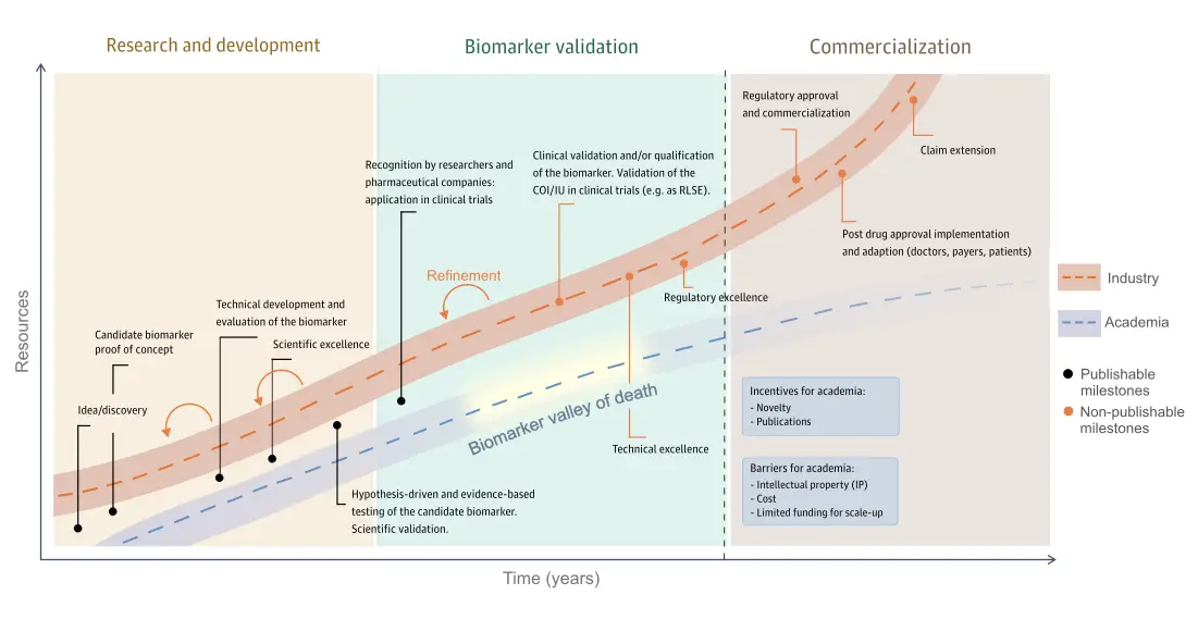
Fig.1 The path to becoming a unicorn biomarker consisting of three stages: Research and development, biomarker validation, and commercialization.

Discovering a promising protein signal is only the beginning. To progress toward clinical utility, a candidate must undergo technical development, scientific and clinical validation, regulatory alignment and, ultimately, implementation on a reliable and scalable platform. Many candidates never progress beyond discovery or early validation, while others stall when the cost and technical requirements increase.

The journey begins with research and development, where a biomarker is discovered through scientific hypothesis testing. Most biomarkers remain in research and development, as advancing further requires investment in technical excellence and quality control. The second stage is the biomarker validation stage where biomarkers are validated in clinical trials and technical performance on the final assay design is rigorously tested. This phase is often called the “biomarker valley of death” because the work is costly, technically demanding, and rarely publishable, causing many candidates to stall.

The final stage is commercialization, which focuses on achieving regulatory approval for specific contexts of use (CoUs) or intended uses (IUs) and implementing marketing strategies to generate a return on the investment. Here, academia and industry collaborate to validate, adopt, market, and implement the biomarker for clinical applications.

What does it take to become Unicorn biomarker?
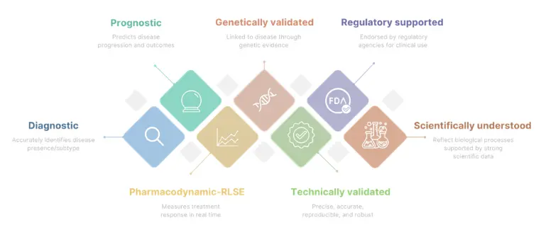
Fig. 2 An outline of the requirements a unicorn biomarker has to fulfill.

Unicorn biomarkers are rare and valuable, as they must fulfill various requirements (Fig. 3). Unlike biomarkers that serve a single purpose, they provide value across multiple stages of research and clinical development. They combine diagnostic utility, prognostic utility and pharmacodynamic utility, which is a combination of requirements that can be challenging because the criteria often is conflicting. While diagnostic and prognostic biomarkers must reliably reflect stable disease characteristics, pharmacodynamic biomarkers need to be sufficiently sensitive and dynamic to detect treatment-induced changes.

Beyond clinical utility, a unicorn biomarker must also be scientifically understood, technically validated to ensure reliable and reproducible results, and regulatory supported to enable its use in clinical development and, ultimately, patient care. Only when these scientific, technical, clinical, and regulatory criteria come together can a biomarker be considered a true unicorn biomarker.

Why the exact protein measurement matters

Proteins exist in different fragments, modified forms, or structural regions, each reflecting different biological processes. As a result, two measurements of the same protein may not necessarily provide the same clinical or biological insight if they target different parts of the molecule.

This is why the exact molecular target matters in biomarker development. The value of a biomarker does not only depend on identifying the right protein, but on measuring the right part of that protein in the right biological and clinical context.
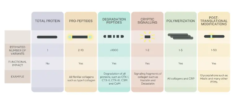
Fig. 3 The structural complexity of proteins and implications for quantification.

This is the principle behind our Nordic ProteinFingerPrint Technology™. By targeting specific protein fragments generated during extracellular matrix remodeling, the technology is designed to measure defined biological processes rather than total protein abundance alone. This enables a more precise understanding of whether tissue is being formed, degraded, or modified by disease-related enzyme activity, helping researchers connect biomarker measurements to disease mechanisms, progression, and treatment response.

PRO-C3: Journey toward Unicorn Biomarker Status

One example approaching this standard is nordicPRO-C3™. PRO-C3 detects a defined neo-epitope generated during type III collagen formation, linking the measurement to active fibrogenesis rather than total collagen abundance.

Its development has extended well beyond initial discovery. PRO-C3 has demonstrated diagnostic utility as part of the ADAPT score, prognostic value in fibrotic disease and pharmacodynamic utility for measuring treatment-related changes in fibrosis activity. It has also been used extensively in pharmaceutical clinical development.

The launch of Elecsys PRO-C3 by Roche Diagnostics transferred the measurement onto the globally established cobas platform as an IVD-labelled assay. This progression from a biologically defined neo-epitope to a clinically studied and commercially deployed assay is why the paper identifies PRO-C3 as a potential unicorn bomarker.

Why this matters for drug developers

Unicorn biomarkers are valuable to drug developers because they link biological mechanisms with measurable treatment effects. When associated to a clear Context of Use, they can support patient enrichment by identifying individuals with relevant disease activity, disease stage, or risk of progression. This can reduce heterogeneity in trials and improve the ability to detect a meaningful treatment effect.

Their pharmacodynamic utility is especially relevant to drug developers, as they can provide earlier evidence that a therapy is affecting the intended biological pathway before traditional clinical outcomes become visible. This can support proof-of-mechanism, dose selection, treatment monitoring, and go/no-go decisions, helping developers make more informed choices before moving into larger and more costly studies. By providing earlier and more mechanistic grounded information, they can reduce uncertainty, improve trial design, and help lower the risk of late-stage failure.

By combining diagnostic, prognostic, and pharmacodynamic utility with strong scientific, technical, and clinical validation, unicorn biomarkers represent an aspirational standard for biomarker development.

PRO-C3 is not an isolated discovery. It represents Nordic Biosciennce’s approach to biomarker development: begin with biologically meaningful protein fragments and continue through the technical, clinical and regulatory work required to make the measurement useful. We build biomarkers to cross the biomarker valley of death.

Myths and misconceptions about biomarkers

  • While some biomarkers may be subject to biological variability, many others such as NT-proBNP for heart failure or prostate-specific antigen (PSA) for prostate cancer have well-characterized reference ranges and robust assay reproducibility. In fact, standardization and quality control measures have significantly improved the reliability of many biomarker assays.

  • Although oncology has led the way, biomarkers are now well-established in many fields, including cardiology (e.g., troponin for myocardial infarction), hepatology (e.g., ELF or FIB-4 for liver fibrosis) [67], diabetes (HbA1c) and rheumatology (e.g., anti-CCP for rheumatoid arthritis). Their utility spans diagnosis, prognosis, and treatment monitoring across multiple disease domains.

  • While regulatory-grade validation is ideal for clinical implementation (particularly for RLSE or VSE CoUs), exploratory biomarkers can and should be incorporated into early-phase trials to guide hypothesis generation and decision-making and generating data that will enable eventual clinical validation for regulatory use later. Waiting for full validation can delay insights into their potential link to drug MoA or clinical efficacy and hinder development [81]. Further, the use of a biomarker during pre-clinical phases of drug development increases the likelihood of final regulatory approval from 8% to 25%.

  • Although biomarker assays introduce additional costs, these are often small compared with the overall expense of clinical trials. Biomarkers can support clinical development by providing mechanistic insights into treatment response and disease biology, thereby informing critical decisions related to efficacy, safety, and proof of mechanism. As a result, biomarkers may contribute to more efficient and informative clinical trials.
  • When biomarkers have a defined CoU, including their role, target population, and setting, they streamline rather than complicate regulatory pathways. In fact, the FDA’s BEST Resource provides a structured framework for qualifying biomarkers.

Discuss your biomarker strategy

Explore how biologically specific biomarkers could support patient enrichment, proof of mechanism, treatment monitoring or development decisions in your clinical program.

    Fibroblast Activity and Tissue Destruction in Autoimmune Disease | Webinar

    Fibroblast Activity and Tissue Destruction in Autoimmune Disease: Understanding Tissue Activity and Its Implications for Drug Development

    Overview

    The body continuously regenerates, which inherently means that tissue is constantly being broken down and formed. In autoimmune diseases we usually focus on inflammation and immune activity, but inflammation is what starts the process, not what causes the lasting harm. The damage comes when that balance tips, fibroblasts become activated, and cartilage, bone, and connective tissue are broken down faster than the body can repair them. 

    Watch the replay of this webinar to learn more about how evaluating tissue activity reveals what is actively happening in the disease biology, whether a treatment is truly working, and how these insights can support the development of better therapies in autoimmunity, PsA, RA, SSc and lupus. 

    Agenda

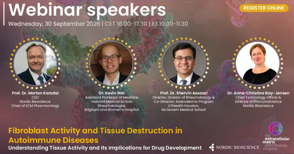
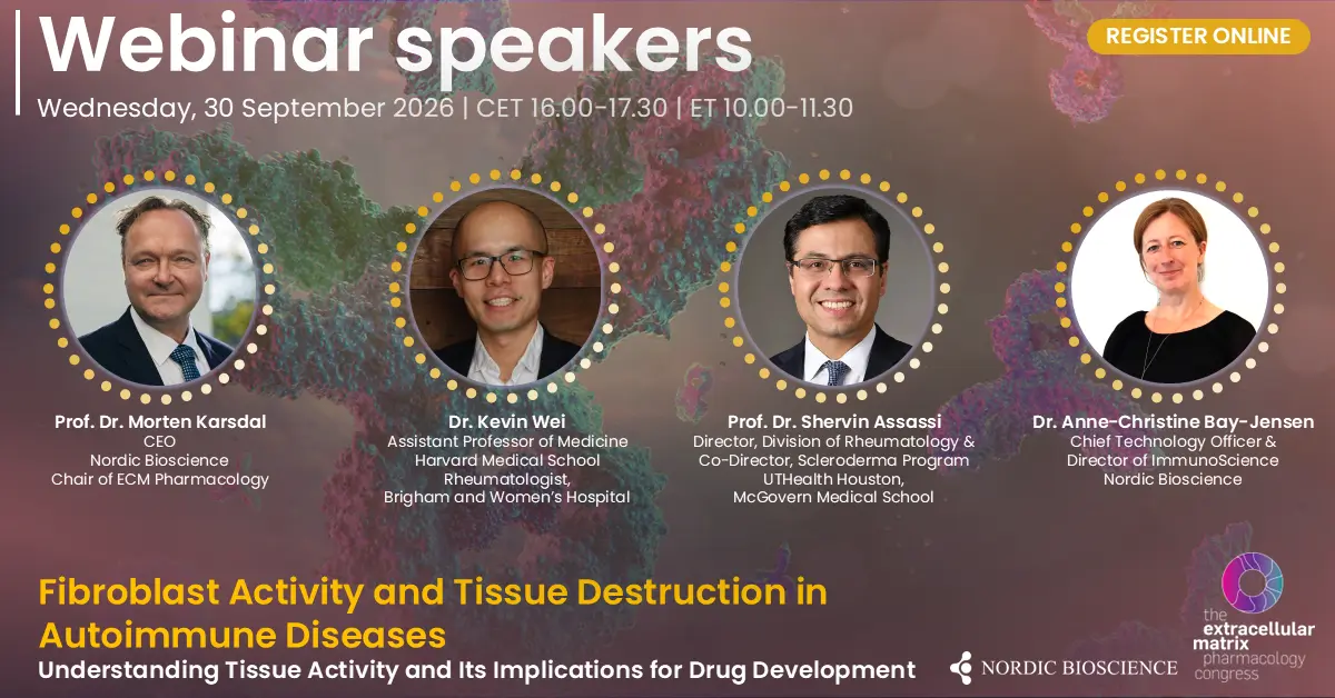
    • Introduction and welcome & Myth busting with Unicorn biomarkers | Prof. Dr. Morten Karsdal
    • When inflammation isn’t enough: Synovial Tissue Fibrosis as a Driver of Treatment-Refractory RA | Dr. Kevin Wei
    • From tissue biology to clinical application: how fibrogenesis and tissue destruction markers are used for prognosis, pharmacodynamics and prediction in RA, PsA and SSc | Dr. Anne-Christine Bay-Jensen
    • Fibrosis and inflammation of the Skin and Lung in Systemic Sclerosis: Tracking tissue remodeling, progression and treatment response with biomarkers | Prof. Dr. Shervin Assassi
    • Q&A

    Scientific topics

    Autoimmune diseases are commonly viewed through the lens of inflammation and immune activity. However, inflammation represents only part of the underlying disease biology. Fibroblast activation, extracellular matrix remodeling, and ongoing tissue destruction can provide complementary information about what is happening within affected tissues and how disease activity changes during treatment. 

    In psoriatic arthritis, tissue turnover biomarkers offer a way to investigate structural and connective tissue activity alongside traditional measures of inflammation. Understanding these processes may help distinguish different aspects of disease activity, monitor the biological effects of treatment, and provide additional insight into therapeutic response. 

    Rheumatoid arthritis is likewise characterized by substantial heterogeneity within the synovium. Distinct synovial pathotypes, including fibroblast-rich or fibroid phenotypes, point to disease mechanisms that may not be adequately described by inflammatory activity alone. Emerging research into fibroblast states, synovial organization, and the regulation of fibrogenesis is therefore changing how we understand persistent disease and differences in treatment response. 

    Across systemic sclerosis, lupus, and autoimmunity more broadly, the same principle applies. Many different disease pathways end up driving the same core process: the breakdown and rebuilding of tissue. This means disease activity can be understood as the balance between how much tissue is being destroyed and how much is being formed. The protein fragments released when tissue breaks down give us the most direct way to measure that balance. 

    By bringing together perspectives on biomarkers, synovial pathobiology, and fibrogenesis, this webinar examines how measuring tissue activity—not only inflammation—could improve patient characterization, pharmacodynamic assessment, and ultimately drug development in rheumatology.

    Speakers

    Dr. Kevin Wei

    • Dr. Kevin Wei is an Assistant Professor of Medicine at Harvard Medical School and a rheumatologist at Brigham and Women’s Hospital. 
    • His research focuses on identifying novel cellular and molecular therapeutic targets in inflammatory diseases using single-cell technologies. 
    • Dr. Wei is a member of the NIH Accelerating Medicines Partnership in Rheumatoid Arthritis and Systemic Lupus Erythematosus (AMP-RA/SLE) consortium. 
    • Within the consortium, he leads the implementation of single-cell technologies to study rheumatoid arthritis joint tissues. 
    • He is Co-Director of the Brigham and Women’s Hospital Center for Cell Profiling Single Cell Multiomics Core. 
    • Dr. Wei also serves as Associate Director of the Harvard Medical School/BWH Joint Biology Consortium Cellular Systems Core. 
    • His work applies advanced cellular profiling and multiomics approaches to better understand disease mechanisms and tissue-specific cellular states in rheumatic diseases. 
    • He has received research awards from the Rheumatology Research Foundation, Burroughs Wellcome Fund, Harvard Catalyst, and the BWH Department of Medicine. 
    • Dr. Wei earned his BS in Biology from Duke University and his MD and PhD from Stanford University School of Medicine. 
    • He completed his Internal Medicine residency and Rheumatology fellowship at Brigham and Women’s Hospital and also serves on the advisory board of STAR Protocols. 

    Prof. Dr. Morten Karsdal

    • Dr. Morten Karsdal joined Nordic Bioscience in 2001 and became CEO in June 2010, leading the company to significant advancements in biomarker development and disease biology.
    • Dr. Karsdal is a KOL in extracellular matrix research, with more than 700 publication, 44,684 citations, and an impressive H-index of 106.
    • Dr. Karsdal is an honorary professor of inflammation research at the University of Southern Denmark, where he continues to supervise PhD students, fostering the next generation of researchers.
    • Dr. Karsdal chairs the Extracellular Matrix Pharmacology Congress, an important forum for advancing drug development by focusing on the extracellular matrix (ECM) as a key factor in most chronic diseases. He is renowned for his deep expertise in fibrosis, rheumatology (including rheumatoid arthritis and osteoarthritis), diabetes, and other chronic conditions, particularly in relation to ECM and biomarker research.
    • Dr. Karsdal has led the development of FDA-approved and supported molecular diagnostics, as well as more than 100 commercialized biomarker assays, including ELISA assays and high precision automated platforms.
    • He has extensive experience in clinical trial design and the clinical application of biochemical markers, often serving as a consultant to major pharmaceutical companies for the use of serological biomarkers in clinical trials.
    • In 2016, he and his research team authored the first edition of “Biochemistry of Collagens, Laminins and Elastin,” published by Elsevier Science. The book, now in its 3rd edition as of 2023, is a key resource on collagens and structural proteins, with a focus on their applications in chronic diseases.

    Prof. Dr. Shervin Assassi

    • Dr. Shervin Assassi is Professor of Medicine and Director of the Division of Rheumatology at UTHealth Houston’s McGovern Medical School. 
    • He is Co-Director of the Scleroderma Program, which includes one of the largest specialized scleroderma clinics in the United States. 
    • His research focuses on systemic sclerosis (scleroderma), integrating clinical features with genetic and gene-expression data to better understand disease mechanisms. 
    • Dr. Assassi’s laboratory aims to identify novel therapeutic targets and clinically useful biomarkers for systemic sclerosis. 
    • His translational research combines primary human samples with experimental models of fibrosis to investigate the molecular mechanisms underlying the disease. 
    • He is a leader of the Scleroderma Research Foundation-funded CONQUER Registry, serving as Chair of its Executive Committee and Director of the CONQUER Biorepository. 
    • Dr. Assassi has co-authored more than 220 peer-reviewed publications in systemic sclerosis, fibrosis, biomarkers, and related areas. 
    • His research program is supported by the National Institutes of Health, the Scleroderma Research Foundation, and industry collaborations. 
    • He holds a Master’s degree in Clinical Research and additional academic appointments in biomedical informatics and biomedical sciences. 
    • Alongside his research and clinical leadership, Dr. Assassi contributes to translational research education and holds leadership positions in professional rheumatology organizations and multicenter research consortia. 

    Dr. Anne-Christine Bay-Jensen 

    • Dr. Anne-Christine Bay-Jensen is Chief Technology Officer (CTO) and Director of Immunoscience at Nordic Bioscience, where she has worked since 2008. 
    • She joined Nordic Bioscience as a postdoctoral researcher and has since progressed into senior scientific and technology leadership roles. 
    • Her research focuses on the development of novel biomarkers for rheumatic and immune-mediated diseases, with approximately two decades of experience in the field. 
    • Dr. Bay-Jensen’s work uses biomarkers to characterize disease activity, tissue remodeling, and treatment response, supporting the development of precision medicine approaches. 
    • Her research has a strong focus on osteoarthritis and rheumatoid arthritis, including disease mechanisms, patient stratification, and therapeutic evaluation. 
    • Her scientific work also spans spondyloarthritis, systemic sclerosis, inflammatory bowel disease, inflammatory mediators, pharmacology, and immunology. 
    • A central theme of her research is understanding how changes in tissues and the extracellular matrix (ECM) can provide measurable insights into chronic inflammatory and connective tissue diseases. 
    • As CTO, she combines scientific leadership with the development and translation of biomarker technologies for use in preclinical research and clinical drug development. 
    • Dr. Bay-Jensen has an h-index of 66, i10-index of 229, and more than 13,600 citations as of September 2026. 
    • Her work bridges biomarker discovery, disease biology, and clinical translation to improve the characterization and treatment of rheumatic and inflammatory diseases. 

    This webinar was co-hosted with the International Society of Extracellular Matrix Pharmacology.