FAP-cleaved Type III Collagen (C3F) is a Potential Marker for Intestinal Fibrosis in CD Patients

Fibroblast Activation Protein (FAP)-Cleaved Type III Collagen (C3F) is a Potential Marker for Intestinal Fibrosis in Patients with Crohn’s Disease

Introduction

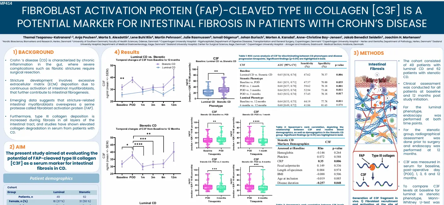
Crohn’s disease (CD) is characterized by chronic inflammation in the gut, where severe complications such as fibrotic strictures require surgical resection. The stricture development involves excessive extracellular matrix (ECM) deposition due to continuous activation of intestinal myofibroblasts, that further contribute to instestinal fibrogenesis. Emerging data suggests that stricture-related intestinal myofibroblasts overexpress a serine protease called fibroblast activation protein (FAP). Furthermore, type III collagen deposition is increased during fibrosis in all layers of the intestinal tract, and studies have shown elevated collagen degradation in serum from patients with CD.

This study aimed to evaluate the potential of FAP-cleaved type III collagen (C3F) as a serum marker for intestinal fibrosis in CD.

Poster

Conclusion

C3F is elevated in patients with stenotic CD compared to luminal CD at baseline and remains elevated after surgical resection, suggesting that C3F reflects fibroblast activity during fibrostenosis and tissue remodeling following surgery. In patients with luminal CD, C3F levels were significantly higher, and early C3F levels (baseline, 3 months and 6 months) were significantly positively associated with ileal stenosis assessed at 12 months. This suggests that increased C3F may be predictive of future endoscopic disease activity or even a potential relapse.

Concluding, the findings of this study highlight the potential use of C3F as a novel biomarker for intestinal fibrosis in CD.

Get in touch

Are you interested in exploring collaboration possibilities?
Enter your information in the form and a representative will contact you shortly.

    Biomarkers of ECM Remodeling Reflect Pharmacodynamic Effects of IMU-856

    Biomarkers of Extracellular Matrix Remodeling Reflect Pharmacodynamic Effects on IMU-856, an Oral Epigenetic Modulator of Barrier Regeneration

    Introduction

    Celiac disease (CeD) is characterized by intestinal inflammation and epithelial damage upon exposure to immunogenic gluten peptide, directly disrupting tissue architecture and by extension the extracellular matrix (ECM). The intestinal tissue is rich in ECM proteins such as collagens, which, during inflammation, undergo enzymatic remodeling due to increased proteolytic activity.

    IMU-856 is an orally available and systematically acting small-molecule modulator of sirtuin 6 (SIRT6), a histone/non-histone protein deacetylase and transcriptional regulator that aims to restore intestinal barrier function and regenerate bowel epithelium.

    This study aimed to investigate biomarkers of ECM remodeling as potential indicators of disease activity and pharmacodynamic response to IMU-856 in patients with CeD.

    Poster

    Conclusion

    Biomarkers of ECM remodeling reflect histological inflammation and mucosal architecture parameters, offering a direct insight into intestinal barrier integrity. These biomarkers potentially reflect treatment-induced improvement in intestinal tissue remodeling upon treatment with IMU-856.

    Get in touch

    Are you interested in exploring collaboration possibilities?
Enter your information in the form and a representative will contact you shortly.

      CPa9-HNE is Associated with Transmural Healing in Pediatric Crohn’s Disease

      CPa9-HNE, a biomarker of neutrophil activity is associated with transmural healing in pediatric Crohn’s disease

      Introduction

      CPa9-HNE, a calprotectin fragment released by human neutrophil estate during NETosis, has been shown to strongly correlate with endoscopic disease activity in adults with inflammatory bowel disease (IBD). CPa9-HNE also serves as an indicator of pharmacodynamic response. This study aimed to investigate the association of CPa9-HNE with endoscopic disease activity and transmural healing in patients with pediatric CD.

      Poster

      Conclusion

      This study showed that CPa9-HNE, a biomarker of neutrophil activity, is associated with mucosal and radiologic mucosal damage in pediatric CD. These findings highlight the potential use of CPa9-HNE as a complementary tool for the endoscopic or radiographic assessment of intestinal mucosal damage/healing and provide an opportunity for further studies in children with CD.

      Get in touch

      Are you interested in exploring collaboration possibilities?
Enter your information in the form and a representative will contact you shortly.

        ECM-derived Biomarkers of Mucosal Damage and Neutrophil Activity in Ulcerative Colitis Patients

        ECM-derived biomarkers of mucosal damage and neutrophil activity are associated with the failure of VZD

        Introduction

        Ulcerative colitis (UC) is a chronic relapsing inflammatory disorder affecting the colon. A hallmark of UC is increased immune cell activity and dysregulated extracellular matrix remodeling, particularly of the mucosal collagens type III and IV, leading to mucosal damage. Anti-TNF agents have significantly improved the management of UC-still, up to 40% of patients experience treatment failure, with the anti-α4β7 integrin agent vedolizumab (VDZ) being frequently initiated following anti-TNF failure. This presents the unmet need for biomarkers enabling early assessment of treatment success.

        This study investigates whether biomarkers of neutrophil activity, type III, and IV collagen remodeling could serve as early indicators of VDZ failure in anti-TNF experienced patients with UC.

        Poster

        Conclusion

        The early increase in CTX-III, reflecting fibrosis resolution, was associated with the absence of VDZ failure, whereas increased neutrophil activity (CPa9-HNE), IAF-mediated collagen type III degradation (C3F) and mucosal damage (C3M, C4M) were associated with the failure of VZD. These findings suggest that biomarkers reflecting neutrophil activity and inflammatory events in the mucosa may serve as promising tools for the early and dynamic assessment of later VDZ treatment outcomes in anti-TNF experienced patients with UC.

        Get in touch

        Are you interested in exploring collaboration possibilities?
Enter your information in the form and a representative will contact you shortly.

          Biomarkers of Immune Cell Activity can Identify Patients with Confirmed Crohn’s Disease

          Biomarkers of neutrophil and macrophage activity discriminate non-IBD from CD and associate with endoscopic disease activity

          Introduction

          Crohn’s disease (CD) is a chronic inflammatory bowel diseases (IBD) characterized by immune cell infiltration and increased proteolytic activity, driving pathological changes in the structure and function of the intestines. Ileocolonoscopy (IC) is the gold standard for diagnosing and monitoring patients with CD but the invasive nature of IC renders it as least acceptable from a patient perspective. Pan-enteric endoscopy (PCE) is an attractive and less invasive method.

          This study aimed to investigate whether biomarkers of immune cell activity could identify patients with suspected CD, and if they associated with endoscopic disease activity at IC and PCE.

          Poster

          Conclusion

          CPa9-HNE [neutrophil activity] and VICM [macrophage activity] could identify patients with confirmed CD and were associated with endoscopic disease activity. Both biomarkers provided stronger discriminative performance when assessing disease activity by utilizing PCE. Biomarkers of neutrophil and macrophage activity may therefore provide additonal information to endoscopic assessment in the diagnosis and monitoring of CD.

          Get in touch

          Are you interested in exploring collaboration possibilities?
Enter your information in the form and a representative will contact you shortly.

            Markers of ECM Remodeling and Wound Healing are Associated with Fibrostenotic Crohn’s Disease

            Markers of extracellular matrix remodeling and wound healing are associated with fibrostenotic Crohn’s disease

            Introduction

            Crohn’s disease (CD) is a chronic inflammatory condition of the gastrointestinal tract, characterized by excessive extracellular matrix (ECM) remodeling that leads to fibrosis and stricture formation. Strictures are primarily detected using imaging techniques, such as computed tomography and magnetic resonance imaging. However, no validated biomarkers are currently available to assess the presence of strictures. The discovery of such biomarkers would improve early diagnosis and facilitate more effective patient management using anti-fibrotic and anti-inflammatory treatments.

            This study aimed to investigate the association of biomarkers reflecting ECM remodeling, fibroblast activity, and neutrophil activity with CD phenotypes.

            Poster

            Conclusion

            The fibrosis marker (PRO-C6) was associated with stenosis and was elevated in CD patients with high levels of global stricture scores. Markers of mucosal damage (C7M, C3M/PROC3) and inflammation (CPa9-HNE) were associated with patients with stenosis and penetrating disease and showed a positive association with the global stricture score. These data suggest that markers of ECM remodeling could be valuable tools for assessing fibrostenosis in patients with CD.

            Get in touch

            Are you interested in exploring collaboration possibilities?
Enter your information in the form and a representative will contact you shortly.

              Canstatin Is Associated with Mortality Risk in Advanced Atherosclerosis

              Canstatin, a type IV collagen fragment, is associated with risk of cardiovascular and all-cause mortality in patients with advanced atherosclerosis

              Introduction

              Atherosclerosis, a common underlying cause of cardiovascular disease, is defined by the formation of
              plaques in the arterial walls. Changes in the ECM composition impact the risk for plaque rupture, which may cause acute complications (i.e. stroke or myocardial infarction (MI)). Type IV collagen is primarily known as a major component of basement membranes and has previously been reported to promote plaque stability. Canstatin is the non-collagenous C-terminal domain of type IV collagen alpha 2 chain. It is not only a by-product of proteolytic activity, but also a bioactive molecule. This study investigated if canstatin was associated with adverse outcomes in patients with advanced carotid atherosclerosis.

              Poster

              Conclusion

              Higher circulating canstatin levels in patients undergoing carotid endarterectomy predicted cardiovascular mortality and all-cause mortality over 7.5 years. This suggests that canstatin is a potential novel tool for risk stratification in patients with advanced atherosclerosis, warranting further studies.

              Get in touch

              Are you interested in exploring collaboration possibilities?
Enter your information in the form and a representative will contact you shortly.

                Collagen Type I Degradation Biomarkers Are Associated with Risk of Mortality After STEMI

                Collagen type I degradation biomarkers are associated with risk of mortality after ST-elevated
                myocardial infarction.

                Introduction

                Following ST-elevation myocardial infarction (STEMI), there is acute degradation of type I collagen (COL1), the primary structural protein of the myocardium. This process reflects extensive extracellular matrix remodeling, which may contribute to cardiac tissue destabilization and elevate the risk of subsequent adverse events. To better understand this pathological remodeling, we aimed to quantify COL1 using specific plasma biomarkers, including a novel signaling fragment of COL1 (C1SIG) and a more established COL1 degradation marker (C1M). Additionally, we investigated the prognostic value of these biomarkers for predicting all-cause mortality following a STEMI event.

                Poster

                Conclusion

                C1M and C1SIG are independently prognostic for mortality in STEMI patients after 1 year, in a multivariate
                model based on the Framingham Score. Assessing acute extracellular matrix processing in STEMI patients
                using COL1 biomarkers could be beneficial for predicting mortality and identifying a patient subset at
                increased risk of long-term outcome.

                Get in touch

                Are you interested in exploring collaboration possibilities?
Enter your information in the form and a representative will contact you shortly.

                  ACR70 Response and Its Dependence on Deep Tissue Efficacy

                  Introduction

                  Over the past decade, drugs developed for rheumatoid arthritis (RA) have been approved based on an ACR20 response rate of 60%. There is an increasing need for new RA drugs to achieve an ACR100 response in a significant portion of patients. Achieving ACR100 can profoundly improve patients’ quality of life by reducing pain, enhancing physical function, and decreasing fatigue.

                  The key question remains: which patients are likely to achieve ACR100?

                  An important aspect of RA pathogenesis involves the destruction and remodeling of bone, cartilage, and synovial tissue. Serum biomarkers that measure collagen and other extracellular matrix fragments can be used to assess disease activity at the tissue level. Examples include C1M (type I collagen destruction), C4M (type IV collagen destruction), and Osteocalcin (bone formation) [1].

                  The aim of this study was to examine the demographic, clinical, and serum markers of tissue remodeling as predictors linked to ACR20, ACR50 and ACR70 responses to tocilizumab.

                  Poster

                  Conclusion

                  While predictors of moderate response (ACR20) included clinical, demographic, and biomarker factors, predictors of significant response (ACR70) were exclusively biomarkers of extracellular matrix fragmentation. To achieve remission (ACR70 or ACR100) may require therapeutic interventions that specifically target and address tissue remodeling processes.

                  Get in touch

                  Are you interested in exploring collaboration possibilities?
Enter your information in the form and a representative will contact you shortly.

                    Serological Extracellular Matrix Fragments May Serve as Early Kidney Damage Biomarkers

                    Serological extracellular matrix fragments may serve as early kidney damage biomarkers in children with defects in Alport genes

                    Introduction

                    Alport syndrome is a genetic disorder caused by variants in COL4A3, COL4A4, or COL4A5, which encode type IV collagen. The α3.α4.α5(IV) collagen network is a critical structural component of the glomerular and tubular basement membranes within the kidney’s extracellular matrix (ECM). Variants in this network compromise basement membrane integrity, leading to cell injury, inflammation, and fibrotic remodeling of the surrounding interstitial matrix, which contributes to progressive kidney dysfunction.

                    We aim to identify serological protease-generated fragments of the ECM as potential early biomarkers of kidney damage.

                    Poster

                    Conclusion

                    The following biomarkers were altered in children with Alport syndrome, compared to healthy donors, before the onset of detectable proteinuria:


                    These biomarkers could potentially indicate early kidney damage and enable earlier initiation of kidney-protective treatments in children with Alport syndrome.

                    Get in touch

                    Are you interested in exploring collaboration possibilities?
Enter your information in the form and a representative will contact you shortly.