Vitiligo

Biomarkers for vitiligo

Vitiligo is a chronic depigmenting disorder characterized by the progressive loss of melanocytes from the epidermis. While autoimmune mechanisms are central to disease pathogenesis, the extracellular matrix (ECM) also contributes to melanocyte dysfunction and impaired skin homeostasis.

Despite recent advances, several unmet needs remain in vitiligo. Biomarkers may help improve assessment of disease activity and progression, identify distinct disease subtypes, and support patient stratification and treatment selection. In addition, biomarkers could facilitate the development of more targeted therapeutic approaches.

Current treatment selection in vitiligo is largely guided by clinical assessment and physician experience, as reliable predictive biomarkers for treatment response are lacking. As treatment outcomes vary substantially between patients, biomarkers may support a more personalized and targeted therapeutic approach.

Type III collagen in vitiligo

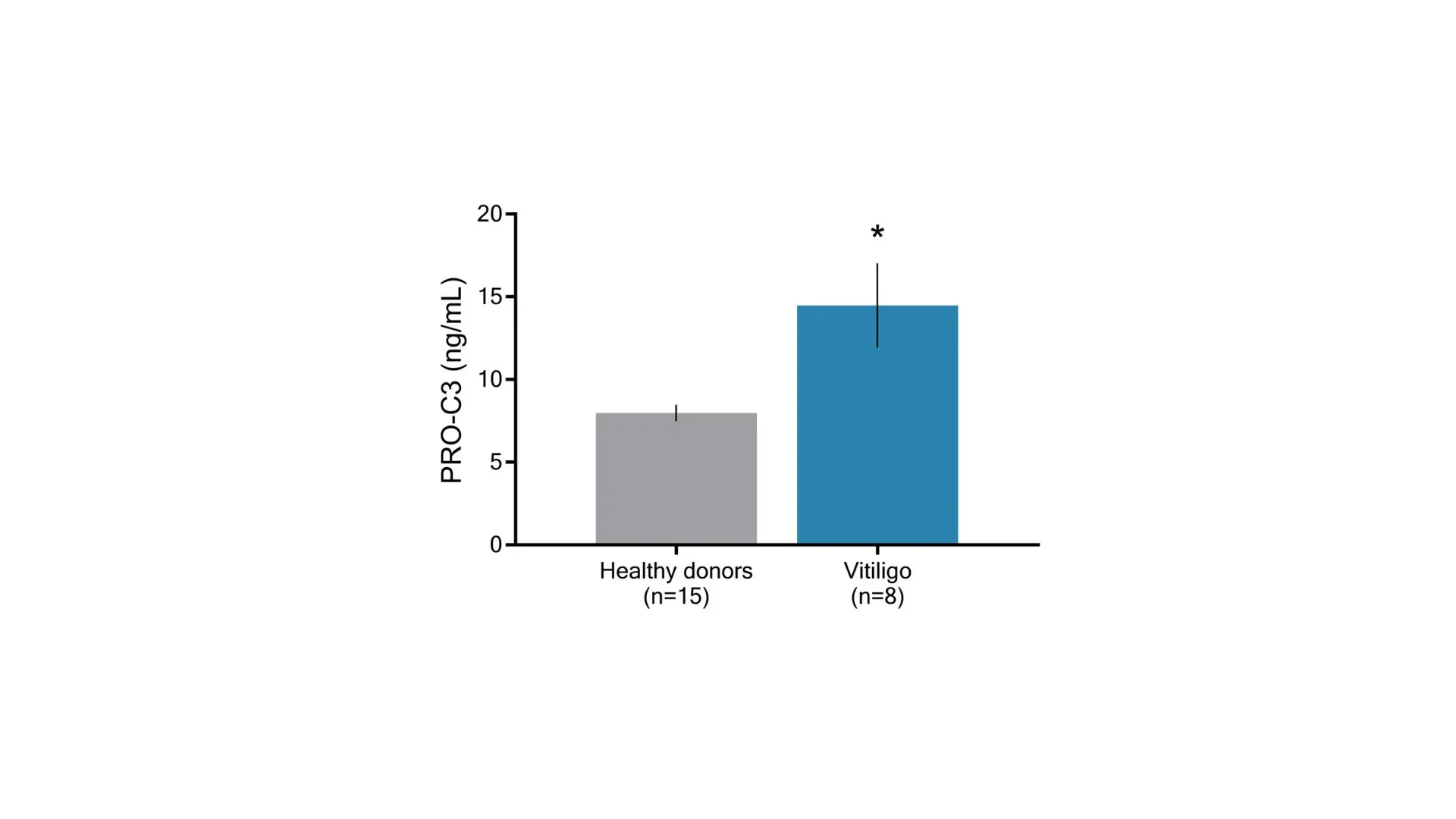
Type III collagen is a key fibrillar collagen in dermal ECM, where it is involved in tissue remodeling and wound healing. Our research indicates that PRO-C3, a Protein FingerPrintTM biomarker of tissue formation, is elevated in vitiligo patients compared to healthy donors (Figure 1).

Figure 1. The PRO-C3 biomarker is upregulated in vitiligo compared to healthy donors.

The Nordic ProteinFingerPrint Technology™ in Vitiligo

Watch this video to learn how our technology can help advance vitiligo research. A simple blood sample can be quantified by the Protein Fingerprint Technology and be used as a sophisticated biomarker tool for tailoring treatment solutions, predicting and monitoring disease progression and treatment response.

About vitiligo

  • Vitiligo is a common, chronic skin disorder characterized by development of depigmented patches resulting from the loss of melanocytes in the skin. Vitiligo affects approximately 0.5–2% of the global population and can occur in individuals of all ages, sexes, and ethnic backgrounds.


    The condition may develop at any age but most commonly begins before the age of 30. Vitiligo often follows an unpredictable course, with periods of stability alternating with episodes of progression. Depigmented patches most frequently appear on the face, hands, feet, and around body openings, although any area of the body may be affected.

  • Vitiligo is primarily diagnosed through clinical examination, assessment of the distribution of depigmented lesions, and medical history. In some cases, a skin biopsy or laboratory testing may be performed to confirm the diagnosis or evaluate for associated autoimmune diseases.


    Treatment aims to halt disease progression, restore pigmentation, and improve quality of life. First-line therapies commonly include topical corticosteroids and topical calcineurin inhibitors for localized disease. Recently, Janus kinase (JAK) inhibitors, including topical formulations and selected systemic therapies, have emerged as targeted treatment options for some patients.

  • The skin is a dynamic tissue composed by multiple cell types embedded within an extracellular matrix (ECM) that provides structural support and regulates cellular communication. Continuous remodeling of the ECM is essential for maintaining skin homeostasis, tissue repair, and immune regulation.


    In vitiligo, an autoimmune response selectively targets melanocytes, resulting in their destruction and the development of depigmented skin lesions. This inflammatory process is associated with alterations in the skin microenvironment, including immune activation, oxidative stress, and changes in extracellular matrix remodeling that may contribute to disease progression and tissue dysfunction.


    Nordic ProteinFingerPrint™ biomarkers can quantify tissue remodeling and disease-related changes directly in a serum sample, providing a minimally invasive approach that may serve as a liquid biopsy for monitoring biological processes associated with vitiligo. These biomarkers have the potential to support disease characterization, monitor treatment response, and improve understanding of the underlying mechanisms driving disease activity.

Get in touch

Are you interested in exploring collaboration possibilities?
Enter your information in the form and a representative will contact you shortly.