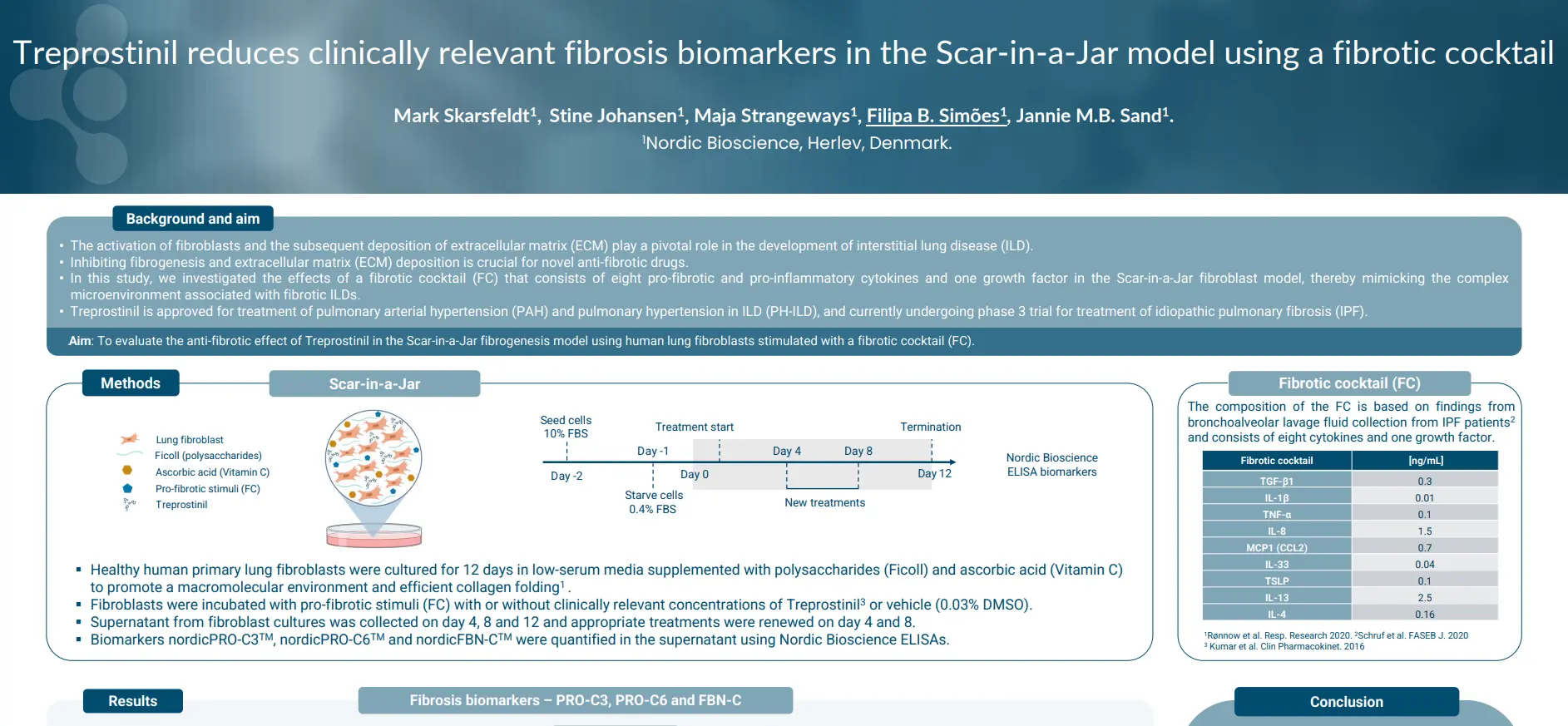
Treprostinil Reduces Fibrosis Biomarkers in the Scar-in-a-Jar Model Using a Fibrotic Cocktail

Treprostinil reduces clinically relevant fibrosis biomarkers in the Scar-in-a-Jar model using a fibrotic cocktail

Introduction

The activation of fibroblasts and the subsequent deposition of extracellular matrix (ECM) play a pivotal role in the development of interstitial lung disease (ILD), making it crucial for novel anti-fibrotic drugs.

In this study, we investigated the effects of a fibrotic cocktail (FC) that consists of eight pro-fibrotic and pro-inflammatory cytokines and one growth factor in the Scar-in-a-Jar fibroblast model, thereby mimicking the complex microenvironment associated with fibrotic ILDs.

Poster

Conclusion

The FC effectively stimulated fibrogenesis in the Scar-in-a-Jar in-vitro model, leading to elevated
levels of the fibrosis biomarkers nordicPRO-C3™, nordicPRO-C6™ and FBN-C™. Treprostinil significantly inhibited fibrogenesis and collagen deposition quantified by ELISA. In conclusion, the Scar-in-a-Jar model is a useful tool for screening novel anti-fibrotic drugs.

Get in touch

Are you interested in exploring collaboration possibilities?
Enter your information in the form and a representative will contact you shortly.

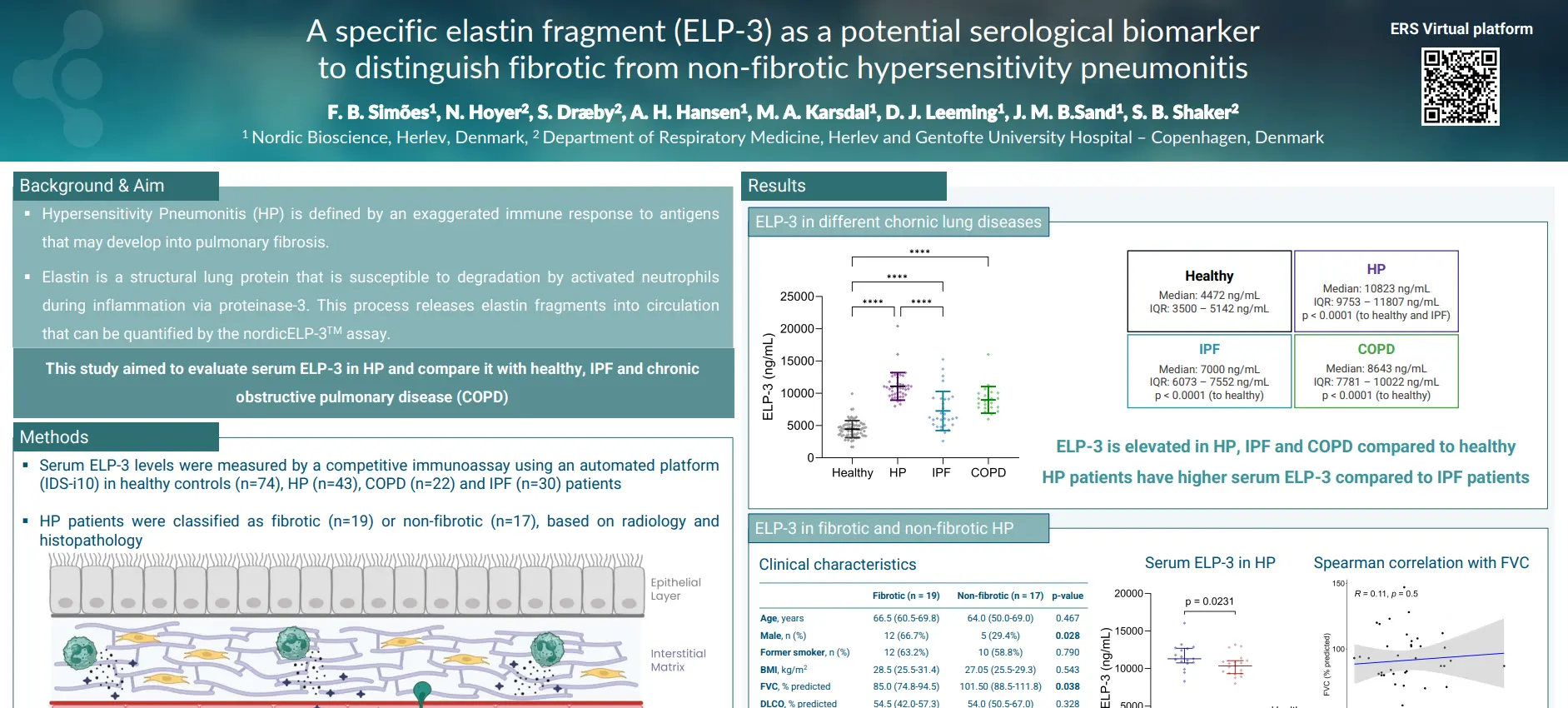
    ELP-3 as a Potential Serological Biomarker to Distinguish Fibrotic from Non-Fibrotic HP

    A specific elastin fragment (ELP-3) as a potential serological biomarker to distinguish fibrotic from non-fibrotic hypersensitivity pneumonitis

    Introduction

    Hypersensitivity Pneumonitis (HP) is defined by an exaggerated immune response to antigens that may develop into pulmonary fibrosis. Elastin, a structural lung protein, is susceptible to degradation by activated neutrophils during inflammation via proteinase-3. This process releases elastin fragments into circulation that can be quantified by the ELP-3 assay.

    This study aimed to evaluate serum ELP-3 in HP and compare it with healthy, IPF and chronic obstructive pulmonary disease (COPD).

    Poster

    Conclusion

    The serum ELP-3 is elevated in patients with different chronic lung diseases, particularly in HP. Notably, ELP-3 was able to separate non-fibrotic from fibrotic HP patients. These findings highlight the potential value of ELP-3 as a biomarker that provides additional clinical information beyond conventional inflammation markers.

    Get in touch

    Are you interested in exploring collaboration possibilities?
Enter your information in the form and a representative will contact you shortly.

      CC16-HNE Is Associated with Mortality and Pulmonary Hypertension in IPF

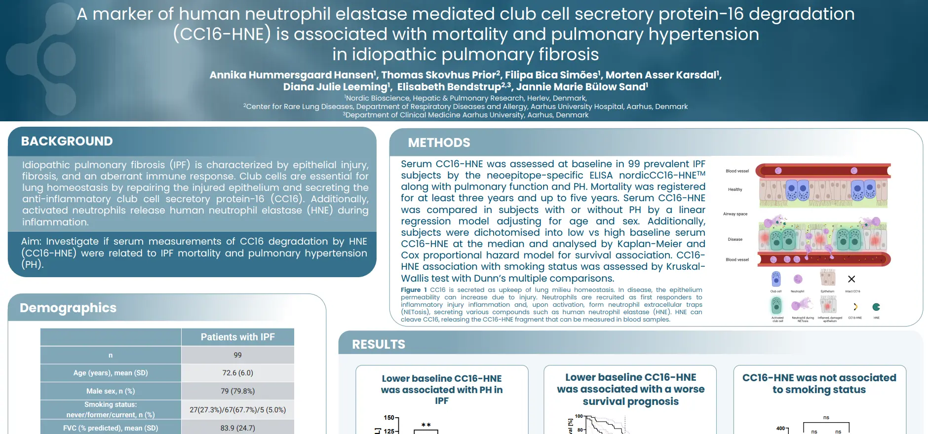
      A marker of human neutrophil elastase mediated club cell secretary protein-16 degradation in (CC16-HNE) is associated with mortality and pulmonary hypertension in idiopathic pulmonary fibrosis

      Introduction

      Idiopathic pulmonary fibrosis (IPF) is characterized by epithelial injury, fibrosis, and an aberrant immune response. Club cells are essential for lung homeostasis by repairing the injured epithelium and secreting the anti-inflammatory club cell secretory protein-16 (CC16). Additionally, activated neutrophils release human neutrophil elastase (HNE) during inflammation. Our aim was to investigate if serum measurements of CC16 degradation by HNE (CC16-HNE) were related to IPF mortality and pulmonary hypertension (PH).

      Poster

      Conclusion

      Low serum CC16-HNE at baseline was associated with increased risk of mortality and a PH complication in IPF. These results indicate that a neutrophil immune response and degradation of CC16 is relevant for disease outcome. CC16 degradation by HNE could serve as a prognostic biomarker for IPF and diagnostic for a PH complication.

      Get in touch

      Are you interested in exploring collaboration possibilities?
Enter your information in the form and a representative will contact you shortly.

        Prolonged Inflammation of Fibroblasts Promotes Formation of Type III Collagen and Fibronectin

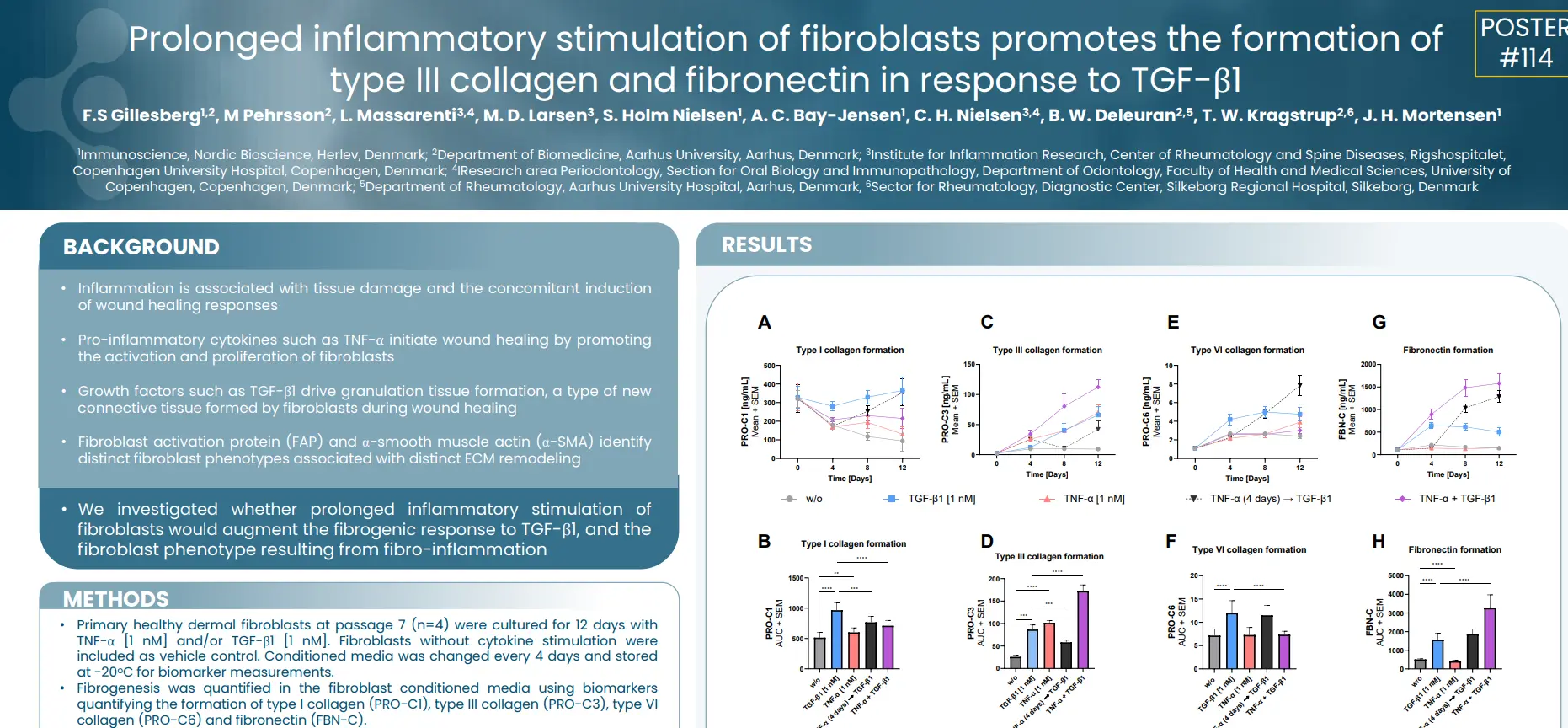
        Prolonged inflammatory stimulation of fibroblasts promotes the formation of type III collagen and fibronectin in response to TGF-β1

        Introduction

        Inflammation is associated with tissue damage and the concomitant induction of wound healing responses. Pro-inflammatory cytokines such as TNF-α initiate wound healing by promoting the activation and proliferation of fibroblasts. Growth factors such as TGF-β1 drive granulation tissue formation, a type of new connective tissue formed by fibroblasts during wound healing. Fibroblast activation protein (FAP) and α-smooth muscle actin (α-SMA) identify distinct fibroblast phenotypes associated with distinct ECM remodeling.

        In this study we investigated whether prolonged inflammatory stimulation of fibroblasts would augment the fibrogenic response to TGF-β1, and the fibroblast phenotype resulting from fibro-inflammation.

        Poster

        Conclusion

        TNF-α shifts TGF-β1 stimulated α-SMA+ fibroblasts towards a fibro-inflammatory FAP+ phenotype. The TGF-β1 + TNF-α stimulation is characterized by increased type III collagen and fibronectin formation, while type I and VI collagen are reduced, compared to TGF-β1 stimulation only. In conclusion, the soluble nordicPRO-C3™ and FBN-C biomarkers may be used to identify FAP + fibroblast activity, associated with fibrogenesis.

        Get in touch

        Are you interested in exploring collaboration possibilities?
Enter your information in the form and a representative will contact you shortly.

          Biomarkers Quantifying Changes in the Hair Follicular ECM Are Elevated in Alopecia Patients

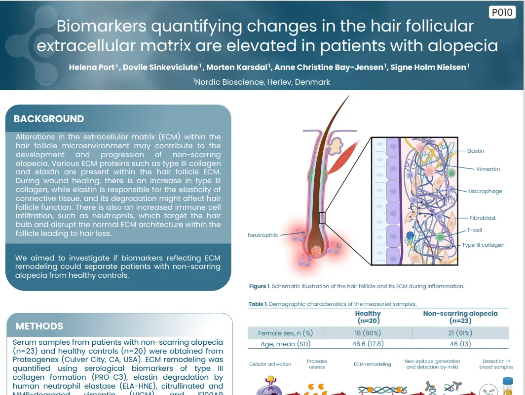
          Biomarkers quantifying changes in the hair follicular extracellular matrix are elevated in patients with alopecia

          Introduction

          Alterations in the extracellular matrix (ECM) within the hair follicle microenvironment may contribute to the development and progression of non-scarring alopecia. Various ECM proteins such as type III collagen
          and elastin are present within the hair follicle ECM. During wound healing, there is an increase in type III
          collagen, while elastin is responsible for the elasticity of connective tissue, and its degradation might affect hair follicle function. There is also an increased immune cell infiltration, such as neutrophils, which target the hair bulb and disrupt the normal ECM architecture within the follicle leading to hair loss.


          We aimed to investigate if biomarkers reflecting ECM remodeling could separate patients with non-scarring alopecia from healthy controls.

          Poster

          Conclusion

          Patients with alopecia had an increase in type III collagen formation and elastin degradation, indicating the wound healing processes as well as the loss of elasticity within the surrounding ECM of the hair follicle. Objective biomarkers quantifying changes the hair follicle and its surrounding tissues could serve as potential biomarkers of diagnosis and disease monitoring in alopecia.

          Get in touch

          Are you interested in exploring collaboration possibilities?
Enter your information in the form and a representative will contact you shortly.

            PRO-C22 Is Associated with Disease Severity, Activity, and Systemic Inflammation in HS

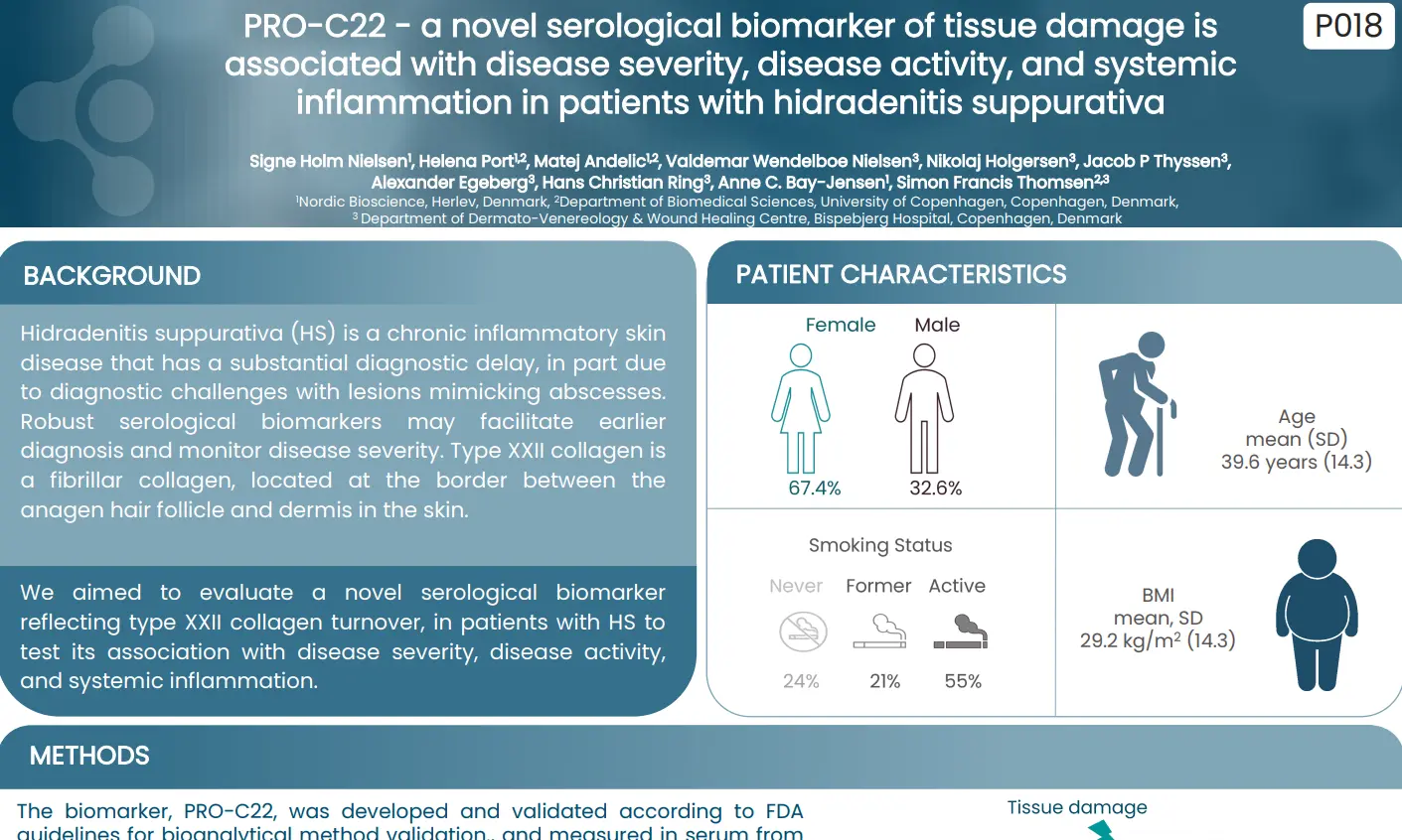
            PRO-C22 – A novel serological biomarker of tissue damage is associated with disease severity, disease activity, and systemic inflammation in patients with hidradenitis suppurativa

            Introduction

            Hidradenitis suppurativa (HS) is a chronic inflammatory skin disease that has a substantial diagnostic delay, in part due to diagnostic challenges with lesions mimicking abscesses. Utilizing robust serological biomarkers could aid in earlier diagnosis and better monitoring of disease severity. One such potential biomarker is type XXII collagen, a fibrillar collagen located at the interface between the anagen hair follicle and the dermis in the skin.

            In this study we aimed to evaluate a novel serological biomarker reflecting type XXII collagen turnover, in patients with HS to test its association with disease severity, disease activity, and systemic inflammation.

            Poster

            Conclusion

            We developed and evaluated the novel biomarker PRO-C22 reflecting tissue damage, which was associated to disease severity, activity and correlated with systemic inflammation in patients with HS. Such biomarker may be useful in early patient stratification and efficacy of treatment marker.

            Get in touch

            Are you interested in exploring collaboration possibilities?
Enter your information in the form and a representative will contact you shortly.

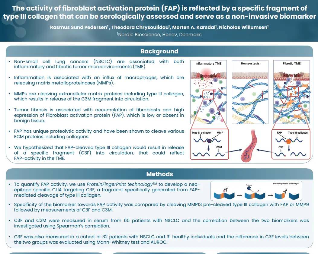
              FAP Activity Is Reflected by a Specific Fragment of Type III Collagen

              The activity of fibroblast activation protein (FAP) is reflected by a specific fragment of type III collagen that can be serologically assessed and serve as a non-invasive biomarker

              Introduction

              Non-small cell lung cancers (NSCLC) are associated with both inflammatory and fibrotic tumor microenvironments (TME). The inflammatory component involves an influx of macrophages, which release matrix metalloproteinases (MMPs) that cleave extracellular matrix proteins, including type III collagen, leading to the release of the C3M fragment into circulation. In parallel, tumor fibrosis is marked by the accumulation of fibroblasts and high expression of Fibroblast Activation Protein (FAP), which is typically low or absent in benign tissue. FAP possesses unique proteolytic activity and has been shown to cleave various ECM proteins, including collagens.

              Based on this, we hypothesized that FAP-mediated cleavage of type III collagen would generate a distinct fragment, C3F, that could enter the circulation and serve as a potential marker of FAP activity within the TME.

              Poster

              Conclusion

              FAP activity can be assessed by targeting a FAP-cleaved fragment of type III collagen, C3F and function as a non-invasive biomarker for patients with NSCLC. By showing that this fragment differs from the MMP-generated fragment C3M, this study supports the notion that different biological processes are reflected in the proteolytic degradation of the ECM and can be reflected by specific circulating protein fragments.

              Get in touch

              Are you interested in exploring collaboration possibilities?
Enter your information in the form and a representative will contact you shortly.

                Type XII Collagen Is Elevated in Patients with Solid Tumors

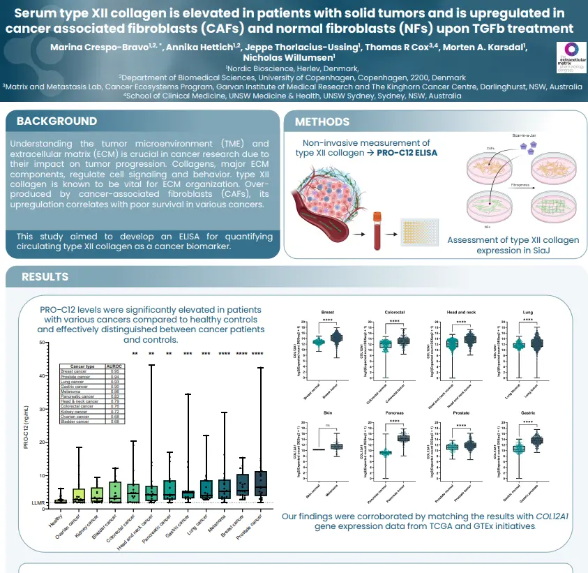
                Serum type XII collagen is elevated in patients with solid tumors and is upregulated in cancer associated fibroblasts (CAFs) and normal fibroblasts (NFs) upon TGFb treatment

                Introduction

                Understanding the tumor microenvironment (TME) and extracellular matrix (ECM) is crucial in cancer research due to their impact on tumor progression. Collagens, major ECM components, regulate cell signaling and behavior, with type XII collagen being particularly vital for ECM organization. This collagen type is often overproduced by cancer-associated fibroblasts (CAFs), and its upregulation has been linked to poor survival outcomes in various cancers. In light of these findings, this study aimed to develop an ELISA for quantifying circulating type XII collagen as a potential cancer biomarker.

                Poster

                Conclusion

                This study unveils a promising avenue for harnessing PRO-C12 as a non-invasive serum biomarker, enabling the quantification of type XII collagen fragments in cancer patients. Further investigations are warranted to explore the potential of PRO-C12 across different cancer types and disease stages, shedding light on its multifaceted role in cancer development.

                Get in touch

                Are you interested in exploring collaboration possibilities?
Enter your information in the form and a representative will contact you shortly.

                  Lessons Learned from the DALIAH Trial

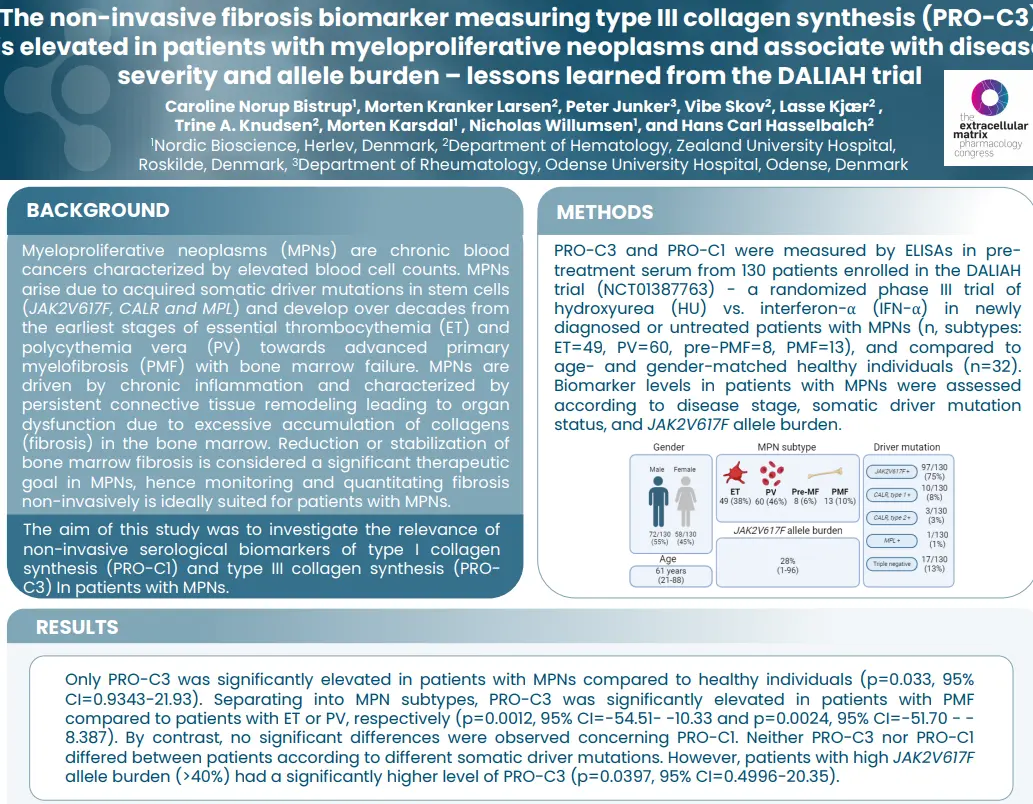
                  The non-invasive fibrosis biomarker measuring type III collagen synthesis (PRO-C3) is elevated in patients with myeloproliferative neoplasms and associate with disease severity and allele burden – lessons learned from the DALIAH trial

                  Introduction

                  Myeloproliferative neoplasms (MPNs) are chronic blood cancers characterized by elevated blood cell counts. MPNs arise due to acquired somatic driver mutations in stem cells (JAK2V617F, CALR and MPL) and develop over decades from the earliest stages of essential thrombocythemia (ET) and polycythemia vera (PV) towards advanced primary myelofibrosis (PMF) with bone marrow failure. MPNs are driven by chronic inflammation and characterized by persistent connective tissue remodeling leading to organ
                  dysfunction due to excessive accumulation of collagens (fibrosis) in the bone marrow. Reduction or stabilization of bone marrow fibrosis is considered a significant therapeutic goal in MPNs, hence monitoring and quantitating fibrosis non-invasively is ideally suited for patients with MPNs.


                  The aim of this study was to investigate the relevance of non-invasive serological biomarkers of type I collagen synthesis (PRO-C1) and type III collagen synthesis (nordicPRO-C3™) in patients with MPNs.

                  Poster

                  Conclusion

                  The non-invasive fibrosis associated biomarker nordicPRO-C3™, that reflects type III collagen synthesis, is elevated in MPN patients and associate with the severity of the disease and with the JAK2V617F allele burden. NordicPRO-C3™ has potential as a non-invasive bone marrow fibrosis biomarker to stratify and monitor patients with MPNs.

                  Get in touch

                  Are you interested in exploring collaboration possibilities?
Enter your information in the form and a representative will contact you shortly.

                    Variations in Fibrotic Activity of CAFs Measured Using Noninvasive Biomarkers

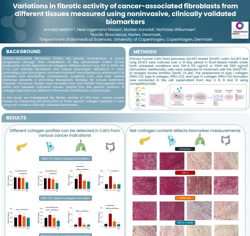
                    Variations in fibrotic activity of cancer-associated fibroblasts from different tissues measured using noninvasive, clinically validated biomarkers

                    Introduction

                    Cancer-associated fibroblasts (CAFs) are pivotal orchestrators of tumor progression through their modulation of the extracellular matrix (ECM), particularly via the deposition of collagens. Different stimuli (e.g. TGF-β, PDGF-AB, IL-1α) can activate fibroblasts and induce phenotypic alterations in CAFs, promoting a pro-tumorigenic microenvironment characterized by enhanced ECM synthesis and remodeling. Consequently, targeting CAFs and their related pathways presents a promising therapeutic strategy for cancer treatment. However, numerous studies have revealed that CAFs exhibit heterogeneity both within and between individual tissues. Despite this, the specific patterns of collagen deposition by different CAFs remain insufficiently characterized.

                    In this study, we investigated the fibrotic activity of CAFs from various cancer tissues by measuring the production of three specific collagen peptides in vitro using non-invasive, clinically validated biomarkers.

                    Poster

                    Conclusion

                    These findings underscore the heterogeneity in collagen production and fibrotic activity among CAFs from different indications, providing valuable insights into the ECM dynamics within distinct TMEs.
                    Collagen-based non-invasive biomarkers demonstrate the capability to differentiate between the fibrotic activity of CAFs isolated from different tissues. In conclusion, this model proves to be a useful tool for anti-fibrotic drug screening.

                    Get in touch

                    Are you interested in exploring collaboration possibilities?
Enter your information in the form and a representative will contact you shortly.