Minor Collagen Hypothesis in Cancer

The Minor Collagen Hypothesis

Collagen is a crucial protein that provides structural support and strength to tissues throughout the body. However, there is a group of highly specialized minor collagens that may play a critical role in cancer progression. Most research on the biology of collagens has focused on the abundant collagens, but the highly specialized minor collagens that are expressed only at certain developmental stages may be deregulated in cancer.

The Hypothesis

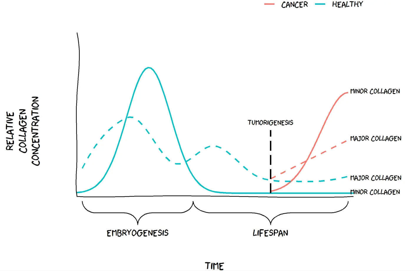
Minor collagens are widely expressed during embryogenesis, as they organize the extracellular matrix (ECM) to support tissue development. However, in adult tissues, minor collagens become restricted to a few specific organs and are expressed at very low levels.

Recent studies have suggested that minor collagens may become deregulated in cancer, where they are highly upregulated in tumor tissues. This upregulation is thought to occur as the ECM is reorganized during tumor development and progression. We have chosen to call this hypothesis “The Minor Collagen Hypothesis”. We hypothesize that minor collages, which are normally restricted to embryonic tissues, may become active again in adult tissues as cancer progresses. The Minor Collagen Hypothesis proposes that this reactivation may be a key component in in tumor growth and invasion by altering the composition of the ECM.
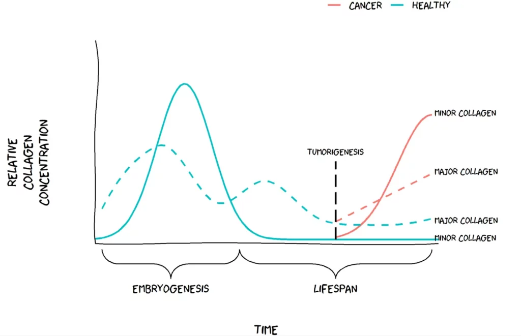
Visualization of the minor collagen hypothesis. Figure by Jeppe Thorlacius-Ussing.

Applying The Minor Collagen Hypothesis in clinical research

Biomarkers are important for indicating the presence or severity of a disease, and in cancer, they are crucial for early detection, monitoring of cancer progression, and guiding treatment decisions.

In general, several collagen biomarkers are overexpressed in cancer. However, minor collagens may offer a more specific biomarker for certain types of cancer. For example, recent research has shown that certain subtypes of cancer-associated fibroblasts (CAFs) express specific minor collagens. These collagens can be detected in blood, making them potential targets for drug development aimed at CAFs.

This hypothesis offers researchers new possibilities and highlights the need for more specific biomarkers for cancer diagnosis and treatment that could lead to better patient outcomes. Identifying biomarkers that detect specific minor collagens could be a breakthrough in cancer research and might shed light on an overlooked aspect of cancer progression.

Minor collagens are typically expressed during embryogenesis or tissue development and are expressed at low levels afterward. In cancer, however, these specialized collagens are expressed again to form an environment conducive to cancer growth. The abundant major collagens are also expressed during tissue development and growth spurts in childhood and adolescence but tapers off in adulthood. The relative induction as a function of cancer is greater for the minor collagens compared to the major collagens. This could mean better biomarker performance. 
Get in touch

Are you interested in exploring collaboration possibilities?
Enter your information in the form and a representative will contact you shortly.

    Tumor Fibrosis in Cancer: Fibroblast Activities and New Biomarkers

    Watch this insightful webinar on tumor fibrosis, where we will explore the latest advancements in cancer research with leading experts in the field.

    This webinar will provide a unique opportunity to hear from experts in the field and gain valuable insights into the latest advancements in tumor fibrosis research. Join us to learn about cutting-edge approaches to developing new therapeutic targets, and the use of biomarkers for patient stratification and evaluating treatment efficacy.

    Agenda

    Opening of the session – Dr. Morten A. Karsdal

    Tumor Fibrosis and Clinical Outcomes: Underlying Mechanisms – Dr. Saurabh Gupta

    The Collagen Landscape in Cancer: Fibroblast Activities and New Biomarkers – Dr. Nicholas Willumsen 

    Unique and unexpected role of collagens in cancer – Dr. Raghu Kalluri 

    Question from the chat

    Expert line-up

    Dr. Raghu Kalluri (Professor and Chairman, Department of Cancer Biology, University of Texas MD Anderson Cancer Center), is an expert in the cellular microenvironment and will discuss, among others, the unique and unexpected role of collagens in cancer.

    Dr. Saurabh Gupta (Senior Director, Clinical Biomarkers Oncology, Precision Medicine), Bristol-Myers Squibb, is a clinical drug development and biomarker research expert and will provide valuable insights into the underlying mechanisms of tumor fibrosis and the association with clinical outcomes.

    Dr. Nicholas Willumsen (Director, Oncology, Nordic Bioscience), will focus on the development and validation of blood-based biomarkers to quantify tumor fibrosis in serum from cancer patients and discuss how this may inform on cancer-associated fibroblast (CAF) activity and subtyping.

    Get in touch

    Are you interested in exploring collaboration possibilities?
Enter your information in the form and a representative will contact you shortly.

      Bile duct ligation in animal models

      Understanding Fibrosis Remodeling in Animal Models

      Animal testing is a crucial aspect of drug development, mandated by the FDA for all premature drugs in development. However, a critical question arises: how can we be sure that the pathology observed in rodents is reflective of the same pathology in humans? This is a significant concern for the medical community because of the potential for negative consequences if clinical trials are based on inaccurate preclinical data as we see in humans. Why then is it so important to measure biomarker formations in rodents? Because it matters whether it mimics the right pathology.

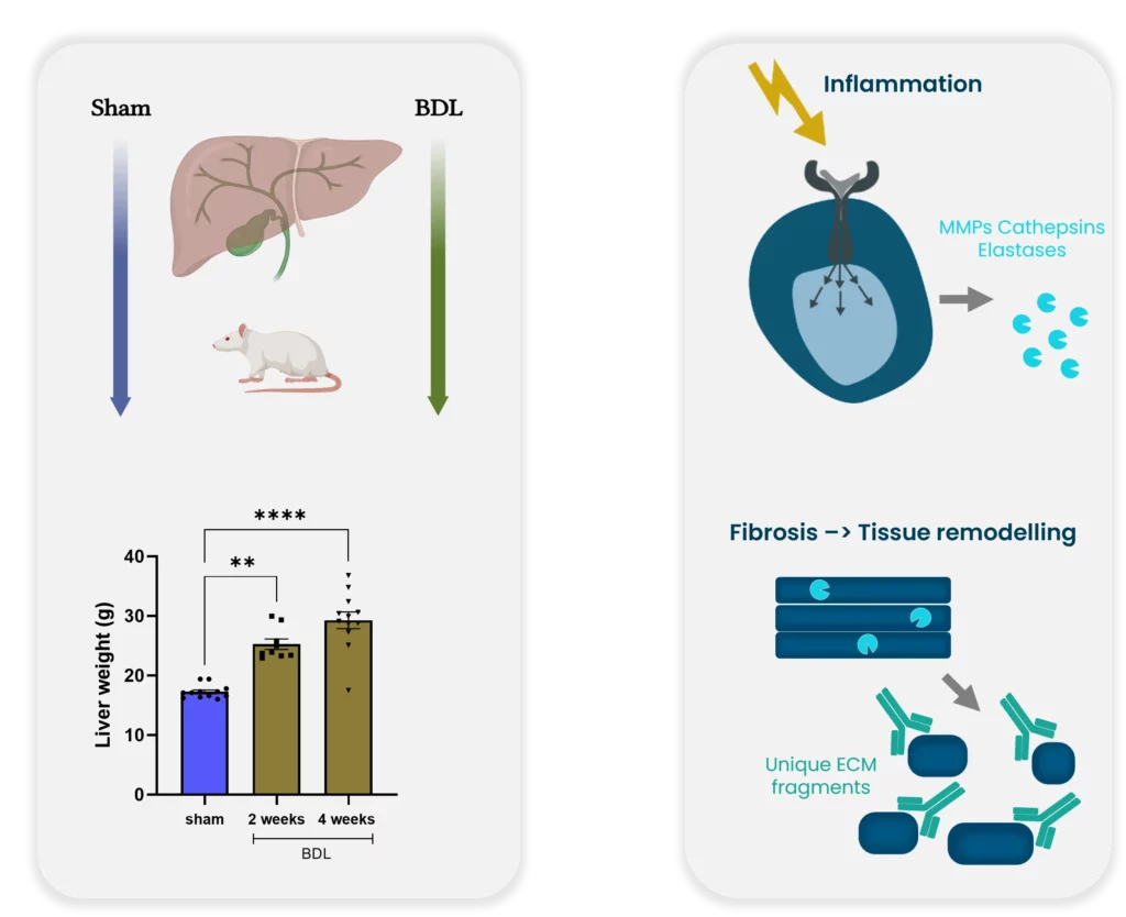
      Example for bridging preclinical and clinical practice. Bile duct ligation: a preclinical model of liver fibrosis.

      The process of transitioning from preclinical to clinical trials is critical to success. Histological stages are built on human tissue sections and used for comparison to create an appropriate animal model. However, none of the existing rodent models currently develop nonalcoholic steatohepatitis (NASH) as seen in humans. This discrepancy raises concerns regarding the usefulness of rodent models for preclinical studies.

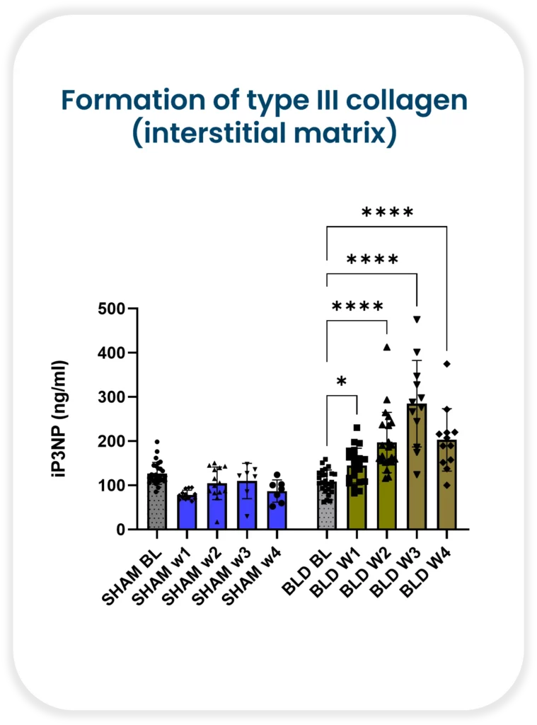
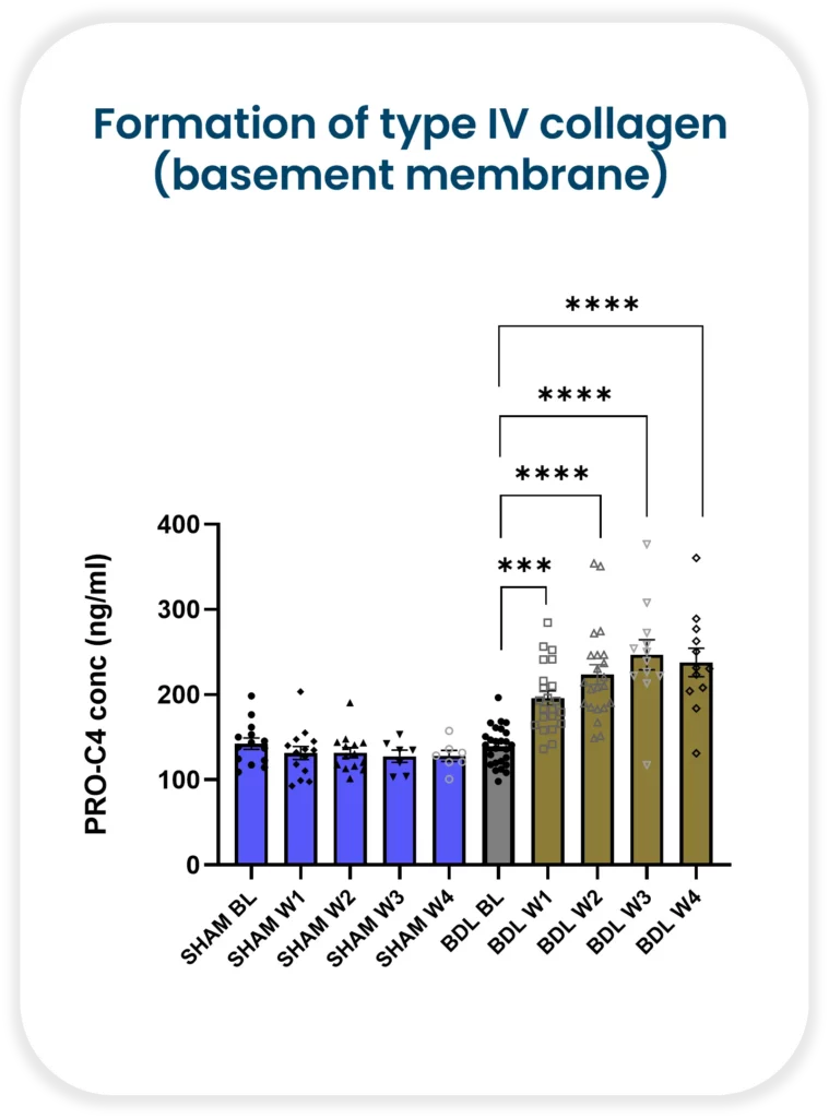
      Despite these concerns, it is still essential to measure biomarker formations in rodents. The reason being, it matters whether the biomarker mimics the correct pathology. Nordic Bioscience has demonstrated increasing formation of both type III collagen (interstitial matrix) and type IV collagen (basement membrane) with continued injury in a rat bile duct ligation model. Collagen III has been shown to be upregulated in human liver fibrosis, including NASH, and is associated with increased severity. On the other hand, collagen IV is a measure of early fibrosis remodeling.

      Bile duct ligation: a preclinical model of liver fibrosis

      It is critical to understand fibrosis remodeling during disease progression in both animals and humans. With Nordic Bioscience’s biomarkers, researchers can improve translation between the preclinical and clinical practice. The use of biomarkers enables researchers to track disease progression and evaluate the effectiveness of potential treatments accurately. Moreover, they can also help identify patients who are likely to respond positively to a particular therapy.

      Animal testing remains a critical aspect of drug development, it is essential to understand rodent model limitations. Biomarkers offer an effective way to track the progression of disease accurately. We can identify patients likely to benefit from a specific treatment and improve the transition from preclinical to clinical trials. By leveraging biomarkers in both animal and human models, we can make sure that the right therapy is provided to the right patient—at the right time.

      Get in touch

      Are you interested in exploring collaboration possibilities?
Enter your information in the form and a representative will contact you shortly.

        HFpEF and Extracellular Matrix Dysregulation: Novel Treatment Strategies

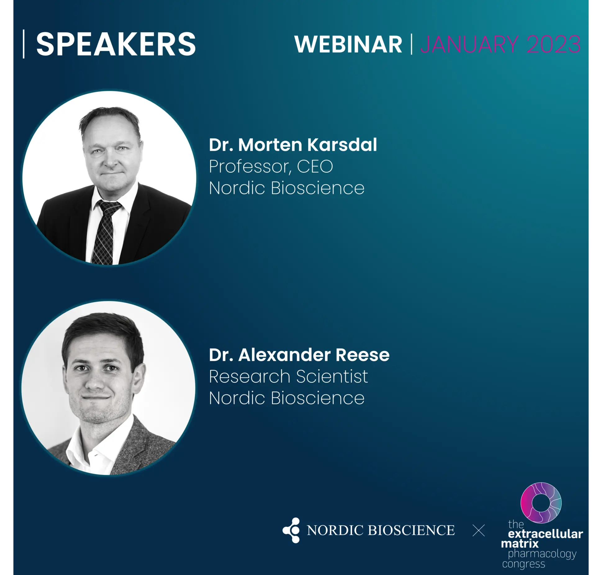
        The session will include a stimulating talk by Dr. Morten Karsdal on understanding the mysteries of the different types of collagens. Dr. Alexander Lynge Reese-Petersen will then spotlight collagen type VI and its relationship to various pathologies, as well as the pharmacological and biological properties of the dangerous hormone Endotrophin.

        Scientific topics

        Extracellular matrix (ECM) dysregulation is the founding cause of tissue fibrosis, a process driving more than 50 pathologies and most chronic diseases. Fibrosis may develop both consequent to either an increase in tissue formation or a decrease in tissue degradation.

        ​​​​​​​Understanding the ECM and the central components of the basement membrane and interstitial ECM will lead to a better understanding of disease mechanisms and to novel treatment strategies. This may be a central part in precision medicine strategies to target the right patients.

        Dr. Morten Karsdal will talk about:

        • ECM remodeling is the common denominator of all fibro-inflammatory disorders
        • Collagens have signaling molecules that we need to understand to understand how the ECM affects cells in fibrosis and tissue turnover
        • Endotrophin and other collagen fragments (Vastatin, Tumstatin and Endostatin) are potential signaling molecules derived from the processing of the collagens in the ECM
        • Understanding ECM turnover may lead to better insight into disease mechanisms and better biological understanding of individual patients

        Dr. Alexander Reese’s presentation will cover:

        • ECM biomarkers are elevated in chronic pathologies and have prognostic significance for outcome. How can we best use this to guide drug development?
        • Endotrophin, a collagen hormone in multi-organ diseases
        • Endotrophin in HFpEF, a potentially treatable high-risk endotype
        • Use of the tissue turnover profile for patient selection

        Get in touch

        Are you interested in exploring collaboration possibilities?
Enter your information in the form and a representative will contact you shortly.

          The CPa9-HNE (NordicCPa9™) biomarker in Crohn’s and Colitis

          CPa9-HNE ELISA has emerged as a novel serum calprotectin biomarker

          Status quo: Crohn’s and Colitis biomarkers

          Conventional serum calprotectin biomarkers are often not as clinically useful as the fecal versions, which is related to the half-life of calprotectin in blood (only 5-6 hours), leading to dissociation of calprotectin protein. The short half-life of calprotectin in blood reduces the window in which the current serum calprotectin ELISA assay can detect calprotectin dimer protein, which is composed of monomers S100A8 and S100A9.

          By applying the Protein Fingerprint technology, we have identified a neo-epitope of the S100A9 monomer of calprotectin derived from proteolytic cleavage by human neutrophil elastase (HNE). This ELISA assay is referred to as the CPa9-HNE (nordicCPa9™) assay and is a new and innovative method for quantification of calprotectin in serum developed by Nordic Bioscience.

          Inflammatory Bowel Disease (IBD) flares in the intestines.

          A new tool to measure… calprotectin

          Patients with Crohn’s disease and ulcerative colitis have been found to show significantly higher levels of CPa9-HNE (nordicCPa9™) in their serum than healthy subjects, with the AUC reaching 0.98 (CI: 0.97-1.00, p<0.0001) for CD and 0.96 (CI: 0.92-1.00, p<0.0001) for UC, proving the assay’s efficacy in distinguishing between disease states.

          In comparison, the MRP8/14 serum calprotectin assay from Bühlmann Lab, while also used to identify IBD, showed poorer separation between patients and healthy subjects, with AUCs of 0.72 (CI: 0.59-0.86, p=0.0025) for UC and 0.70 (CI: 0.56-0.84, p=0.0046) for CD, respectively.

          The nordicCPa9™ biomarker showed a better correlation with the endoscopic score for Crohn’s disease (SES-CD) and ulcerative colitis (MES) than the FCP. At the same time, the MRP8/14 assay and neutrophil count showed no significant correlation with endoscopic scores for IBD patients.

          To investigate the accuracy of the nordicCPa9™, we performed a received operator characteristic curve, demonstrating nordicCPa9™ with acceptable accuracy to identify patients with IBD in remission vs. active disease.

          Finally, we demonstrated that the nordicCPa9™ biomarker could detect the calprotectin neo-epitope only in the supernatant of in vitro activated primary human neutrophils, but not in inactive primary human neutrophils. This was in contrast to the MRP8/14 serum CP assay of the Bühlmann lab, as both inactive and activated primary human neutrophils secreted detectable levels of calprotectin.

          NordicCPa9™ ELISA proved to be a novel serum calprotectin biomarker with significant clinical potential as a biomarker for patients with IBD to monitor disease activity and neutrophil activity.

          However, circulating calprotectin metabolites released from locally inflamed mucosa are not be dissociated further. They can be readily quantified using an ELISA-based technique called Protein Fingerprint technology. This refined ELISA technique quantifies protein metabolites or neo-epitopes derived from proteolysis that reflect local tissue inflammation and remodeling.

          Read the full article here.

          Get in touch

          Are you interested in exploring collaboration possibilities?
Enter your information in the form and a representative will contact you shortly.

            NEJM Evidence study shows PRO-C6 to Segregate HFpEF

            PRO-C6 Findings In NEJM Evidence to Help Patient Segregation in HFpEF

            A persistent problem remains a challenge in HFpEF – patient heterogeneity.

            We need the right patients for the right treatment, but how do we get it to them? We believe that one approach to solving this problem is to improve patient segregation and endotyping.


            In collaboration with Bristol Myers Squibb and University of Pennsylvania, we recently identified a subset of patients at a very high risk of adverse outcomes, all characterized by increased fibroblast activity. Using our PRO-C6 biomarker assay we can accurately quantify this risk profile and differentiate patients based on how likely they are to be re-hospitalized or have all-cause mortality due to heart failure.

            These findings were recently published in NEJM Evidence.

            So how do we deal with patient heterogeneity?

            We believe it is time to give physicians and clinicians a better tool than what is available now.

            This is endotrophin endotyping, heart failure risk stratification redefined.

            Get in touch

            Are you interested in exploring collaboration possibilities?
Enter your information in the form and a representative will contact you shortly.

              Good versus bad fibroblasts

              Are fibroblasts good or are they bad?

              We need to talk about the good and the bad fibroblasts – the dangerous fibroblasts that overgrow organs and destroy organ function, but also the necessary specialized fibroblasts in bone, the osteoblasts. 

              Fibroblasts are an important type of cell in the human body that play a crucial role in tissue repair and maintenance. However, not all fibroblasts are created equal – there are both good and bad fibroblasts that have vastly different effects on the body.

              The dangerous fibroblasts, known as myofibroblasts, are responsible for pathological tissue remodeling in various diseases, such as fibrosis. These fibroblasts overgrow organs and destroy organ function. While myofibroblasts play a role in wound healing, their persistence and unchecked growth can lead to excessive scarring and tissue damage.

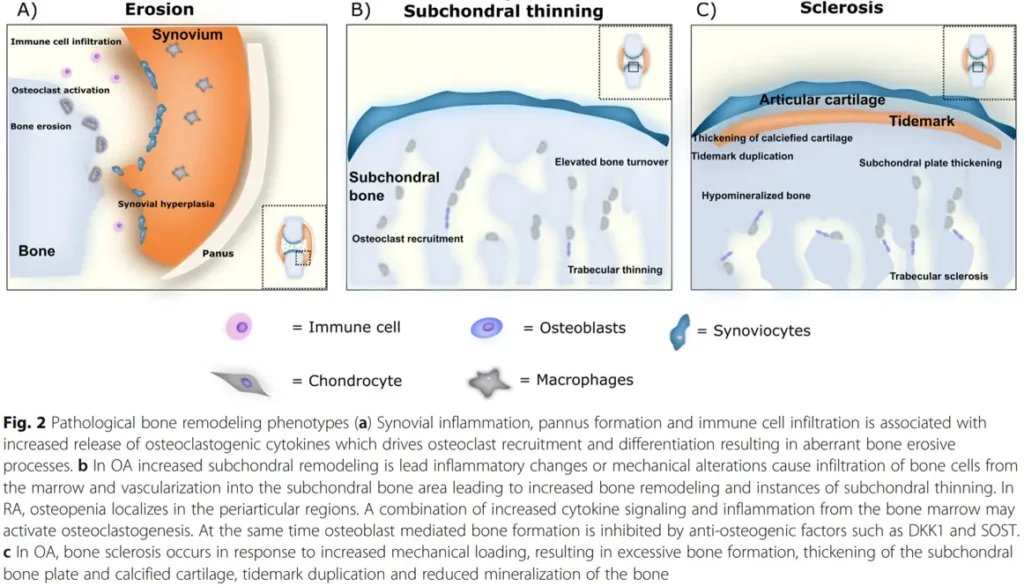
              Pathological bone remodeling phenotypes

              On the other hand, there are specialized fibroblasts, such as osteoblasts, that are necessary for maintaining bone health. Osteoblasts are responsible for producing the extracellular matrix that makes up bone tissue, and are crucial in the process of bone formation.

              Unfortunately, the success in biomedical sciences such as genomics and proteomics is not paralleled in the medical product development methods, resulting in a lack of translation into improved drug safety and efficacy. This can lead to some antifibrotic treatments having deleterious effects on bone fibroblasts, such as osteoblasts, and may cause unwanted side effects.

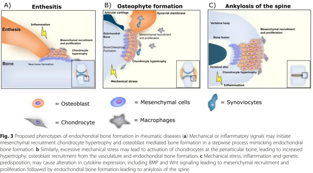
              Proposed phenotypes of endochondral bone formation in rheumatic diseases

              In rheumatic diseases, bone inflammation and remodeling phenotypes are proposed to be a result of endochondral bone formation. Studies have identified bone as a possible endocrine organ, and the availability of valid biochemical bone markers suggests that assessing bone turnover should also play an important role in general safety pharmacology.

              To address this issue, there is a need for improved methods to assess the effects of treatments on different types of fibroblasts. This is where bone inflammation panels come in – these panels can be used to assess whether a treatment is having a deleterious effect on bone fibroblasts, such as osteoblasts.

              In conclusion, while fibroblasts play a crucial role in tissue repair and maintenance, it is important to distinguish between the good and bad fibroblasts. Unchecked growth of myofibroblasts can lead to tissue damage and organ dysfunction, while osteoblasts are necessary for maintaining bone health. Improved methods of assessing the effects of treatments on different types of fibroblasts, such as bone inflammation panels, can help ensure that treatments are safe and effective.

              With our bone inflammation panel, you can assess whether your treatment is having a deleterious effect.

              Related publications:

              Should biochemical markers of bone turnover be considered standard practice for safety pharmacology?

              Biochemical markers in osteoarthritis with lessons learned from osteoporosis

              Role of biochemical markers in the management of osteoporosis

              Get in touch

              Are you interested in exploring collaboration possibilities?
Enter your information in the form and a representative will contact you shortly.

                The effect of copper depletion therapy on breast cancer patients

                The affect on the collagen microenvironment in breast cancer patients

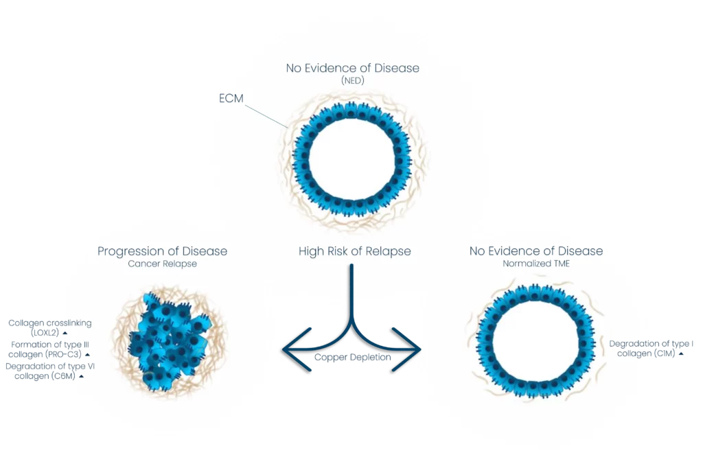
                In a recently published study based on a collaboration between Nordic Bioscience, Memorial Sloan Kettering Cancer Center, Weill Cornell Medicine, and Nancy E. and Peter C. Meinig School of Biomedical Engineering, we have examined the effect of copper depletion therapy on the collagen microenvironment in breast cancer patients with a high risk of relapse.

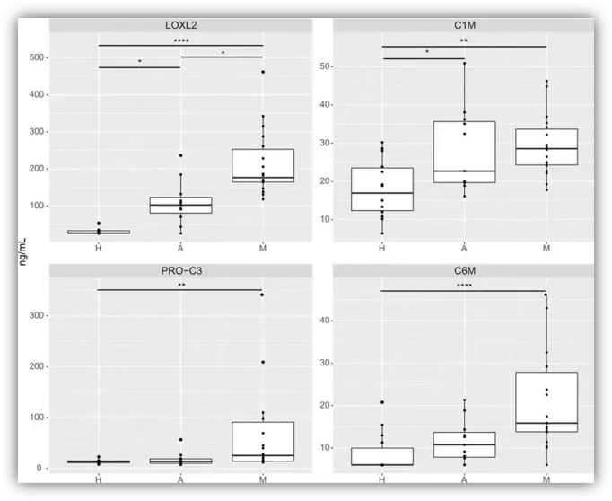
                Tetrathiomolybdate (TM) is a novel, copper-depleting compound associated with promising survival in a phase II study of patients with high-risk and triple-negative breast cancer. In the study, levels of all collagen biomarkers were higher in those with the disease, metastatic and adjuvant, as compared with healthy controls. 

                Baseline biomarkers of collagen crosslinking (LOXL2), collagen formation (PRO-C3), and collagen degradation (C1M and C6M) in the exploratory cohort

                We propose a novel mechanism for preventing metastases through altered collagen processing in the tumor microenvironment. We hypothesize that decreased collagen cross-linking and increased degradation caused by the treatment may alter the immune response in the pre-metastatic sites and thereby decrease the metastatic potential.

                The effect of copper depletion therapy on collagen turnover in breast cancer patients

                Preclinical studies revealed decreased collagen deposition, lower levels of myeloid-derived suppressor cells, and higher CD4+ T-cell infiltration in TM-treated mice compared with controls.

                In conclusion, the study showed novel mechanisms of TM targeting the TME and immune response with potential applications across cancer types.

                Get in touch

                Are you interested in exploring collaboration possibilities?
Enter your information in the form and a representative will contact you shortly.

                  Can we see the future of kidney function with a simple urine test?

                  The future of the kidney with a simple urine test

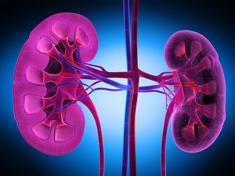
                  Autosomal dominant polycystic kidney disease (ADPKD) is a hereditary condition that causes the development of cysts in the kidneys. This condition affects millions of people worldwide and can lead to kidney failure if left untreated. One of the biggest challenges in treating ADPKD is identifying biomarkers that can predict disease progression and response to treatment.

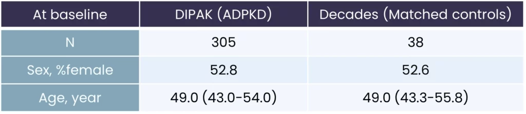
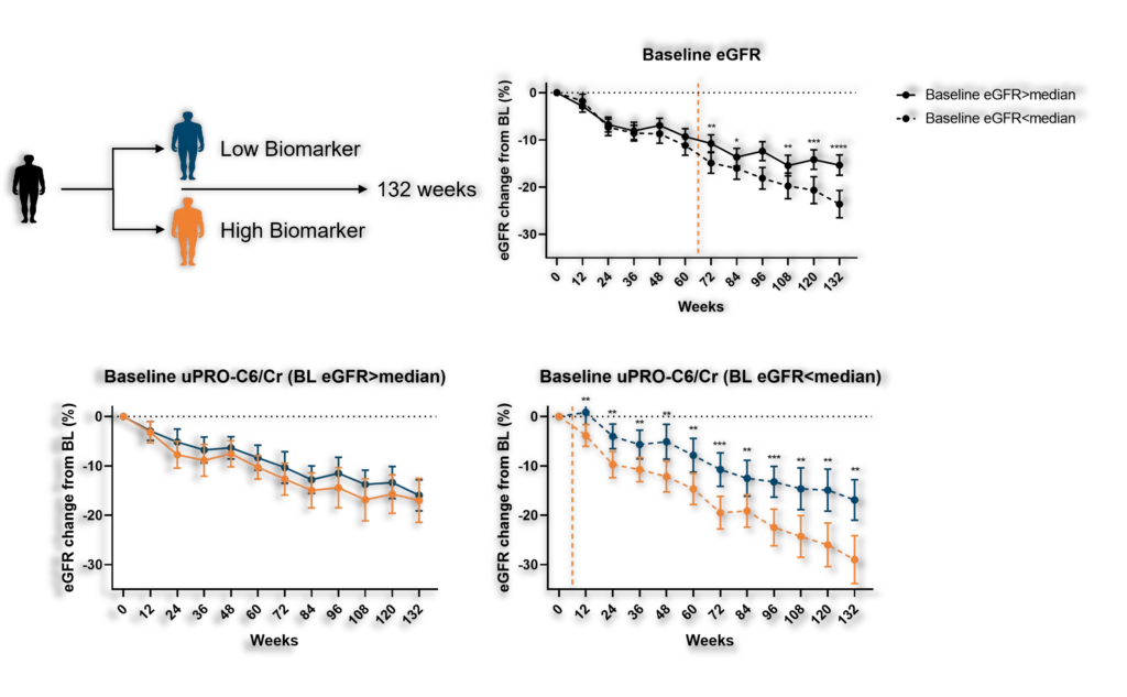
                  Our Renal and Cardiovascular Research team showed that biomarkers of collagen remodeling measured in urine and circulation at baseline are associated with the rate of decline in kidney function in patients with ADPKD. In the study (DIPAK-1), we measured the effect of lanreotide on patients with stage 3 chronic kidney disease. The following demographics were involved:

                  DIPAK-1 demographics

                  To assess the effect of lanreotide, the team selected a number of biomarkers that were best suited to assess the effect of the drug on interstitial matrix turnover and basement membrane turnover. We have selected a number of biomarkers:

                  • PRO-C3, measuring collagen type III formation in serum and urine (interstitial matrix turnover)
                  • C3M, measuring collagen type III degradation in serum and urine (interstitial matrix turnover)
                  • PRO-C6, measuring collagen type VI formation in serum and urine. Also measures the release of the bioactive fragment endotrophin, associated with pro-fibrotic and pro-inflammatory processes (interstitial matrix turnover)
                  • LG1M, measuring Laminin gamma 1 degradation in serum and urine (basement membrane turnover)
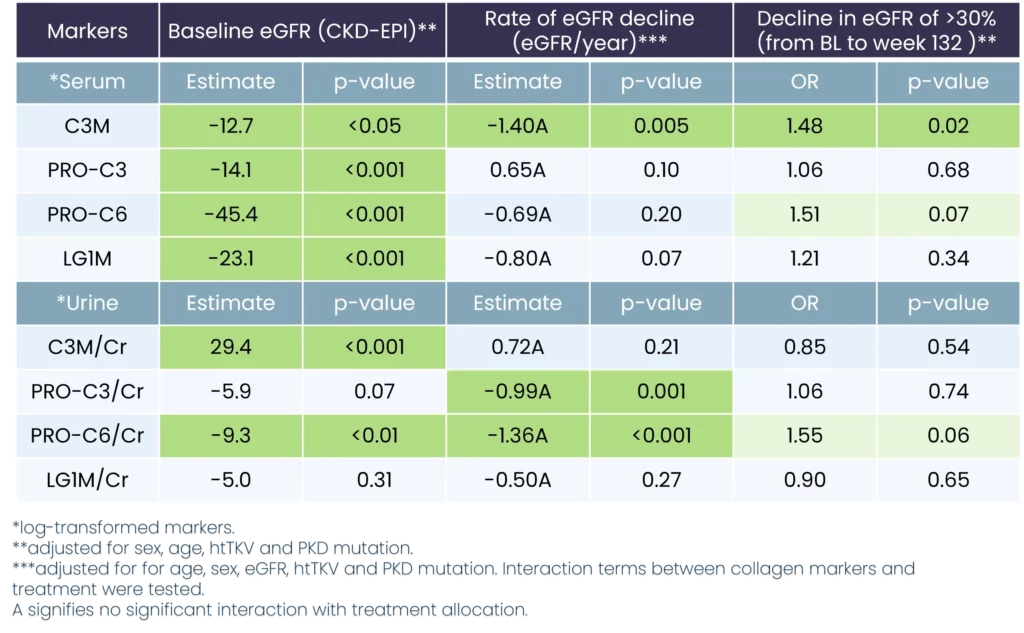
                  Kidney biomarker data compared to baseline values
                  Predicting decline in kidney function with prognostic biomarkers

                  The data we found implies that fibrogenesis may be an important pathophysiological process driving ADPKD disease progression. In addition, the use and development of drugs that interfere with fibrogenesis may be promising to halt disease progression in ADPKD. However, we still need to validate in an independent cohort, preferably in early-stage disease.

                  The findings of the study are significant because they offer new insights into the potential for biomarkers to predict disease progression in ADPKD. This information may help clinicians to identify patients who are at risk of developing kidney failure and to develop more effective treatments to slow or halt disease progression.

                  We have shown that biomarkers of collagen remodeling are associated with the rate of decline in kidney function in patients with ADPKD. These findings suggest that fibrogenesis may be an important pathophysiological process driving ADPKD disease progression and that the use of drugs that interfere with fibrogenesis may be promising to halt disease progression. Further research is needed to validate these findings in an independent cohort, but the results of this study offer new hope for patients with ADPKD.

                  We believe it is time to put kidney fibrosis more in the center – even in diseases where other aspects (such as kidney volume) are the focus of attention. At Nordic Bioscience, we have the tools to do so. 

                  Get in touch

                  Are you interested in exploring collaboration possibilities?
Enter your information in the form and a representative will contact you shortly.

                    Load absorption with osteoarthritis

                    How much load can the cartilage in the joints absorb?

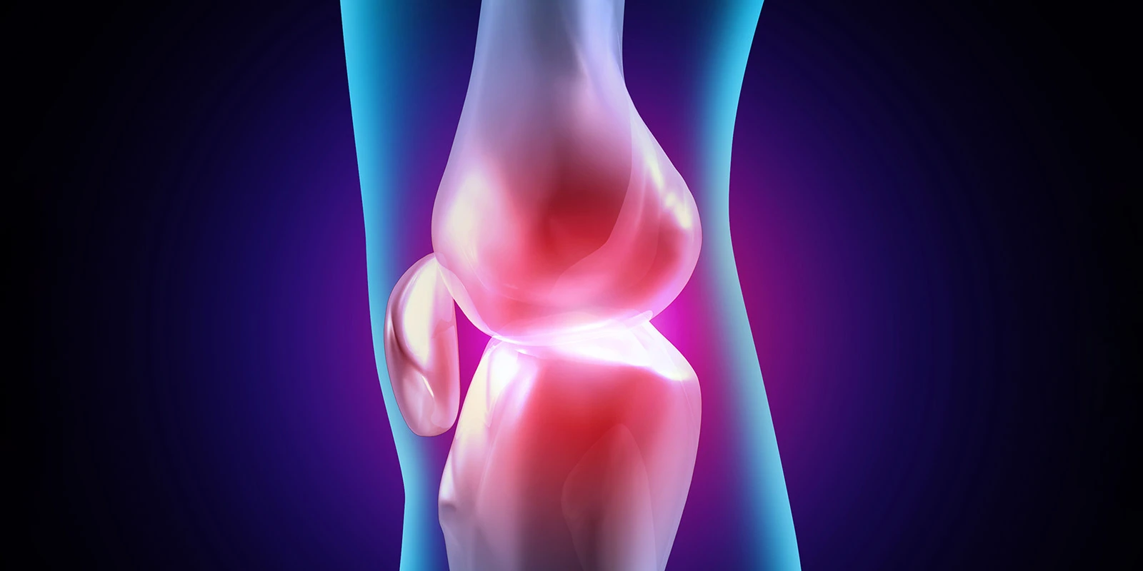
                    Mechanical loading is an essential part of the function and maintenance of the joint. Despite the importance of intermittent mechanical loading, this factor is rarely considered in preclinical models of cartilage, limiting their translatability.

                    The cartilage of the joints of patients with osteoarthritis is negatively affected in their capacity to absorb the load. Despite this pivotal role, mechanical load is rarely a component of translational drug screening assays when testing novel OA treatments.

                    We showcased our novel translational cartilage loading model in a publication in Applied Sciences.

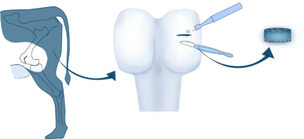
                    Cartilage explant isolation

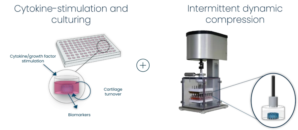
                    In a culture plate format well suited for lead candidate screening we investigated the effect of growth factors on ex-vivo cartilage remodeling and the interaction with dynamic intermittent loading. Cartilage remodeling was investigated in the presence of IGF-1 or TGF-β1, as well as a TGF-β receptor 1 (ALK5) kinase inhibitor and assessed with biomarkers for type II collagen formation and fibronectin degradation (FBN-C).

                    Dynamic compression and cartilage remodeling in Bovine Cartilage Explants

                    Amongst others, PRO-C2 has shown that not only does mechanical loading preserve cartilage, but it also positively modulates the effect of known growth factors, such as IGF-1.

                    Screening potential drug candidates in physiological loading conditions could provide a more accurate translation to push forward the urgent medical need for better treatments of osteoarthritis.

                    Get in touch

                    Are you interested in exploring collaboration possibilities?
Enter your information in the form and a representative will contact you shortly.