Serum immunoassays identify cartilage acidic protein (CRTAC1) as a biomarker of osteoarthritis
Introduction
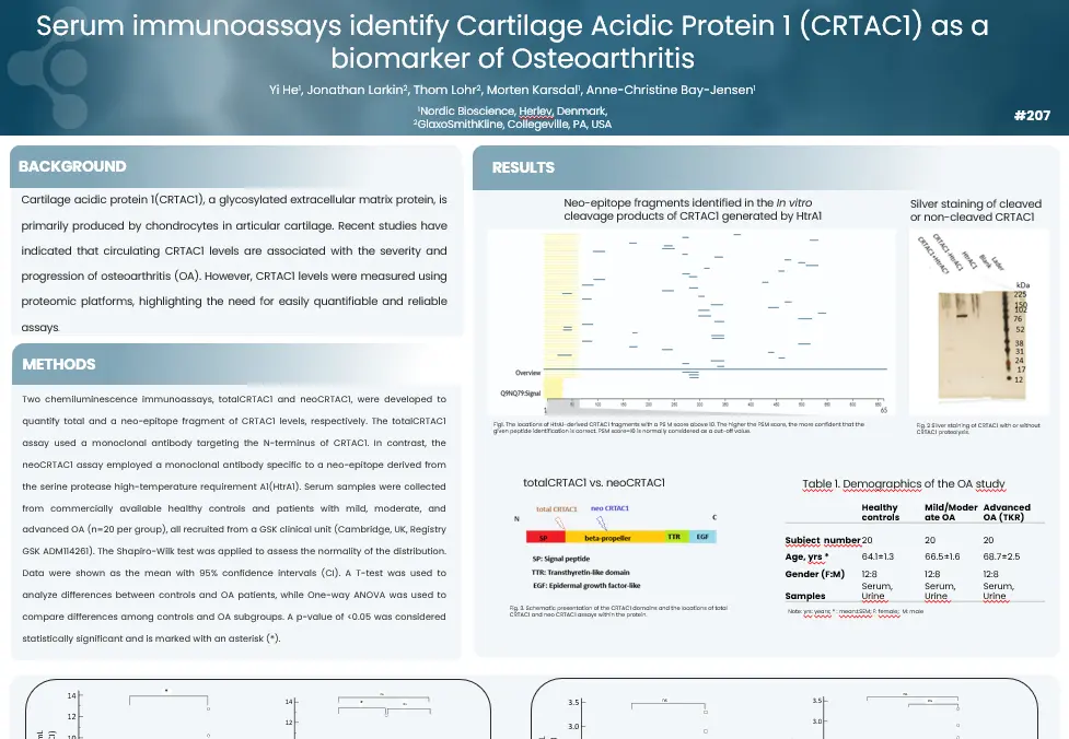
Cartilage acidic protein 1 (CRTAC1), a glycosylated extracellular matrix protein, is primarily produced by chondrocytes in articular cartilage. Recent studies have indicated that circulating CRTAC1 levels are associated with the severity and progression of osteoarthritis (OA). However, CRTAC1 levels were measured using proteomic platforms, highlighting the need for easily quantifiable and reliable assays.
This study validated CRTAC1 as a promising new biomarker for OA, utilizing easily quantifiable serum immunoassays. Differences in diagnostic performance between the total and neo-epitope CRTAC1 assays were observed. In conclusion, this study offers valuable insights into the role of CRTAC1 and its degradation process in Osteoarthritis.
Get in touch
Are you interested in exploring collaboration possibilities? Enter your information in the form and a representative will contact you shortly.
Developing a blood-based biomarker targeting α-synuclein fragments for the early diagnosis of PD
Introduction
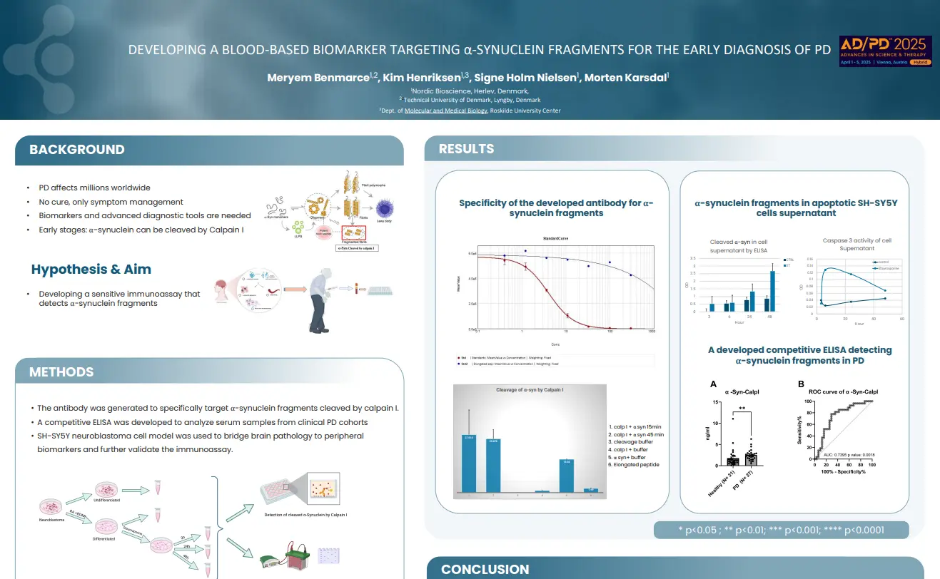
Parkinson’s disease (PD) affects millions worldwide and currently has no cure. Treatments focus only on managing symptoms, explaining why there is a pressing need for biomarkers and advanced diagnostic tools to enable earlier detection and better disease management. In the early stages of PD, α-synuclein—can be cleaved by Calpain I, presenting a potential target for biomarker development.
The aim of this study was to develop a sensitive immunoassay that detects α-synuclein fragments.
α-Synuclein fragments cleaved by calpain I are key early drivers of Parkinson’s disease pathology. This blood-based biomarker holds promise for enabling early diagnosis and identifying patients who are likely to respond to treatment.
Get in touch
Are you interested in exploring collaboration possibilities? Enter your information in the form and a representative will contact you shortly.
Endotrophin, quantified by a novel biomarker, is prognostic of outcome in heart failure patients
Introduction
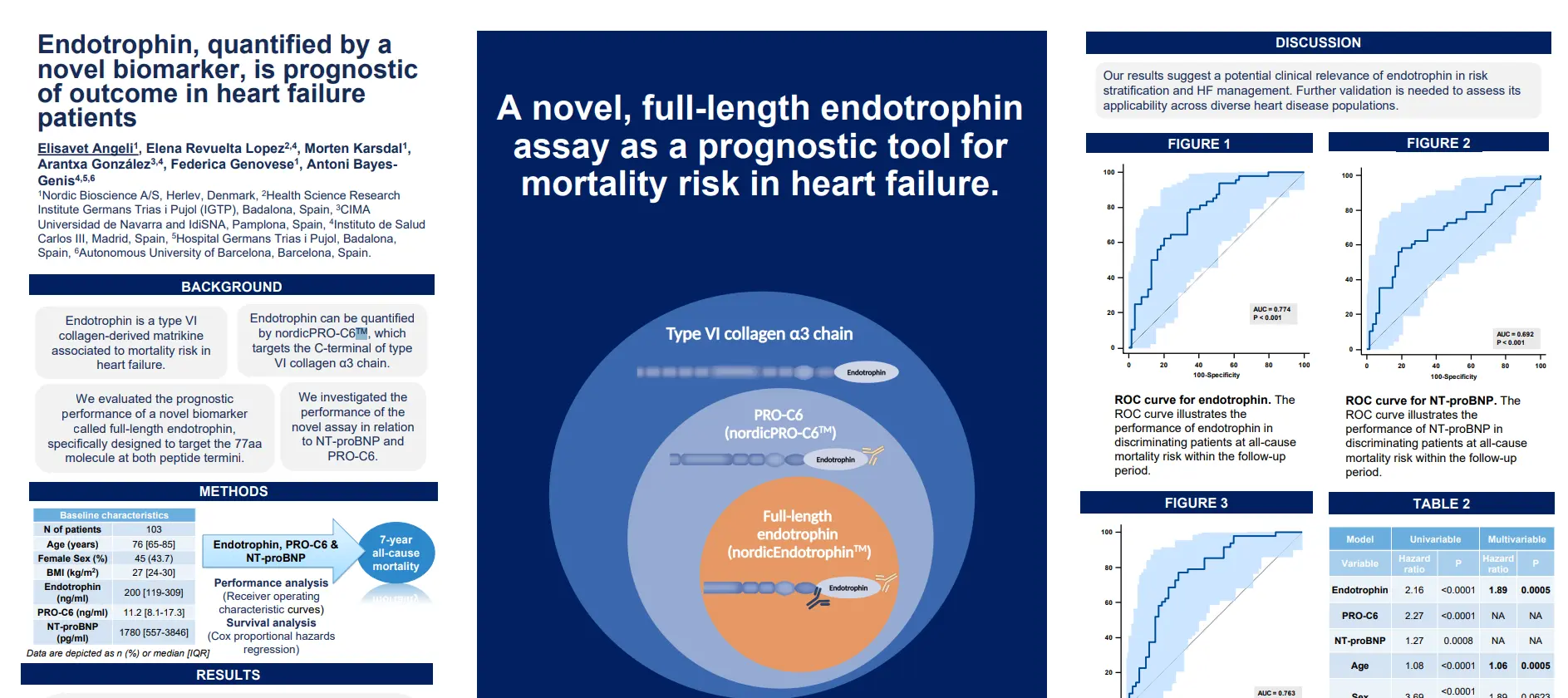
NordicEndotrophin™ is a type VI collagen-derived matrikine associated to mortality risk in heart failure. It can be quantified by nordicPRO-C6™, which targets the C-terminal of type VI collagen α3 chain. In this study, we evaluated the prognostic performance of a novel biomarker called full-length nordicEndotrophin™, specifically designed to target the 77aa molecule at both peptide termini. We investigated the performance of the novel assay in relation to NT-proBNP and nordicPRO-C6™.
This study presents the novel biomarker of full-length nordicEndotrophin™ demonstrating comparable performance to NT-proBNP and nordicPRO-C6™ in predicting mortality risk in HF patients. The biomarker’s prognostic value remains robust even after adjusting for relevant risk factors.
Get in touch
Are you interested in exploring collaboration possibilities? Enter your information in the form and a representative will contact you shortly.
A novel urinary marker of collagen type 1 degradation reflects kidney disease severity and fibrosis in IgA nephropathy.
Introduction
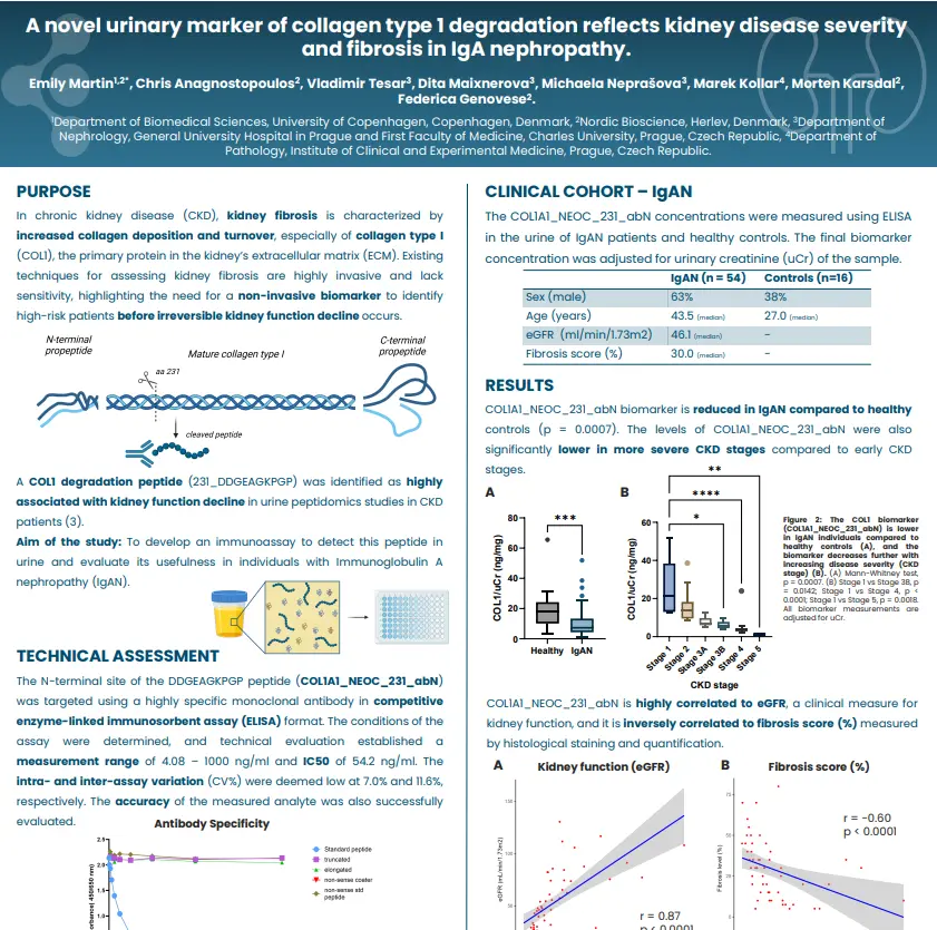
In chronic kidney disease (CKD), kidney fibrosis is characterized by increased collagen deposition and turnover, especially of collagen type I (COL1), the primary protein in the kidney’s extracellular matrix (ECM). Existing techniques for assessing kidney fibrosis are highly invasive and lack sensitivity, highlighting the need for a non-invasive biomarker to identify high-risk patients before irreversible kidney function decline occurA COL1 degradation peptide (231_DDGEAGKPGP) was identified as highly associated with kidney function decline in urine peptidomics studies in CKD patients (3).
The aim of this study was to develop an immunoassay to detect this peptide in urine and evaluate its usefulness in individuals with Immunoglobulin A nephropathy (IgAN).
We developed a novel and robust urinary assay which showed potential as a non-invasive biomarker of kidney disease severity and fibrosis in IgAN. It will be of interest to evaluate its prognostic potential in appropriate kidney disease cohorts and further in other organ diseases to understand the specificity of the biomarker to CKD.
Get in touch
Are you interested in exploring collaboration possibilities? Enter your information in the form and a representative will contact you shortly.
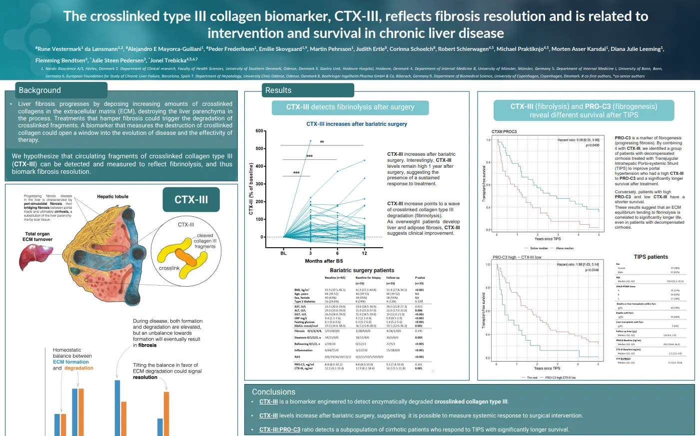
The crosslinked type III collagen biomarker, CTX-III, reflects fibrosis resolution and is related to intervention and survival in chronic liver disease
Introduction
Liver fibrosis progresses by deposing increasing amounts of crosslinked collagens in the extracellular matrix (ECM), destroying the liver parenchyma in the process. Treatments that hamper fibrosis could trigger the degradation of crosslinked fragments. A biomarker that measures the destruction of crosslinked collagen could open a window into the evolution of disease and the effectivity of therapy. We hypothesize that circulating fragments of crosslinked collagen type III (nordicCTX-III™) can be detected and measured to reflect fibrinolysis, and thus biomark fibrosis resolution.
NordicCTX-III™ is a biomarker engineered to detect enzymatically degraded crosslinked collagen type III. Its levels have been shown to increase after bariatric surgery, suggesting it is possible to measure systemic response to surgical intervention. Additionally, the nordicCTX-III™: nordicPRO-C3™ ratio detects a subpopulation of cirrhotic patients who respond to TIPS with significantly longer survival.
Get in touch
Are you interested in exploring collaboration possibilities? Enter your information in the form and a representative will contact you shortly.
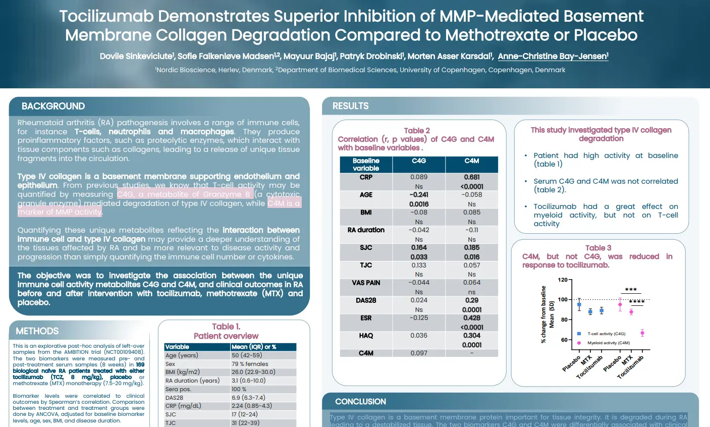
Tocilizumab demonstrates superior inhibition of MMP-mediated basement membrane collagen degradation compared to methotrexate or placebo
Introduction
Rheumatoid arthritis (RA) pathogenesis involves a range of immune cells, for instance T-cells, neutrophils and macrophages. They produce proinflammatory factors, such as proteolytic enzymes, which interact with tissue components such as collagens, leading to a release of unique tissue fragments into the circulation. Type IV collagen is a basement membrane supporting endothelium and epithelium. From previous studies, we know that T-cell activity may be quantified by measuring C4G, a metabolite of Granzyme B (a cytotoxic granule enzyme) mediated degradation of type IV collagen, while C4M is a marker of MMP activity. Quantifying these unique metabolites reflecting the interaction between immune cell and type IV collagen may provide a deeper understanding of the tissues affected by RA and be more relevant to disease activity and progression than simply quantifying the immune cell number or cytokines.
The aim of this study was to investigate the association between the unique immune cell activity metabolites C4G and C4M, and clinical outcomes in RA before and after intervention with tocilizumab, methotrexate (MTX) and placebo.
Type IV collagen is a basement membrane protein important for tissue integrity. It is degraded during RA leading to a destabilized tissue. The two biomarkers studied, C4G and C4M, were differentially associated with clinical outcome measures. Importantly, only C4M, a marker of MMP-derived tissue destruction, could be inhibited by tocilizumab. None of the markers were modulated by MTX.
Get in touch
Are you interested in exploring collaboration possibilities? Enter your information in the form and a representative will contact you shortly.
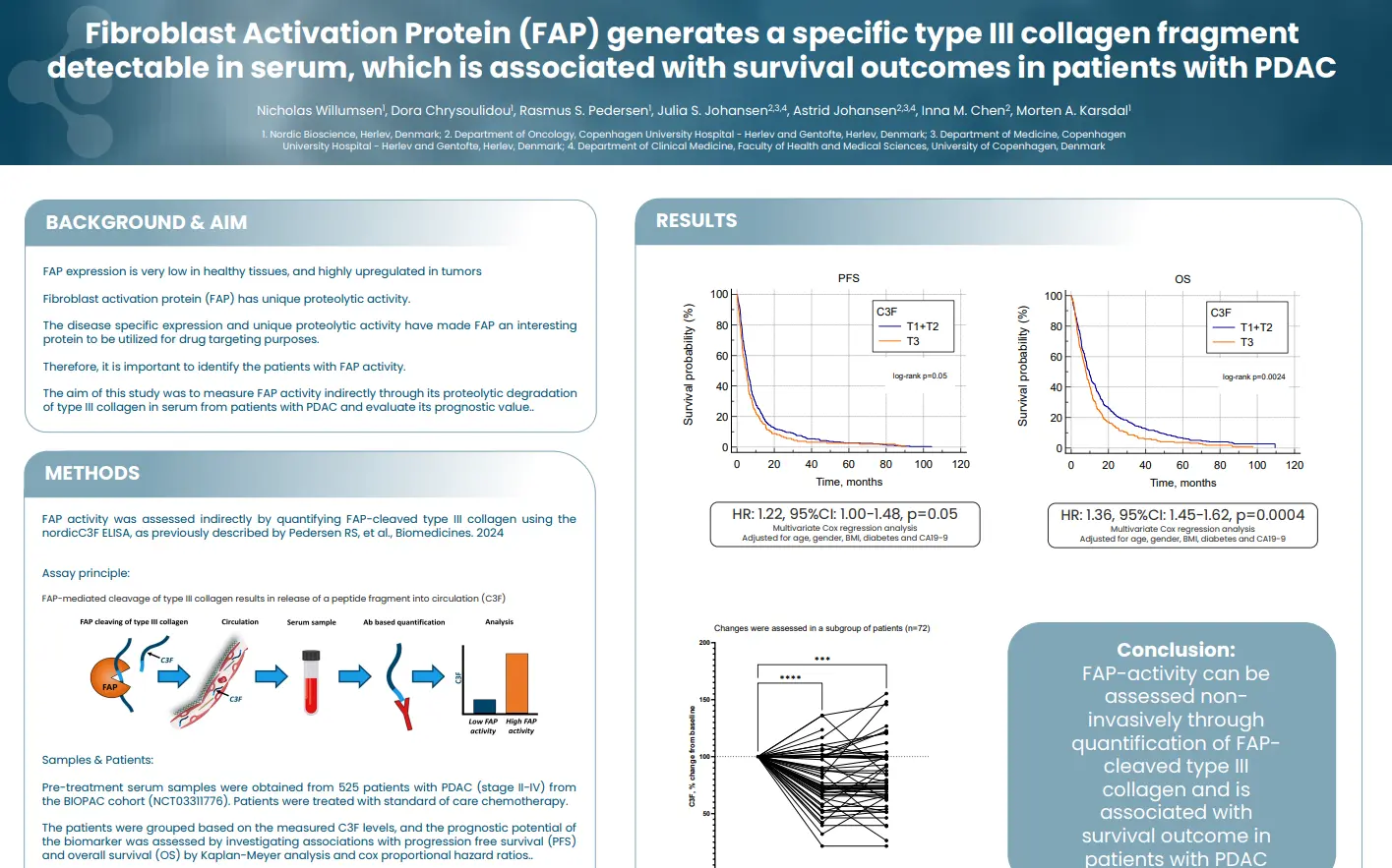
Fibroblast Activation Protein (FAP) generates a specific type III collagen fragment detectable in serum, which is associated with survival outcomes in patients with PDAC
Introduction
FAP expression is very low in healthy tissues, and highly upregulated in tumors Fibroblast activation protein (FAP) has unique proteolytic activity. The disease specific expression and unique proteolytic activity have made FAP an interesting protein to be utilized for drug targeting purposes. Therefore, it is important to identify the patients with FAP activity.
In this study we aimed to measure FAP activity indirectly through its proteolytic degradation of type III collagen in serum from patients with PDAC and evaluate its prognostic value.
FAP-activity can be assessed non-invasively through quantification of FAP-cleaved type III collagen and is associated with survival outcome in patients with PDAC.
Get in touch
Are you interested in exploring collaboration possibilities? Enter your information in the form and a representative will contact you shortly.
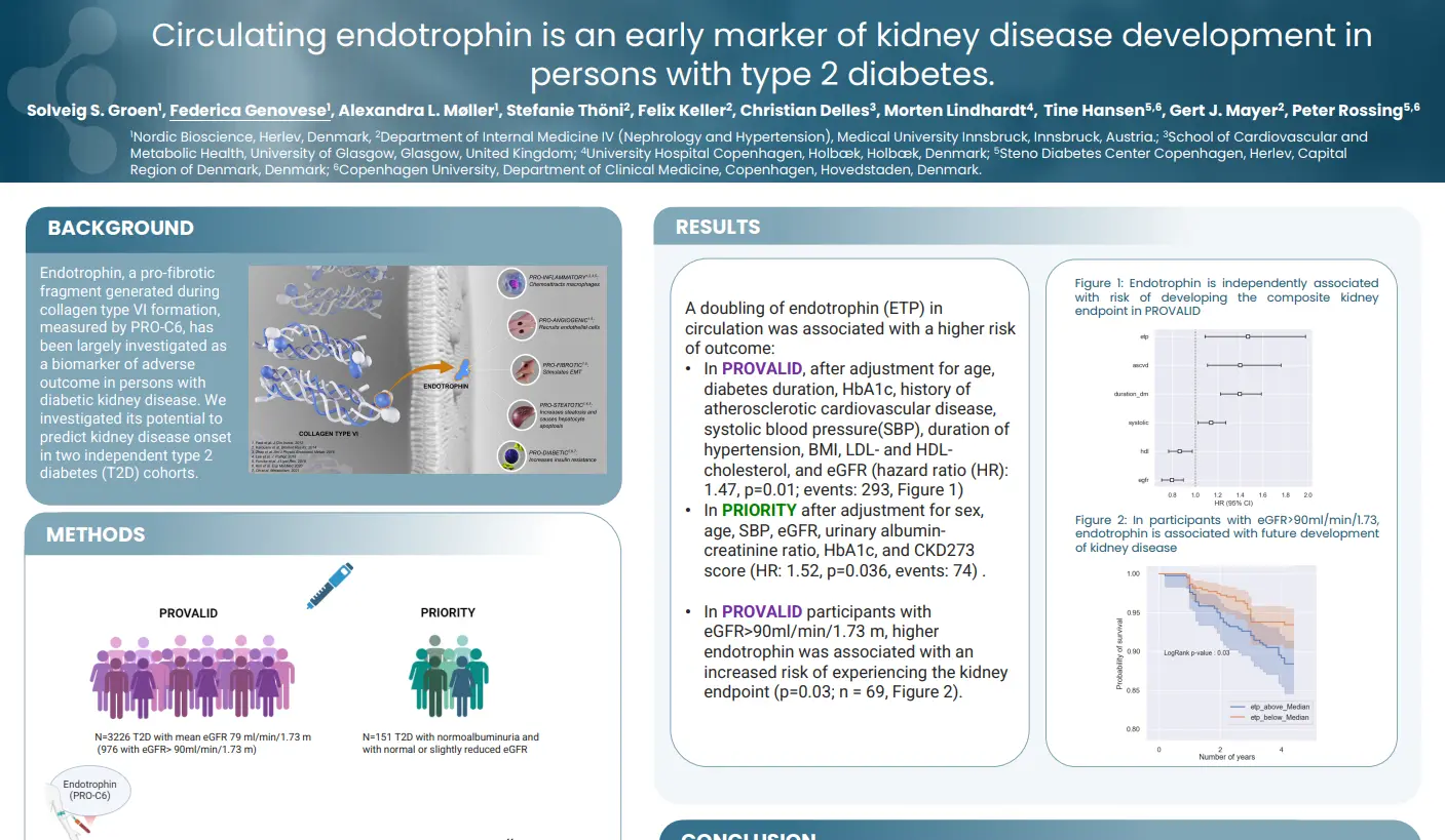
Circulating endotrophin is an early marker of kidney disease development in persons with type 2 diabetes
Introduction
NordicEndotrophin™ (Endotrophin), a pro-fibrotic fragment generated during collagen type VI formation, measured by nordicPRO-C6™, has been largely investigated as a biomarker of adverse outcome in persons with diabetic kidney disease. We investigated its potential to predict kidney disease onset in two independent type 2 diabetes (T2D) cohorts.
Circulating nordicEndotrophin™, measured by the nordicPRO-C6™ assay, was a risk marker for kidney outcomes in people with T2D without or with early kidney disease. This adds to the evidence that nordicEndotrophin™is a relevant biomarker of kidney complications in T2D, even in persons with no or mild kidney disease at baseline.
Get in touch
Are you interested in exploring collaboration possibilities? Enter your information in the form and a representative will contact you shortly.
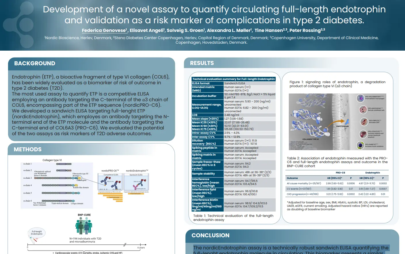
Development of a novel assay to quantify circulating full-length endotrophin and validation as a risk marker of complications in T2D
Introduction
NordicEndotrophin™ (ETP), a bioactive fragment of type VI collagen (COL6), has been widely evaluated as a biomarker of risk of outcome in type 2 diabetes (T2D). The most used assay to quantify ETP is a competitive ELISA employing an antibody targeting the C-terminal of the α3 chain of COL6, encompassing part of the ETP sequence (nordicPRO-C6™).
We developed a sandwich ELISA targeting full-lenght ETP (nordicEndotrophin™), which employes an antibody targeting the N-terminal end of the ETP molecule and the antibody targeting the C-terminal end of COL6A3 (nordicPRO-C6™). We evaluated the potential of the two assays as risk markers of T2D adverse outcomes.
The nordicEndotrophin™ assay is a technically robust sandwich ELISA quantifying the full-lenght endotrophin molecule in circulation. This biomarker presents a similar, or possibly higher prognostic power for complications of T2D than the competitive ELISA nordicPRO-C6™, used so far to quantify nordicEndotrophin™ in circulation.
Get in touch
Are you interested in exploring collaboration possibilities? Enter your information in the form and a representative will contact you shortly.
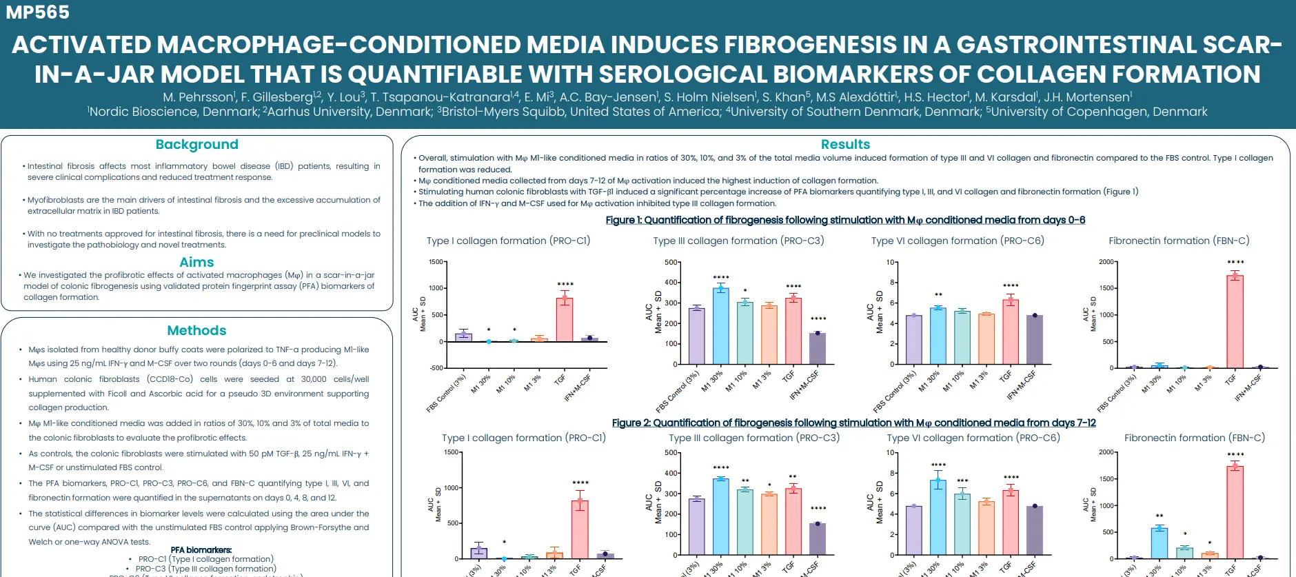
Activated macrophage-conditioned media induces fibrogenesis in a gastrointestinal scar-in-a-jar model that is quantifiable with serological biomarkers of collagen formation
Introduction
Intestinal fibrosis affects most inflammatory bowel disease (IBD) patients, resulting in severe clinical complications and reduced treatment response. Driven primarily by myofibroblasts, this condition is characterized by the excessive accumulation of extracellular matrix in the intestines. With no treatments approved for intestinal fibrosis, there is a need for preclinical models to investigate the pathobiology and novel treatments.
In this study we investigated the profibrotic effects of activated macrophages (Mφ) in a scar-in-a-jar model of colonic fibrogenesis using validated protein fingerprint assay (PFA) biomarkers of collagen formation.
The noninvasive PFA biomarkers can be used to objectively quantify fibrogenesis in the in vitro Scar-in-a-Jar model, providing a valuable tool for investigating the underlying mechanisms of fibrogenesis.
Get in touch
Are you interested in exploring collaboration possibilities? Enter your information in the form and a representative will contact you shortly.