Non-invasive biomarkers of ECM turnover are prognostic for combinations of checkpoint inhibition immunotherapy in solid tumors
Introduction
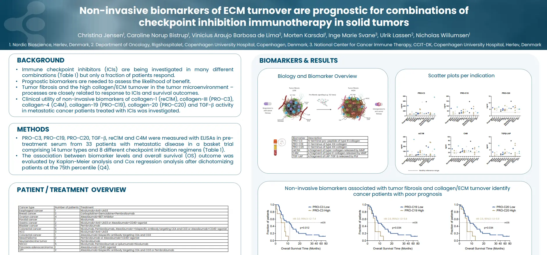
Immune checkpoint inhibitors (ICIs) are being investigated in many different combinations (Table 1) but only a fraction of patients respond. This highlights the need for prognostic biomarkers that can help identify patients most likely to respond. Tumor fibrosis and the high collagen/ECM turnover in the tumor microenvironment – processes are closely related to response to ICIs and survival outcomes.
Across a diverse cohort of patients with metastatic cancer treated with different checkpoint inhibition regimens, non-invasive biomarkers associated with tumor fibrosis and collagen/ECM turnover (nordicPRO-C3™, PRO-C19, PRO-C20, TGF-β, reC1M and C4M) could identify cancer patients with poor prognosis.
Get in touch
Are you interested in exploring collaboration possibilities? Enter your information in the form and a representative will contact you shortly.
Unraveling collagen signatures in cancer-associated fibroblasts: A biomarker-driven approach
Introduction
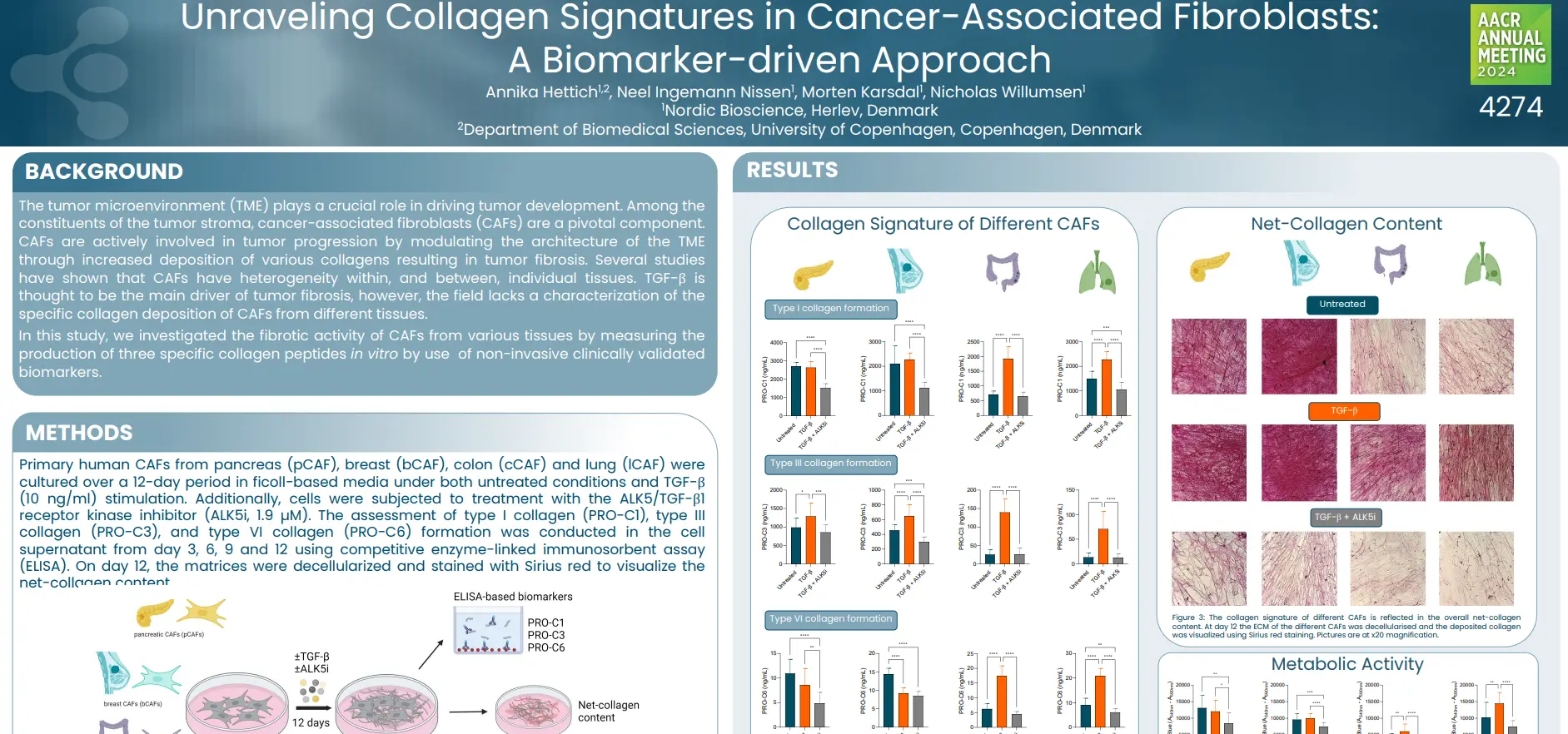
The tumor microenvironment (TME) plays a crucial role in driving tumor development. Among the constituents of the tumor stroma, cancer-associated fibroblasts (CAFs) are a pivotal component. CAFs are actively involved in tumor progression by modulating the architecture of the TME through increased deposition of various collagens resulting in tumor fibrosis. Several studies have shown that CAFs have heterogeneity within, and between, individual tissues. TGF-β is thought to be the main driver of tumor fibrosis, however, the field lacks a characterization of the specific collagen deposition of CAFs from different tissues.
In this study, we investigated the fibrotic activity of CAFs from various tissues by measuring the production of three specific collagen peptides in vitro by use of non-invasive clinically validated biomarkers.
These findings underscore the heterogeneity in collagen production among CAFs from different indications, providing valuable insights into the ECM dynamics within distinct TMEs. Collagen-based non-invasive biomarkers further demonstrate the capability to differentiate between the fibrotic activity of CAFs isolated from different tissues. These insights support the utility of this model as a useful tool for anti-fibrotic drug screening.
Get in touch
Are you interested in exploring collaboration possibilities? Enter your information in the form and a representative will contact you shortly.
Novel tissue turnover biomarkers are associated with prolonged QTc and pulmonary arterial hypertension in patients with systemic sclerosis
Introduction
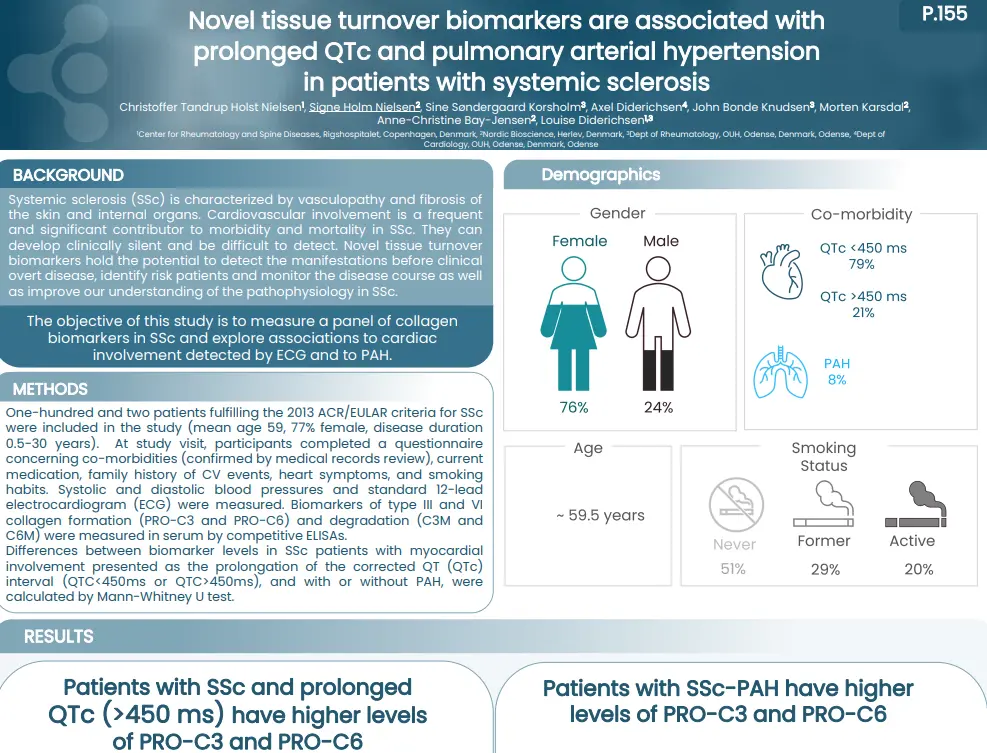
Systemic sclerosis (SSc) is characterized by vasculopathy and fibrosis of the skin and internal organs. Cardiovascular involvement is a frequent and significant contributor to morbidity and mortality in SSc. They can develop clinically silent and be difficult to detect. Novel tissue turnover biomarkers hold the potential to detect the manifestations before clinical overt disease, identify risk patients and monitor the disease course as well as improve our understanding of the pathophysiology in SSc.
The objective of this study is to measure a panel of collagen biomarkers in SSc and explore associations to cardiac involvement detected by ECG and to PAH.
Patients with SSc and prolonged ECG presented an altered tissue turnover, by an increased level of nordicPRO-C3™ and nordicPRO-C6™. In addition, SSc patients with presence of PAH had increased levels of nordicPRO-C3™ and nordicPRO-C6™ as well. presented an altered tissue turnover in presence of PAH, and with a QTc>450 ms. Our study indicates that they could serve as biomarkers of these manifestations and warrant further studies in cardiac disease in SSc.
Get in touch
Are you interested in exploring collaboration possibilities? Enter your information in the form and a representative will contact you shortly.
TNF-α and TGF-β Synergistically Promote Fibrogenesis in a In Vitro Model of Fibro-inflammation
Introduction
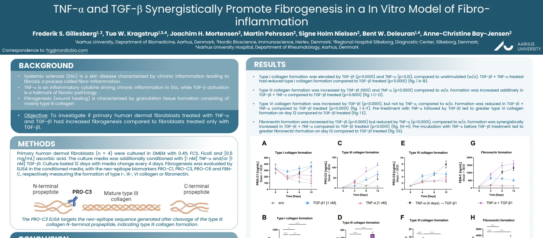
Systemic sclerosis (SSc) is a skin disease characterized by chronic inflammation leading to fibrosis, a process called fibro-inflammation. TNF-α is an inflammatory cytokine driving chronic inflammation in SSc, while TGF-β activation is a hallmark of fibrotic pathology. Fibrogenesis (wound healing) is characterized by granulation tissue formation consisting of mainly type III collagen.
The aim of this study was to investigate if primary human dermal fibroblasts treated with TNF-α and TGF-β1 had increased fibrogenesis compared to fibroblasts treated only with TGF-β1.
Inflammatory TNF-α stimulation increases TGF-β driven fibrogenesis in dermal fibroblasts, by promoting their formation of type III collagen and fibronectin. Consequently, biomarkers of type III collagen formation and fibronectin formation may be markers of early fibrosis in fibro-inflammatory skin disease.
Get in touch
Are you interested in exploring collaboration possibilities? Enter your information in the form and a representative will contact you shortly.
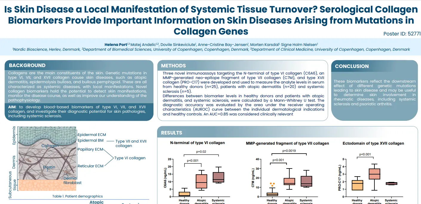
Is Skin Disease a Local Manifestation of Systemic Tissue Turnover? Serological Collagen Biomarkers Provide Important Information on Skin Diseases Arising from Mutations in Collagen Genes
Introduction
Collagens are the main constituents of the skin. Genetic mutations in type VI, VII, and XVII collagen cause skin diseases, such as atopic dermatitis, epidermolysis bullosa, and bullous pemphigoid. These are all characterized as systemic diseases, with local manifestations. Novel collagen biomarkers hold the potential to detect skin manifestations, monitor the disease course, as well as improve our understanding of the pathophysiology.
The aim of this study was to develop blood-based biomarkers of type VI, VII, and XVII collagen, and investigate their diagnostic potential for skin pathologies, including systemic sclerosis.
These biomarkers reflect the downstream effect of different genetic mutations leading to skin disease and may be useful to determine skin involvement in rheumatic diseases, including systemic sclerosis and psoriatic arthritis.
Get in touch
Are you interested in exploring collaboration possibilities? Enter your information in the form and a representative will contact you shortly.
Hi-drad-uh-NIE-tis sup-yoo-ruh-TIE-vuh – also known as hidradenitis suppurutiva (HS) – is a pathologically complicated skin condition, where chronic skin inflammation leads to abscesses and scarring. It is a systemic disease with local manifestations, meaning that the chronic insult to the skin is systemic, but it is physically located where skin rubs against skin, such as the armpits, groins and under the breasts. It is well known that immune cells, such as neutrophils and mast cells are involved, but what do we know about tissue remodeling?
When the beautiful collagens of the skin become a part of disease pathogenesis
Patients with HS not only experience pain from the neutrophil-rich tunnels but also from excessive tissue remodeling that causes scarring of the skin. These patients have an imbalance in tissue formation and tissue repair, partly due to the excessive activity of immune cells, which release enzymes that degrade the skin.
One group of tissue-degrading enzymes are matrix metalloproteinases, abbreviated as MMPs. These are released by macrophages, the most numerous inflammatory cells found in HS patients. MMPs infiltrate and contribute to HS pathology, signaling that increased activity of MMPs degrades the proteins of the skin tissue, such as collagens. This process can be quantified by specific blood-based biomarker assays targeting this pathological process.
Pathological fragments in HS may be used to identify disease types
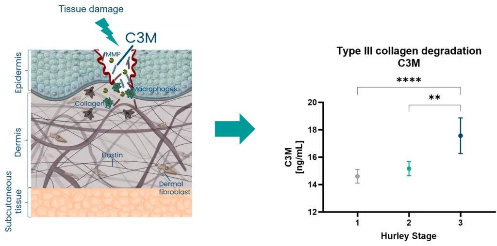
In HS, biomarkers of tissue remodeling such as type III collagen degraded by MMPs (C3M), are associated with disease severity (Hurley Staging).
Figure 1. Biomarkers of tissue remodeling associate with Hurley Stages
C3M is released upon MMP activation and measures dermal tissue remodeling. This raises the question – can we use C3M to identify subtypes of patients based on their disease activity, and potentially molecular endotypes to help select the right treatment for the patients?
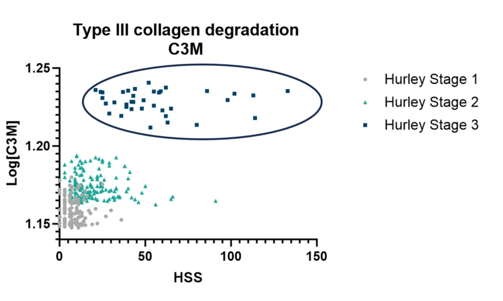
To address this, the levels of C3M are different depending on how active the disease is when divided into the Sartorious Score (HSS).
Finding the patients with high C3M levels reflects high disease activity, and may indicate a different subtype of patients needing a different treatment type.
Figure 2. Type II collagen degradation biomarker C3M as a patient stratification tool
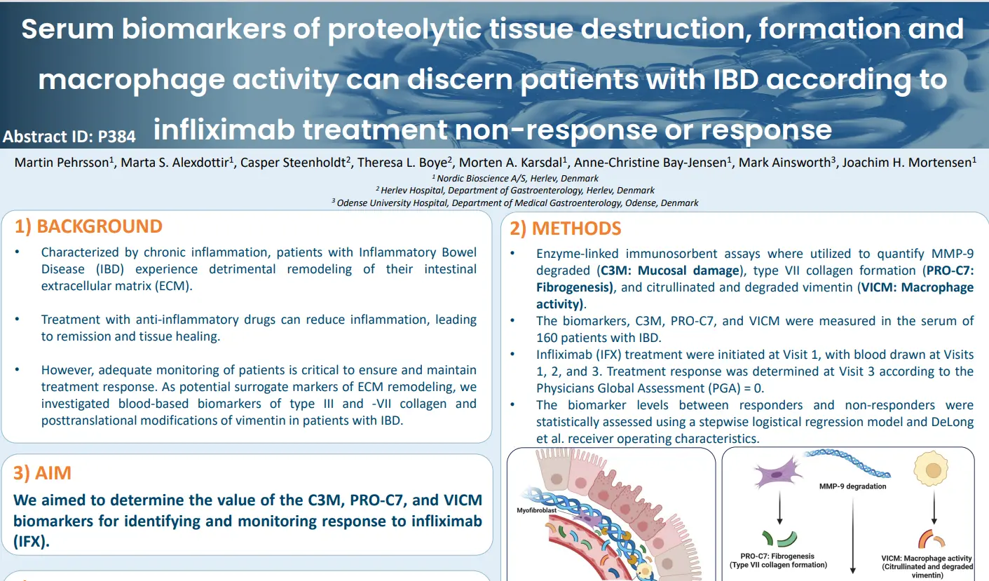
Serum biomarkers of proteolytic tissue destruction, formation and macrophage activity can discern patients with IBD according to infliximab treatment non-response or response
Introduction
Characterized by chronic inflammation, patients with Inflammatory Bowel Disease (IBD) experience detrimental remodeling of their intestinal extracellular matrix (ECM). Treatment with anti-inflammatory drugs can reduce inflammation, leading to remission and tissue healing. However, adequate monitoring of patients is critical to ensure and maintain treatment response.
As potential surrogate markers of ECM remodeling, we investigated blood-based biomarkers of type III and -VII collagen and posttranslational modifications of vimentin in patients with IBD. Our aim was to determine the value of the C3M, PRO-C7, and VICM biomarkers for identifying and monitoring response to infliximab (IFX).
Quantifying a combination of non-invasive biomarkers of ECM remodeling and macrophage activity provided AUCs of 0.684 to 0.797 identifying responders to IFX treatment. Each biomarker provided value at the three different visits (Visit 1, 2, and 3). Combining all three biomarkers measured at each visit resulted in an AUC of 0.797 identifying responders to IFX treatment.
Get in touch
Are you interested in exploring collaboration possibilities? Enter your information in the form and a representative will contact you shortly.
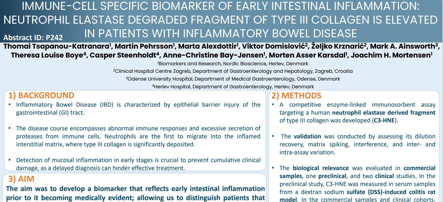
Immune-cell specific biomarker of early intestinal inflammation: Neutrophil elastase degraded fragment of type III collagen is elevated in patients with inflammatory bowel disease
Introduction
Inflammatory Bowel Disease (IBD) is characterized by epithelial barrier injury of the gastrointestinal (GI) tract and is driven by abnormal immune responses and excessive secretion of proteases from immune cells. Among these, neutrophils are the first to migrate into the inflamed interstitial matrix, where type III collagen is significantly deposited. Early detection of mucosal inflammation is crucial to prevent cumulative clinical damage, as a delayed diagnosis can hinder effective treatment.
In this study we aimed to develop a biomarker that reflects early intestinal inflammation prior to it becoming medically evident; allowing us to distinguish patients that would benefit from an anti-inflammatory treatment.
C3-HNE levels are elevated in patients with IBD compared with HD. This increase is also observed in conditioned media from primary neutrophils activated with lipopolysaccharide (LPS) for six hours. Importantly, C3-HNE reflects the early stages of clinically apparent mucosal damage in experimental models of colitis. As such, this biomarker holds promise for identifying early mucosal injury or acute inflammation in the gastrointestinal tract. Nevertheless, additional studies are needed to evaluate its clinical validity.
Get in touch
Are you interested in exploring collaboration possibilities? Enter your information in the form and a representative will contact you shortly.
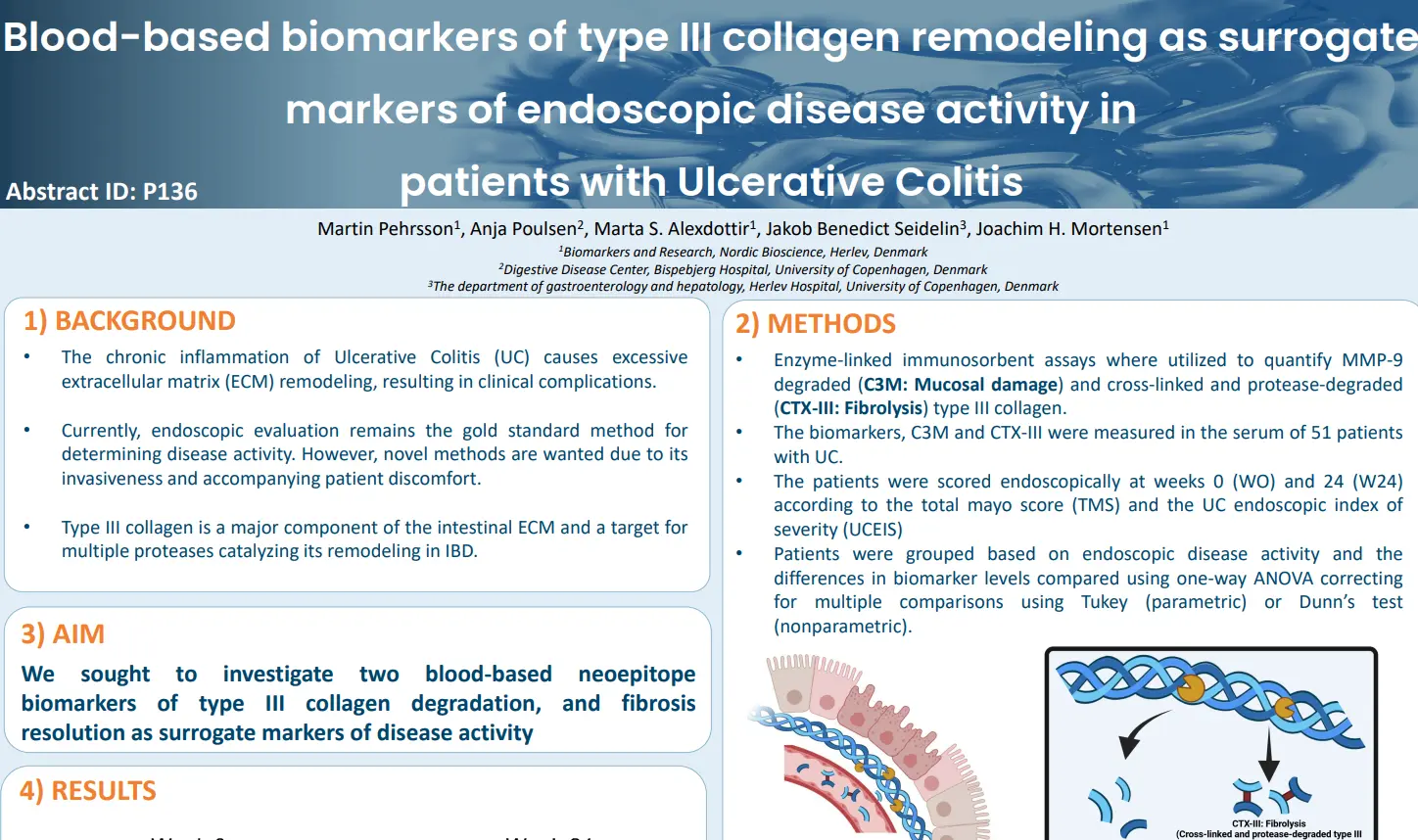
Blood-based biomarkers of type III collagen remodeling as surrogate markers of endoscopic disease activity in patients with ulcerative colitis
Introduction
The chronic inflammation of Ulcerative Colitis (UC) causes excessive extracellular matrix (ECM) remodeling, resulting in clinical complications. Currently, endoscopic evaluation remains the gold standard method for determining disease activity. However, novel methods are wanted due to its invasiveness and accompanying patient discomfort.
As type III collagen is a major component of the intestinal ECM and a target for multiple proteases catalyzing its remodeling in IBD, we sought to investigate two blood-based neoepitope biomarkers of type III collagen degradation, and fibrosis resolution as surrogate markers of disease activity.
C3M was elevated at Week 0 in UC patients with severe endoscopic disease activity according to the Total Mayo Score. The fibrolysis biomarker, nordicCTX-III™, was significantly elevated in patients with moderate endoscopic disease at week 0 and numerically elevated in mild disease compared to severe disease activity.
Get in touch
Are you interested in exploring collaboration possibilities? Enter your information in the form and a representative will contact you shortly.
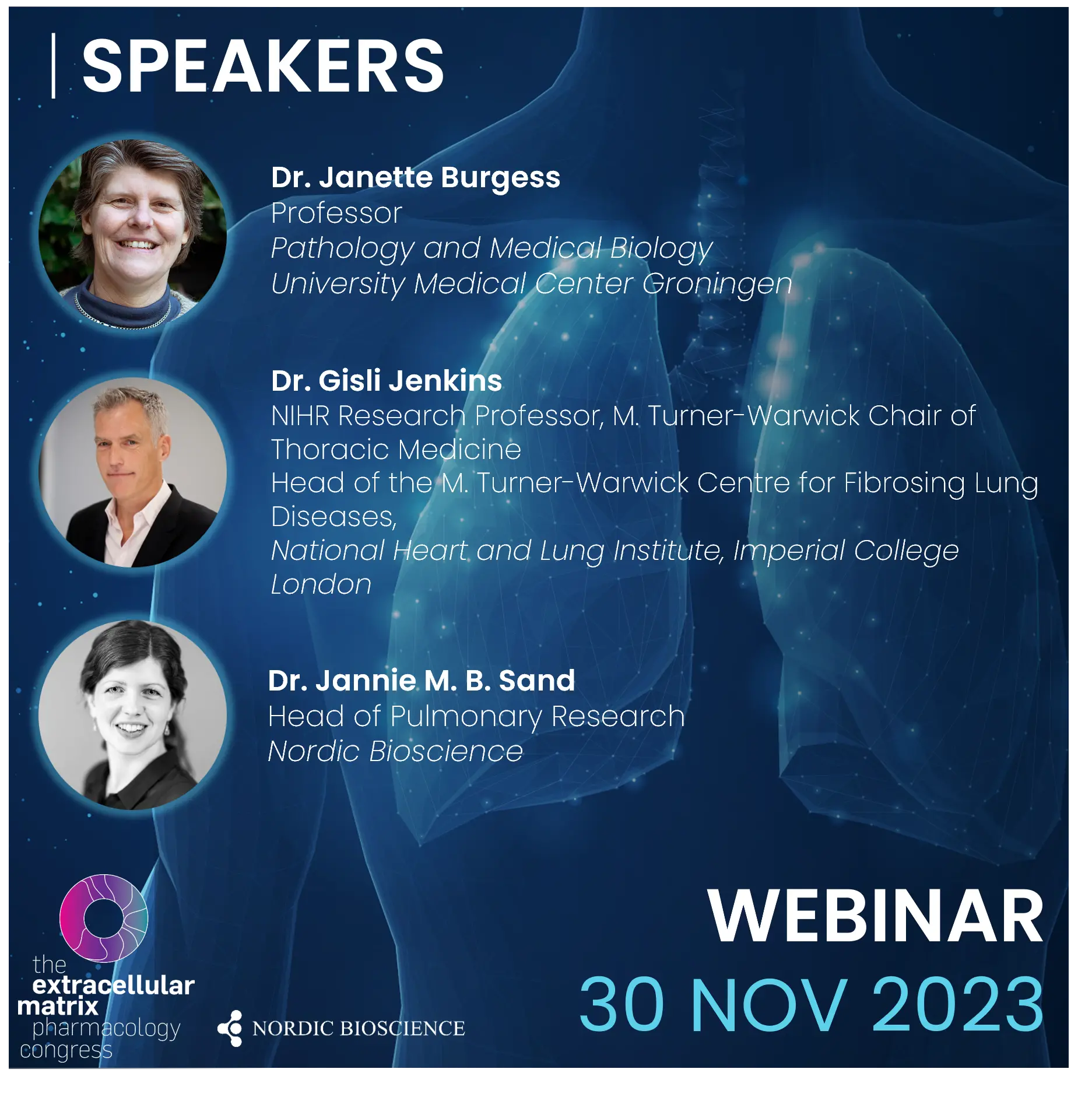
Watch this webinar focused on tissue remodeling, inflammation and biomarkers for pulmonary diseases. Our panel of experts will shed light on the tissue changes occurring in pulmonary diseases including idiopathic pulmonary fibrosis and chronic obstructive pulmonary disease, and how this is affected by the intricate interplay between fibroblasts, epithelial cells, and immune cells.
Extracellular Matrix is an Active Agent in Lung fibrosis – Dr. Janette Burgess
The Role of the Epithelium in Pulmonary Fibrosis – Dr. Gisli Jenking
Prognostic and Pharmacodynamic Biomarkers for ECM Remodeling, Immune Cell Activity, and Endotyping – Dr. Jannie M. B. Sand
General discussion and questions
Scientific topics and speakers
The speakers will take you on a journey from discovering novel biomarkers to implementing them in clinical trials and clinical practice. They will discuss why blood-based biomarkers are needed for pulmonary diseases, and how biomarkers of ECM remodeling and immune cell activity may aid in the identification of novel endotypes and improvement of drug development.
Here’s a sneak peak of Dr. Janette Burgess’ talking points:
How changes in the composition of the ECM in lung fibrosis impact cellular function
How the ECM organization changes in lung fibrosis, and implications for disease driving processes
How matrix stiffness is altered in fibrosis and the cellular responses to these changes
How the above changes can be incorporated in model systems in vitro to expand our understanding of disease underlying mechanisms
Dr. Janette Burgess
Janette Burgess is a Professor of Extracellular Matrix in Disease Pathogenesis at the University Medical Center Groningen in the Netherlands.
She completed her Bachelor of Science (with honours) at the University of Adelaide, Australia, in 1991, and earned her PhD in Hematology at the University of New South Wales in 1998.
After a post-doctoral position at the University of Sydney focusing on the respiratory system’s structural changes in disease, she developed a keen interest in lung pathology.
In 2015, Janette was awarded a Rosalind Franklin Fellowship to join the University Medical Center Groningen, where she is now a tenured Professor.
Her research centers on understanding the role of the extracellular matrix (ECM) in lung pathology, investigating changes in lung tissue and airway structures during disease development.
Using primary human lung cells, tissue samples, and patient clinical information, her work aims to characterize ECM changes during lung diseases and uncover the underlying mechanisms.
Janette Burgess employs novel in vitro cell models, ex vivo human lung tissue models, and advanced microscopy imaging techniques to unravel the complex regulation of the ECM, exploring its potential as a therapeutic target for lung diseases.
Her research bridges basic science with the practical goals of preventing and treating human lung diseases that affect millions of people worldwide.
Dr. Jannie M. B. Sand
Dr. Jannie M. B. Sand is the head of the respiratory research department at Nordic Bioscience, having joined the company in 2010.
She holds a Master of Science in Molecular Biomedicine and a PhD in Clinical Research from the University of Copenhagen.
Dr. Sand’s research is dedicated to biomarker development and understanding lung tissue changes in chronic lung diseases, with a specific focus on pulmonary fibrosis and chronic obstructive pulmonary disease (COPD).
With over a decade of experience in biomarkers of lung tissue remodeling, she has authored over 50 articles, abstracts, and book chapters.
Her team at Nordic Bioscience focuses on developing non-invasive tools for specific lung disease processes, aiming to enhance understanding of pathologies and therapeutic effects.
The tools developed by Dr. Sand’s team include biomarkers for fibrogenesis, lung tissue destruction, basement membrane repair, and immune cell activity.
Their work spans both clinical and preclinical research, with a translational approach to bring tools to the broader community.
The team’s research has successfully identified prognostic and pharmacodynamic biomarkers and explores the identification of novel endotypes in chronic lung diseases.
Dr. Gisli Jenkins
Professor Gisli Jenkins is an NIHR Research Professor and holds the Margaret Turner-Warwick Chair of Thoracic Medicine at Imperial College London.
He serves as the Head of the Margaret Turner-Warwick Centre for Fibrosing Lung Diseases at the National Heart and Lung Institute, located at the Guy Scadding Building at the Brompton Campus.
Dr. Jenkins has honorary contracts with both the Royal Brompton and Harefield NHS Foundation Trust and the Imperial College Healthcare NHS Trust.
His primary research focus is on Interstitial Lung Diseases, with a particular emphasis on Pulmonary Fibrosis.
Prof Jenkins and his team strive to comprehend the biological foundations of pulmonary fibrosis, aiming to translate this understanding into improved outcomes for patients.
He is the Principal Investigator of several longitudinal observational studies, including the PROFILE study, the INJUSTIS Study, the UKILD Post COVID ILD study, and the DEMISTIFI Multi-Morbidity consortium.
Prof Jenkins serves as the pulmonary fibrosis working group lead for the Genomics England Clinical Interpretation Partnership in Respiratory Medicine, the PHOSP-COVID study, and the HEAL COVID platform study.
Recognitions include the ERS Gold Medal in Interstitial Lung Disease in 2020, Fellowship of the European Respiratory Society in 2022, and the role of President of Action for Pulmonary Fibrosis. He was also awarded the BTS Meritorious Service Award for 2022.
Get in touch
Are you interested in exploring collaboration possibilities? Enter your information in the form and a representative will contact you shortly.