Liquid biopsy to track plasma kallikrein-activated TGF-β associates with resistance to combined immunotherapy and radiotherapy in metastatic pancreatic cancer
Introduction
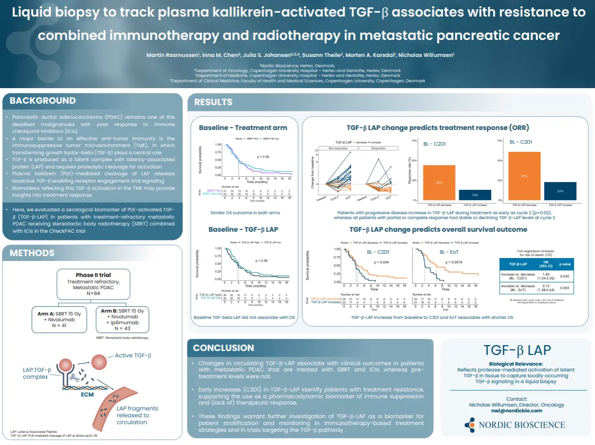
Pancreatic ductual adenocarcinoma (PDAC) remains one of the deadliest malignancies with poor response to immune checkpoint inhibitors (ICIs). A major barrier to an effective anti-tumor immunity is the immunosuppressive tumor environment (TME), in which transforming growth factor-beta (TGF-β) plays a central role. TGF-β is produced as a latent complex with latency-associated protein (LAP) and requires proteolytic cleavage for activation.
Plasma kallikrein (PLK)-mediated cleavage of LAP releases bioactive TGF-β enabling receptor engagement and signaling. Biomarkers reflecting this TGF-β activation in the TME may provide insights into treatment response.
In this study we evaluated a serological biomarker of PLK-activated TGF-β (TGF-β-LAP) in patients with treatment-refractory metastatic PDAC receiving stereotactic body radiotherapy (SBRT) combined with ICIs in the CheckPAC trial.
Poster
Conclusion
Changes in circulating TGF-β-LAP associate with clinical outcomes in patients with metastatic PDAC that are treated with SBRT and ICIs whereas pre-treatment levels were not. Early increases (C2D1) in TGF-β-LAP identify patients with treatment resistance, supporting the use as a pharmacodynamic biomarker of immune suppression and (lack of) therapeutic response. In conclusion, these findings warrant further investigation of TGF-β-LAP as a biomarker for patient stratification and monitoring in immunotherapy-based treatment strategies and in trials targeting the TGF-β pathway.