Machine-learning classification integrating non-invasive biomarkers, clinical characteristics and pulmonary function
Introduction
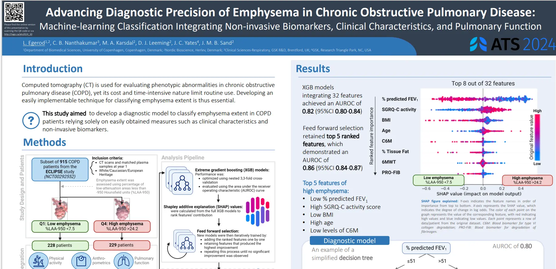
Computed tomography (CT) is used for evaluating phenotypic abnormalities in chronic obstructive pulmonary disease (COPD), yet its cost and time-intensive nature limit routine use. Developing an easily implementable technique for classifying emphysema extent is thus essential.
This study aimed to develop a diagnostic model to classify emphysema extent in COPD patients relying solely on easily obtained measures such as clinical characteristics and non-invasive biomarkers.
Diagnostic models incorporating easily obtainable measures effectively distinguished COPD patients with high emphysema extent from those with low extent. Such models for classifying emphysema patterns have the potential for clinical implementation, aiding in diagnosis or serving as a decision-making tool to determine the necessity of further CT scans.
Get in touch
Are you interested in exploring collaboration possibilities? Enter your information in the form and a representative will contact you shortly.
Data-driven identification and investigation of comorbidity profiles in patients with chronic obstructive pulmonary disease: a multicohort study
Introduction
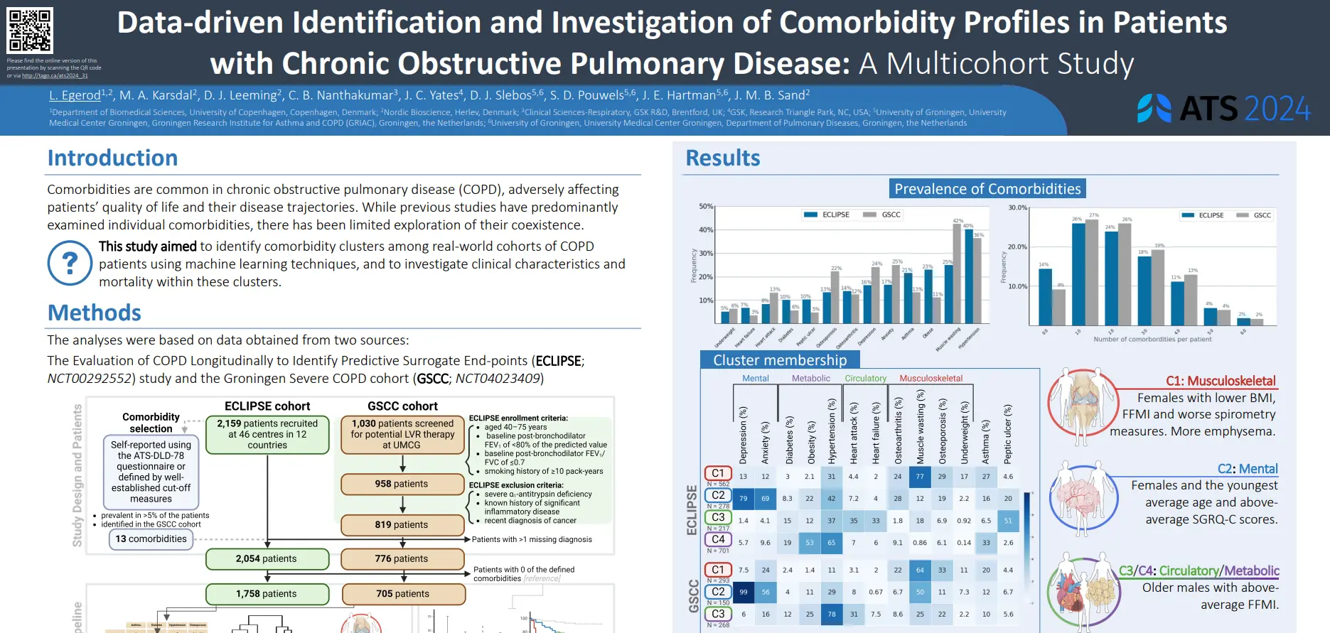
Comorbidities are common in chronic obstructive pulmonary disease (COPD), adversely affecting patients’ quality of life and their disease trajectories. While previous studies have predominantly examined individual comorbidities, there has been limited exploration of their coexistence.
This study aimed to identify comorbidity clusters among real-world cohorts of COPD patients using machine learning techniques, and to investigate clinical characteristics and mortality within these clusters.
This study confirms distinct comorbidity clusters in two well-characterized cohorts of patients with COPD which can be linked to different patient subgroups. In a broad COPD patient population, comorbidity profiles could hold prognostic relevance. The findings of this study enhance the understanding of the comorbidity landscape in COPD and highlights the importance of comorbidity assessment in clinical management.
Get in touch
Are you interested in exploring collaboration possibilities? Enter your information in the form and a representative will contact you shortly.
PRO-C11 and PRO-C16 are markers of intestinal fibrosis and are associated with MRE-confirmed intestinal strictures – Results from the ImageKids study
Introduction
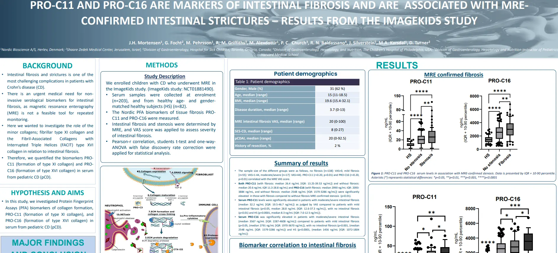
Intestinal fibrosis and strictures is one of the most challenging complications in patients with Crohn’s disease (CD). There is an urgent medical need for non-invasive serological biomarkers for intestinal fibrosis, as magnetic resonance enterography (MRE) is not a feasible tool for repeated monitoring.
In this study we investigated Protein FingerprintAssays (PFA) biomarkers of collagen formation, PRO-C11 (formation of type XI collagen), and PRO-C16 (formation of type XVI collagen) in serum from pediatric CD (pCD).
Based on these biomarker data from the ImageKids study, PRO-C11 and PRO-C16 demonstrate potential as important non-invasive biomarkers reflecting intestinal fibrosis and stenosis.
Get in touch
Are you interested in exploring collaboration possibilities? Enter your information in the form and a representative will contact you shortly.
NordicCPa9™: A neutrophil-derived fragment of calprotectin measured in serum can monitor endoscopic and clinical disease activity in ulcerative colitis
Introduction
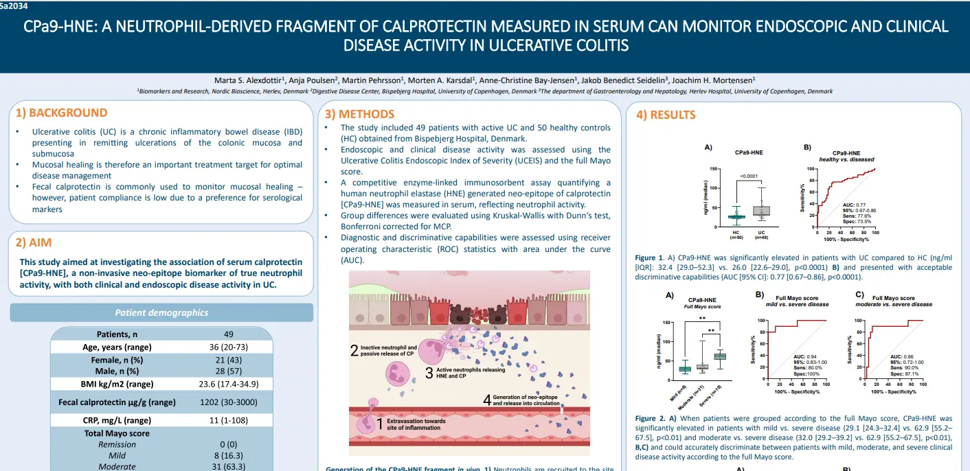
Ulcerative colitis (UC) is a chronic inflammatory bowel disease (IBD) presenting in remitting ulcerations of the colonic mucosa and submucosa. Mucosal healing is therefore an important treatment target for optimal disease management. Fecal calprotectin is commonly used to monitor mucosal healing – however, patient compliance is low due to a preference for serological markers.
In this study we aimed to investigate the association of serum calprotectin [CPa9-HNE], a non-invasive neo-epitope biomarker of true neutrophil activity, with both clinical and endoscopic disease activity in UC.
CPa9-HNE (nordicCPa9™) accurately reflected both clinical and endoscopic disease activity in ulcerative colitis, based on the UCEIS and full Mayo score. These findings highlight the potential use of nordicCPa9™ as a non-invasive tool to monitor both endoscopic and clinical disease activity in UC, with the potential of guiding treatment decisions and better aligning with patient preferences.
Get in touch
Are you interested in exploring collaboration possibilities? Enter your information in the form and a representative will contact you shortly.
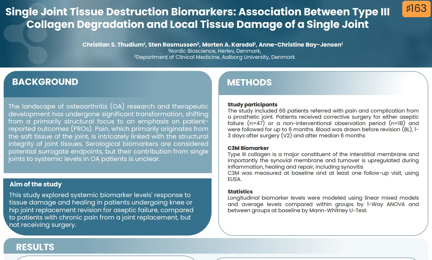
Single joint tissue destruction biomarkers: association between type III collagen degradation and local tissue damage of a single joint
Introduction
The landscape of osteoarthritis (OA) research and therapeutic development has undergone significant transformation, shifting from a primarily structural focus to an emphasis on patient-reported outcomes (PROs). Pain, which primarily originates from the soft tissue of the joint, is intricately linked with the structural integrity of joint tissues. Serological biomarkers are considered potential surrogate endpoints, but their contribution from single joints to systemic levels in OA patients is unclear.
This study explored systemic biomarker levels’ response to tissue damage and healing in patients undergoing knee or hip joint replacement revision for aseptic failure, compared to patients with chronic pain from a joint replacement, but not receiving surgery.
C3M degradation was found to increase in response to tissue insult to the joint from revision surgery, while no change was observed in a non-surgical group with chronic pain of the joint over 6 months. The increase and gradual decrease throughout the study period indicate a relationship between systemic levels of type III collagen degradation fragments and soft-tissue destruction and inflammation of the joint.
Get in touch
Are you interested in exploring collaboration possibilities? Enter your information in the form and a representative will contact you shortly.
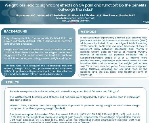
Drug development in the osteoarthritis (OA) field has proven extremely difficult, with a discordance between joint structure and pain. Weight loss has been associated with an effect on pain and function. Several molecular endotypes have been suggested for OA, which could be treatable, such as bone, inflammation, and lately, an overweight endotype.
The aim was to investigate the relationship between obesity, weight loss and patient-reported outcomes (PROs) in patients with persistent pain, and the effect on joint and bone tissue related soluble biomarkers.
Changes in PROs were significantly associated with obesity. Weight loss was associated with an increase in bone and cartilage degradation, as well as lowering in the interstitial matrix degradation. These data indicate that weight loss comes with a risk of increased joint tissue loss, but improvement in tissue inflammation.
Get in touch
Are you interested in exploring collaboration possibilities? Enter your information in the form and a representative will contact you shortly.
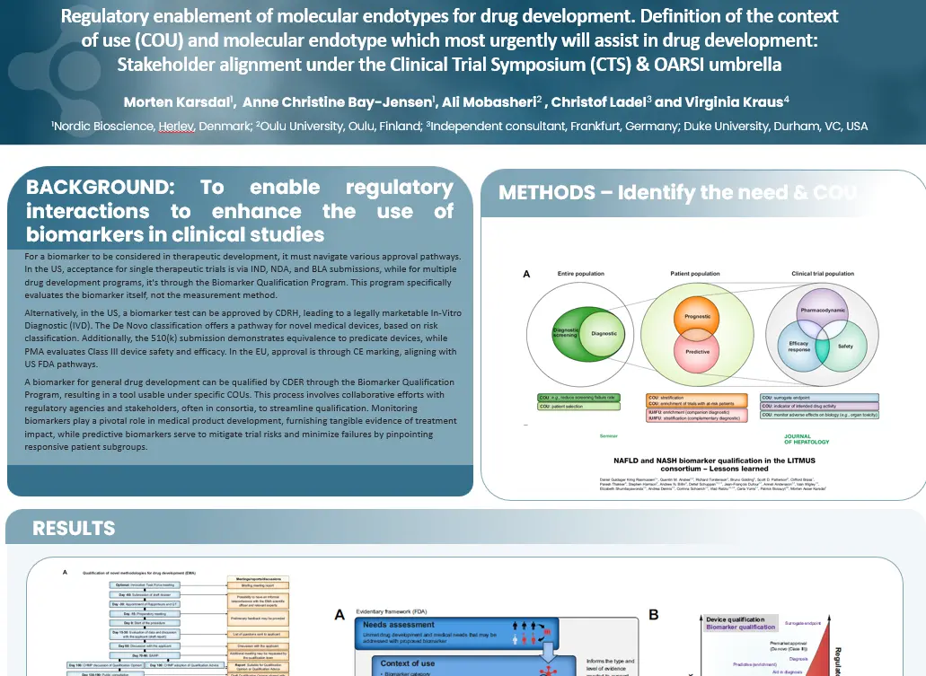
Regulatory enablement of molecular endotypes for drug development. Definition of the context of use (COU) and molecular endotype which most urgently will assist in drug development: Stakeholder alignment under the Clinical Trial Symposium (CTS) & OARSI umbrella
Introduction
For a biomarker to be considered in therapeutic development, it must navigate various approval pathways. In the US, acceptance for single therapeutic trials is via IND, NDA, and BLA submissions, while for multiple drug development programs, it’s through the Biomarker Qualification Program. This program specifically evaluates the biomarker itself, not the measurement method.
Alternatively, in the US, a biomarker test can be approved by CDRH, leading to a legally marketable In-Vitro Diagnostic (IVD). The De Novo classification offers a pathway for novel medical devices, based on risk classification. Additionally, the 510(k) submission demonstrates equivalence to predicate devices, while PMA evaluates Class III device safety and efficacy. In the EU, approval is through CE marking, aligning with US FDA pathways.
A biomarker for general drug development can be qualified by CDER through the Biomarker Qualification Program, resulting in a tool usable under specific COUs. This process involves collaborative efforts with regulatory agencies and stakeholders, often in consortia, to streamline qualification. Monitoring biomarkers play a pivotal role in medical product development, furnishing tangible evidence of treatment impact, while predictive biomarkers serve to mitigate trial risks and minimize failures by pinpointing responsive patient subgroups.
Defining the Context of Use (COU) within real-world or clinical study populations is essential for biomarker development. Following CLSI guidelines ensures technical robustness, while careful risk-benefit evaluation supports clinical relevance. The regulatory enablement of molecular endotypes is essential for drug development.
Get in touch
Are you interested in exploring collaboration possibilities? Enter your information in the form and a representative will contact you shortly.
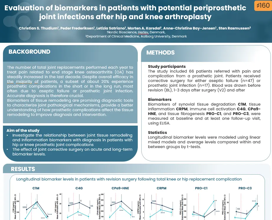
Evaluation of biomarkers in patients with potential periprosthetic joint infections after hip and knee arthroplasty
Introduction
The number of total joint replacements performed each year to treat pain related to end stage knee osteoarthritis (OA) has steadily increased in the last decade. Despite overall efficacy in the majority of patients, a subset of about 20% experience prosthetic complications in the short or in the long run, most often due to aseptic failure or prosthetic joint infection. Accurate diagnosis is therefore crucial. Biomarkers of tissue remodeling are promising diagnostic tools to characterize joint pathological mechanisms, provide a better understanding of how prosthetic complications affect the tissue remodeling to improve diagnosis and intervention
This study aimed to investigate the relationship between joint tissue remodeling and inflammation biomarkers with diagnosis in patients with hip or knee prosthetic joint complications, as well as to assess the effect of joint corrective surgery on acute and long-term biomarker levels.
Get in touch
Are you interested in exploring collaboration possibilities? Enter your information in the form and a representative will contact you shortly.
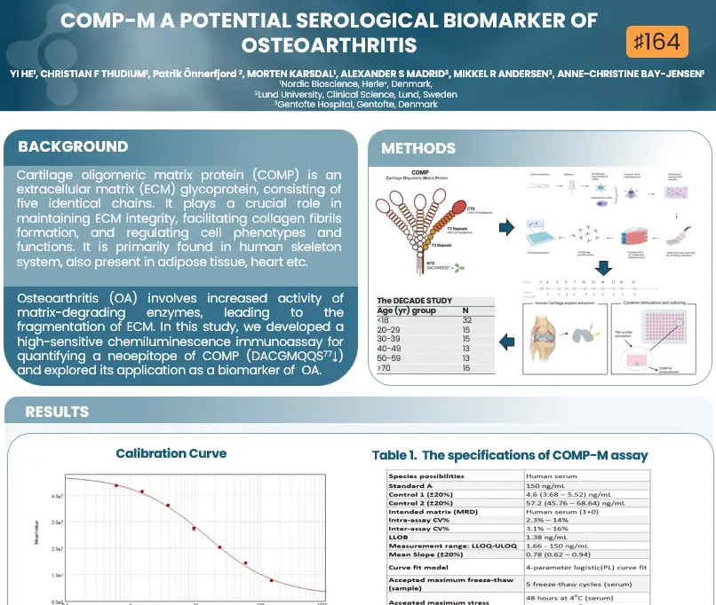
Cartilage oligomeric matrix protein (COMP) is an extracellular matrix (ECM) glycoprotein, consisting of five identical chains. It plays a crucial role in maintaining ECM integrity, facilitating collagen fibrils formation, and regulating cell phenotypes and functions. It is primarily found in human skeleton system, also present in adipose tissue, heart etc. Osteoarthritis (OA) involves increased activity of matrix-degrading enzymes, leading to the fragmentation of ECM.
In this study, we developed a high-sensitive chemiluminescence immunoassay for quantifying a neoepitope of COMP (DACGMQQS77↓)and explored its application as a biomarker of OA.
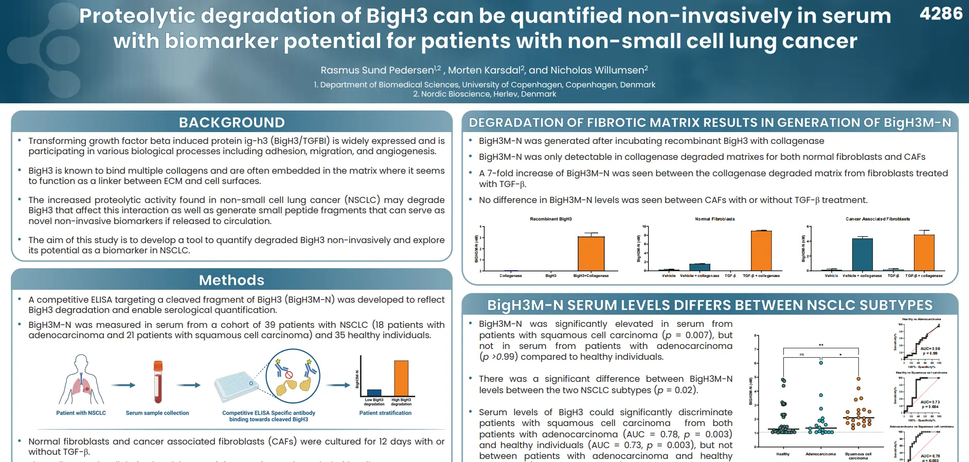
Proteolytic degradation of BigH3 can be quantified non-invasively in serum with biomarker potential for patients with non-small cell lung cancer
Introduction
Transforming growth factor beta induced protein ig-h3 (BigH3/TGFBI) is widely expressed and is participating in various biological processes including adhesion, migration, and angiogenesis. BigH3 is known to bind multiple collagens and are often embedded in the matrix where it seems to function as a linker between ECM and cell surfaces. In non-small cell lung cancer (NSCLC), the increased proteolytic activity found may degrade BigH3, disrupting its ECM interactions and generating peptide fragments that could potentially serve as novel non-invasive biomarkers if released into circulation.
Our aim is to develop a tool to quantify degraded BigH3 non-invasively and explore its potential as a biomarker in NSCLC.
Degradation of BIGH3 can be reflected by non-invasive quantification of the cleaved fragment of BigH3, BigH3M-N, in serum. This suggests BigH3M-N as a promising biomarker in NSCLC with potential for discriminating between subtypes. However, as BigH3M-N is connected to fibroblast matrix biology, the optimal use for this biomarker might be in combination with other ECM biomarkers.
Get in touch
Are you interested in exploring collaboration possibilities? Enter your information in the form and a representative will contact you shortly.