Glioblastoma (GBM) remains a devastating disease with limited treatment options and a lack of reliable, non-invasive biomarkers to guide therapy and predict outcomes.
Tau-A and C4G offer a potential value in patients with glioblastoma
In this study, we identify serum biomarkers (Tau-A and C4G) that are significantly elevated in patients with recurrent GBM and that show a strong association with improved overall survival (OS) when treated with nivolumab and bevacizumab. Unlike conventional imaging or invasive methods, these serum biomarkers provide a non-invasive, dynamic, and easily accessible approach to track disease progression and response to treatment.
Tau-A and C4G offer a potential tool to stratify patients, monitor treatment efficacy, and predict survival outcomes in GBM, addressing a critical gap in current clinical practice.
We therefore encourage clinicians and researchers to explore the validation and integration of Tau-A and C4G into clinical trials and routine practice to unlock new possibilities in personalized GBM management.
Article: Degradation fragments of Tau and type IV collagen as serum biomarkers in patients with recurrent glioblastoma treated with nivolumab and bevacizumab
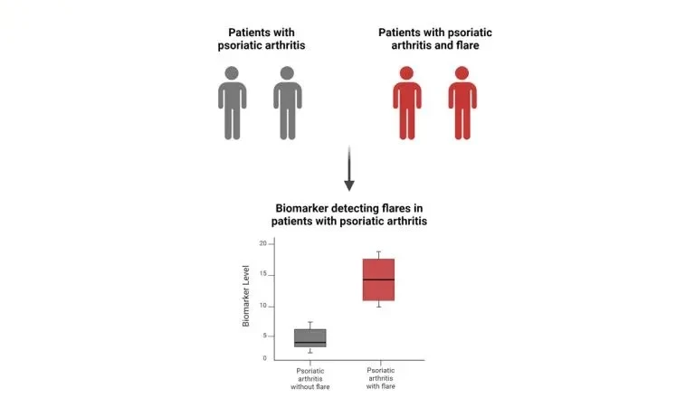
Psoriatic arthritis (PsA) is an inflammatory arthritis associated with psoriasis of the skin. Patients with PsA may experience flares, which are characterized by worsening of disease activity and intensity presenting with swollen and tender joints.
Biomarkers can detect flares in PsA patients
The implementation of biomarkers related to PsA activity and flares, helps identifying the right patients, which will improve patient management and decrease flares. In this study, we evaluated blood-based biomarkers of inflammation, fibrosis, and joint destruction in patients with and without flares, and found that the inflammatory biomarkers VICM, CPa9-HNE, and CRPM, together with the fibrosis biomarker nordicPRO-C3™, showed a good discriminatory performance separating the PsA patients with flares from the patients without.
Such biomarkers may therefore serve as tools for quantitatively monitoring flares in PsA patients and have valuable applications in the detection of disease, as well as the monitoring of health status. This may include diagnosing, staging, predicting progression of the disease, and response to therapy.
This work was performed together with Dr. Vinod Chandran and Dr. Katerina Oikonomopoulou at the Schroeder Arthritis Institute, Krembil Brain Institute in Toronto.
Article: Investigating protease-mediated peptides of inflammation and tissue remodeling as biomarkers associated with flares in psoriatic arthritis
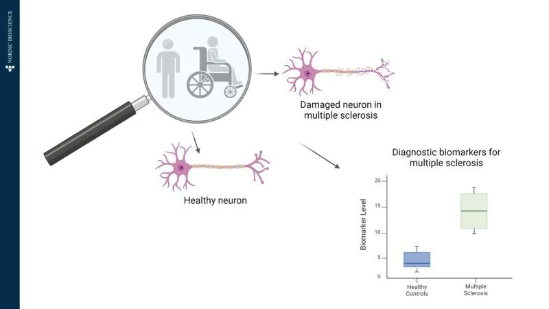
Multiple sclerosis (MS) is a chronic inflammatory disease in which the immune system attacks the myelin sheath, a protective layer surrounding nerves, which disrupts the communication between the brain and the rest of the body. The diagnosis of MS is often delayed because of its symptoms and diagnostic tests overlapping with those of other diseases.
Diagnostic biomarkers for multiple sclerosis
To address this challenge, there is a need for biomarkers that precisely and accurately quantify disease activity, which can provide more accurate results. Blood-based biomarkers quantify fragments of proteins involved in MS pathophysiology, which is why the aim of this this study was to investigate their potential as diagnostic biomarkers.
In this study, we evaluated three such biomarkers: BGM (biglycan degraded by matrix metalloproteinases (MMPs), NIC (cathepsin S-degraded nidogen), and SPARC-M (MMP-degraded secreted protein acidic and rich in cysteine). Testing on healthy donors and patients diagnosed with MS revealed that SPARC-M was the most effective diagnostic biomarker, with an AUC of 0.875.
In clinical management, protein fragments like SPARC-M could provide a specific protein fingerprint for MS. Therefore, blood-based biomarkers such as SPARC-M can aid in disease diagnosis, understanding pathogenesis, monitoring progression, and tailoring treatment strategies in clinical practice.
Article: Diagnostic potential of blood-based biomarkers in multiple sclerosis
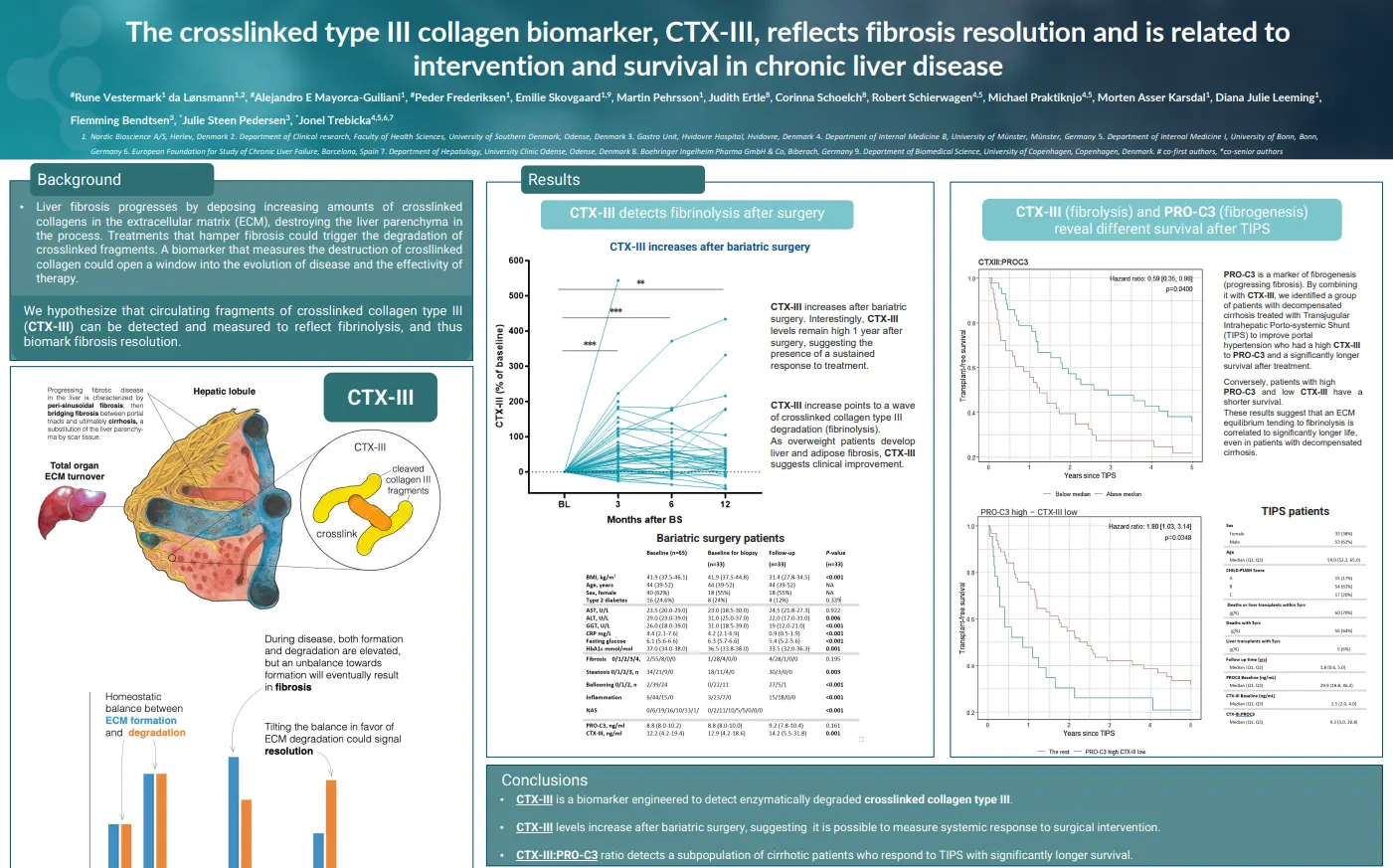
The crosslinked type III collagen biomarker, CTX-III, reflects fibrosis resolution and is related to intervention and survival in chronic liver disease
Introduction
Liver fibrosis progresses by deposing increasing amounts of crosslinked collagens in the extracellular matrix (ECM), destroying the liver parenchyma in the process. Treatments that hamper fibrosis could trigger the degradation of crosslinked fragments. A biomarker that measures the destruction of crosslinked collagen could open a window into the evolution of disease and the effectivity of therapy. We hypothesize that circulating fragments of crosslinked collagen type III (nordicCTX-III™) can be detected and measured to reflect fibrinolysis, and thus biomark fibrosis resolution.
NordicCTX-III™ is a biomarker engineered to detect enzymatically degraded crosslinked collagen type III. Its levels have been shown to increase after bariatric surgery, suggesting it is possible to measure systemic response to surgical intervention. Additionally, the nordicCTX-III™: nordicPRO-C3™ ratio detects a subpopulation of cirrhotic patients who respond to TIPS with significantly longer survival.
Get in touch
Are you interested in exploring collaboration possibilities? Enter your information in the form and a representative will contact you shortly.
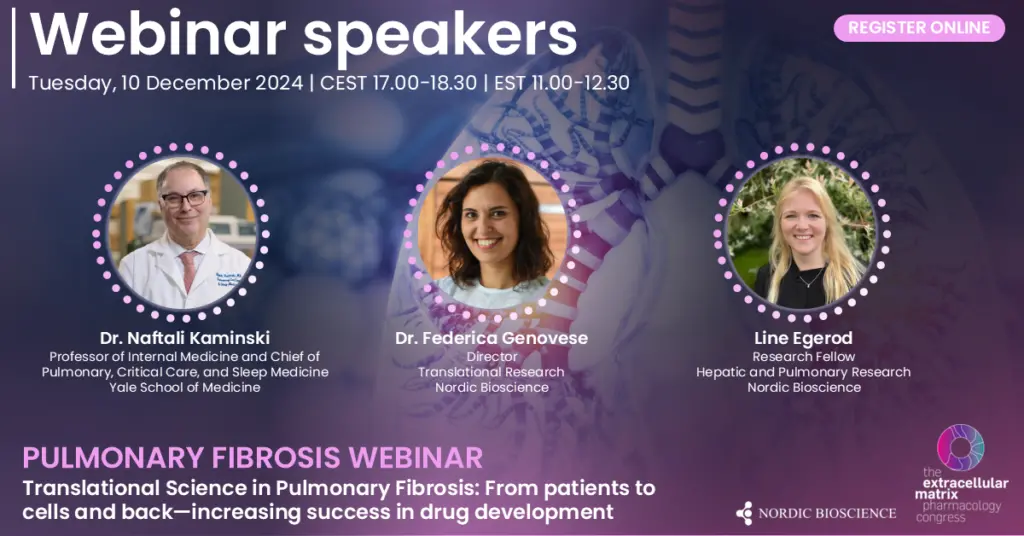
Watch “Translational Science in Pulmonary Fibrosis: From patients to cells and back—increasing success in drug development,” to learn more about what drives pulmonary fibrosis, with insights from machine learning and translational biomarkers.
Leveraging machine learning for patient stratification and advanced analytics – a COPD case study | Line Egerod
Investigating fibrosis mechanisms – what drives fibrosis in the lungs? | Dr. Naftali Kaminski
Utilizing translational biomarkers for drug development in fibrotic lung diseases | Dr. Federica Genovese
Roundtable discussion and Q&A
Scientific topics and speakers
The session will begin with an investigation into the mechanisms driving lung fibrosis, examining key processes that underlie this progressive condition. We will then delve into the role of machine learning in pulmonary research, highlighting its application for patient stratification and advanced analytics through a COPD case study.
Finally, discussions will focus on the use of translational biomarkers in drug development, offering insights into more targeted and effective therapies. This webinar is designed for researchers, clinicians, and industry professionals interested in the latest translational strategies for tackling pulmonary fibrosis.
Dr. Naftali Kaminski
Dr. Naftali Kaminski is the Boehringer-Ingelheim Endowed Professor of Internal Medicine and Chief of Pulmonary, Critical Care, and Sleep Medicine at Yale School of Medicine since 2013, with prior leadership at the University of Pittsburgh.
He is a global leader in genomic research for chronic lung diseases, including Idiopathic Pulmonary Fibrosis (IPF), COPD, severe asthma, and sarcoidosis, pioneering transcript profiling and omics integration for precision medicine.
Dr. Kaminski’s research has identified novel therapeutic targets in IPF, such as metalloproteases (MMP7, MMP19), phosphatases (SHP2, MKP5), and antifibrotic roles for thyroid hormone signaling. His team’s discoveries include the role of microRNAs (e.g., let-7, mir-29, mir-33) in lung fibrosis and the development of predictive blood-based biomarkers for IPF risk stratification and transplant prioritization.
Dr. Kaminski has authored more than 340 peer-reviewed publications in top journals, including Nature Medicine, NEJM, Science Translational Medicine, and Lancet Respiratory Medicine, while consistently securing NIH funding since 2000.
Recognized for his contributions to pulmonary research, he received awards such as the Marvin I. Schwarz Award (2010), the ERS Gold Medal for ILD (2016), and the ATS Amberson Lecture Award (2022), among many others. He is a Fellow of the American Thoracic Society (ATS) and the European Respiratory Society (ERS) and has served in leadership roles within ATS, including as Chair of the Assembly on Respiratory Cell and Molecular Biology.
Dr. Kaminski is passionate about training the next generation of physician-scientists in genomics, bioinformatics, and systems biology, mentoring numerous successful MDs and PhDs in launching independent, well-funded careers. He continues to influence clinical and translational lung disease research as an associate editor for Thorax, BMJ, and through leadership in pulmonary genomic medicine.
Dr. Federica Genovese
Dr. Federica Genovese is the Director of Cardiovascular and Renal (CVR) Research at Nordic Bioscience. She also heads the Translational Research group.
She joined Nordic Bioscience in 2011 and assumed the role of Group leader of Kidney research in 2015 and then became Director of CVR in 2019.
Dr. Genovese focuses on developing serologically assessed markers to evaluate extracellular matrix remodeling in patients with cardiovascular and renal diseases, aiding in prognostic and pharmacodynamic evaluation.
Her team has produced the bulk of data on endotrophin, measured by the PRO-C6 assay, a fibroblast activity marker and a pro-fibrotic molecule, utilized as risk marker of adverse outcomes in multiple fibro-inflammatory diseases.
Dr. Genovese has authored more than 100 peer-reviewed publications, demonstrating her extensive contributions to the field.
Her H-index is 28, her I10-index is 42, and her research has garnered over 3400 citations as of November 2024.
Line Egerod
Line Egerod is a PhD student in the Hepatic and Pulmonary Research Team at Nordic Bioscience based in Copenhagen.
Before joining Nordic Bioscience in 2022, she worked as a machine learning engineer in Oxford, UK, and Silicon Valley, CA, US, gaining experience across various disease areas.
She is the main data steward for the ECLIPSE cohort, one of the largest and most comprehensive COPD studies to date.
Her research focuses on using interpretable machine learning models alongside inflammation and extracellular matrix biomarkers to uncover opportunities for patient stratification and personalized profiling in COPD.
Working closely with pharmaceutical companies worldwide, she and her colleagues have supported clinical research that has contributed to multiple clinical studies and publications in high-ranking scientific journals.
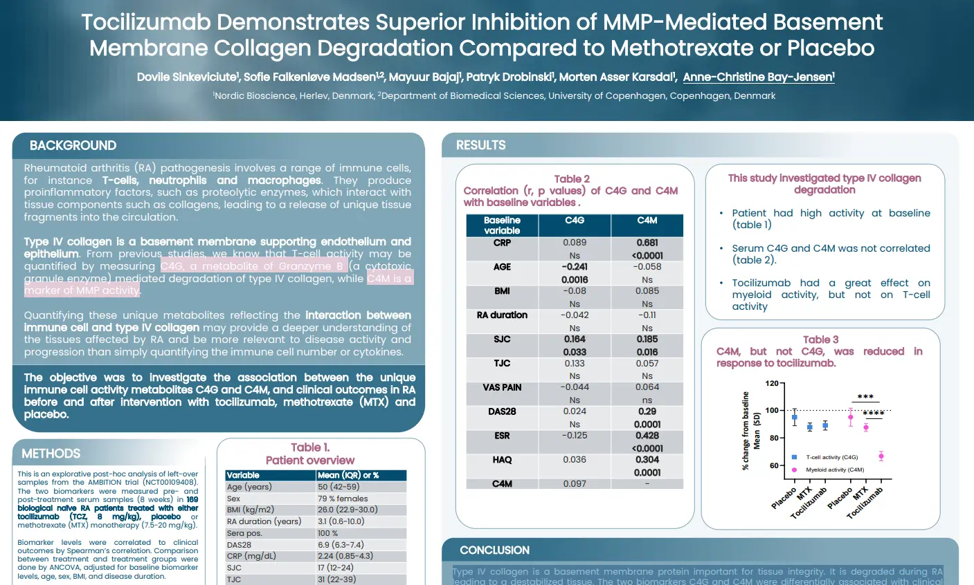
Tocilizumab demonstrates superior inhibition of MMP-mediated basement membrane collagen degradation compared to methotrexate or placebo
Introduction
Rheumatoid arthritis (RA) pathogenesis involves a range of immune cells, for instance T-cells, neutrophils and macrophages. They produce proinflammatory factors, such as proteolytic enzymes, which interact with tissue components such as collagens, leading to a release of unique tissue fragments into the circulation. Type IV collagen is a basement membrane supporting endothelium and epithelium. From previous studies, we know that T-cell activity may be quantified by measuring C4G, a metabolite of Granzyme B (a cytotoxic granule enzyme) mediated degradation of type IV collagen, while C4M is a marker of MMP activity. Quantifying these unique metabolites reflecting the interaction between immune cell and type IV collagen may provide a deeper understanding of the tissues affected by RA and be more relevant to disease activity and progression than simply quantifying the immune cell number or cytokines.
The aim of this study was to investigate the association between the unique immune cell activity metabolites C4G and C4M, and clinical outcomes in RA before and after intervention with tocilizumab, methotrexate (MTX) and placebo.
Type IV collagen is a basement membrane protein important for tissue integrity. It is degraded during RA leading to a destabilized tissue. The two biomarkers studied, C4G and C4M, were differentially associated with clinical outcome measures. Importantly, only C4M, a marker of MMP-derived tissue destruction, could be inhibited by tocilizumab. None of the markers were modulated by MTX.
Get in touch
Are you interested in exploring collaboration possibilities? Enter your information in the form and a representative will contact you shortly.
Understanding Parkinson’s Disease Progression Through Protein Biomarkers
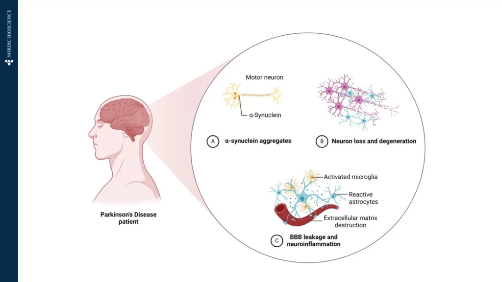
Parkinson’s Disease (PD) is a complex neurodegenerative disorder, primarily affecting the brain’s control over movement, thought, memory, and emotion. Early symptoms often manifest as tremors due to impaired motor skills. Underlying these visible symptoms is a cascade of molecular changes, beginning with alterations in specific proteins—one of which, α-synuclein, plays a critical role in PD pathology. In Figure 1, we have illustrated how α-Synuclein aggregates, which impairs the motor neuron.
Figure 1.Patients diagnosed with Parkinson’s Disease is affected by A) α-Synuclein aggregates affecting the motor neurons, B) Neuron loss and degeneration, and C) Blood-Brain-Barrier (BBB) leakage and neuroinflammation, by activated microglia, reactive astrocytes and extracellular matrix destruction.
In a healthy brain, α-synuclein supports neuronal communication. However, in Parkinson’s, this protein undergoes abnormal processing, driven partly by the enzyme Calpain-1, which cleaves α-synuclein into smaller, altered fragments. This early cleavage disrupts cellular function and promotes the formation of toxic aggregates, which accumulate, kill neurons, and drive disease progression. Intriguingly, these fragmented proteins can cross the blood-brain barrier and enter the bloodstream, providing a potential “window into the brain” for tracking disease activity from a simple blood sample.
At Nordic Bioscience, we have developed an innovative approach to harness this biomarker potential. Using our ProteinFingerPrint Biomarker Technology™, we can detect Calpain-1-cleaved α-synuclein fragments in blood serum with high precision. Our specific assay, α-SYN-C, captures the unique “fragment fingerprint” of PD by quantifying these cleaved fragments, which are significantly elevated in the blood of PD patients compared to healthy individuals,as illustrated in Figure 2.
Figure 2. Patients with Parkinson’s Disease has significant higher levels of α-SYN-C in serum, compared to healthy donors. The α-SYN-C biomarker detects levels of α-Synuclein cleaved by Calpain-1 in serum. The assay is technically validated for measurements in human blood samples.
This biomarker offers a non-invasive, accessible tool for monitoring Parkinson’s Disease progression and evaluating therapeutic responses. By examining α-SYN-C levels in blood samples, our technology not only provides insights into PD mechanisms but also opens doors for developing targeted therapies that address the disease’s underlying pathology. Through this work, we aim to support more accurate PD diagnostics and more effective, individualized treatments in the fight against neurodegeneration.
Get in touch
Are you interested in exploring collaboration possibilities? Enter your information in the form and a representative will contact you shortly.
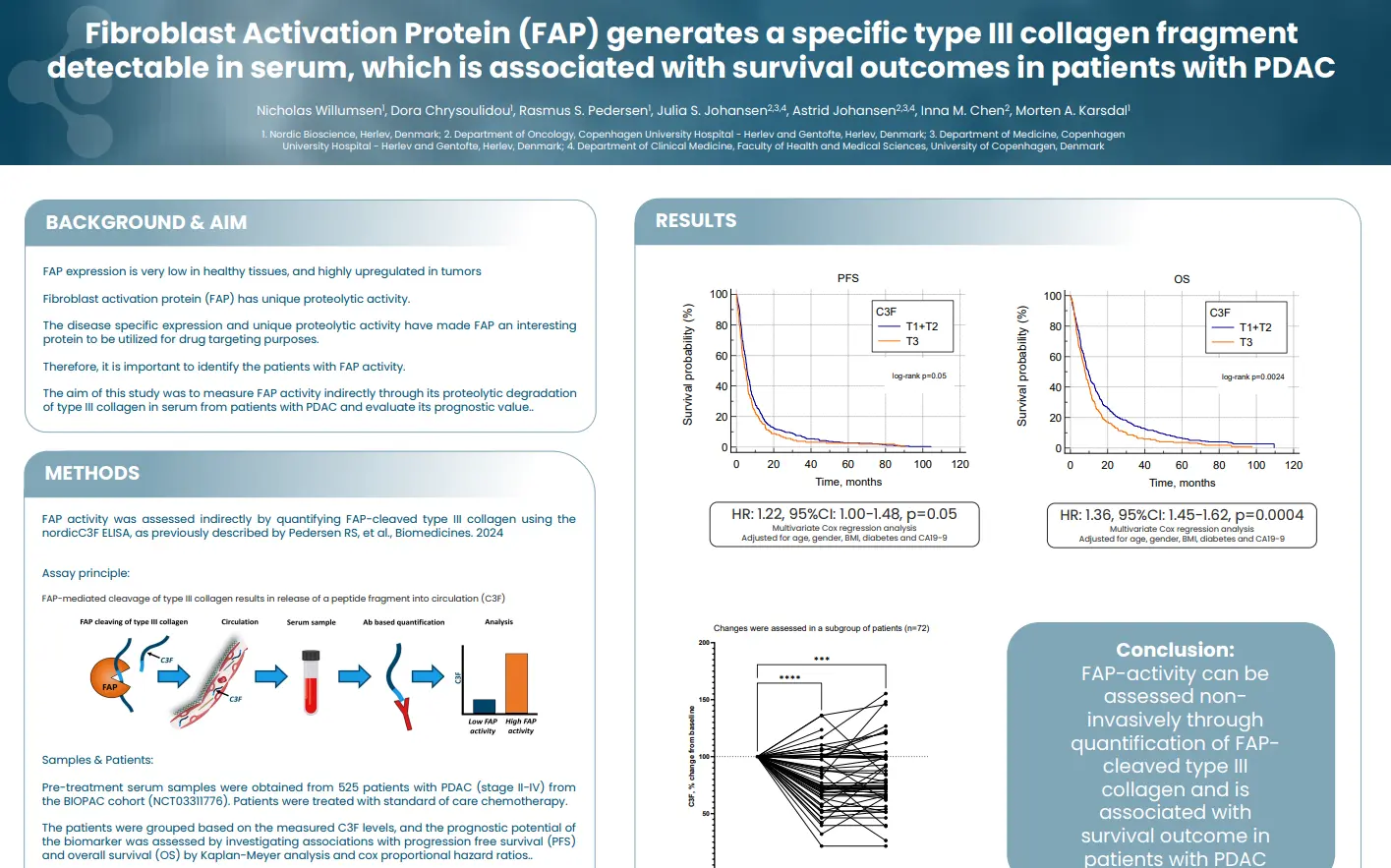
Fibroblast Activation Protein (FAP) generates a specific type III collagen fragment detectable in serum, which is associated with survival outcomes in patients with PDAC
Introduction
FAP expression is very low in healthy tissues, and highly upregulated in tumors Fibroblast activation protein (FAP) has unique proteolytic activity. The disease specific expression and unique proteolytic activity have made FAP an interesting protein to be utilized for drug targeting purposes. Therefore, it is important to identify the patients with FAP activity.
In this study we aimed to measure FAP activity indirectly through its proteolytic degradation of type III collagen in serum from patients with PDAC and evaluate its prognostic value.
FAP-activity can be assessed non-invasively through quantification of FAP-cleaved type III collagen and is associated with survival outcome in patients with PDAC.
Get in touch
Are you interested in exploring collaboration possibilities? Enter your information in the form and a representative will contact you shortly.
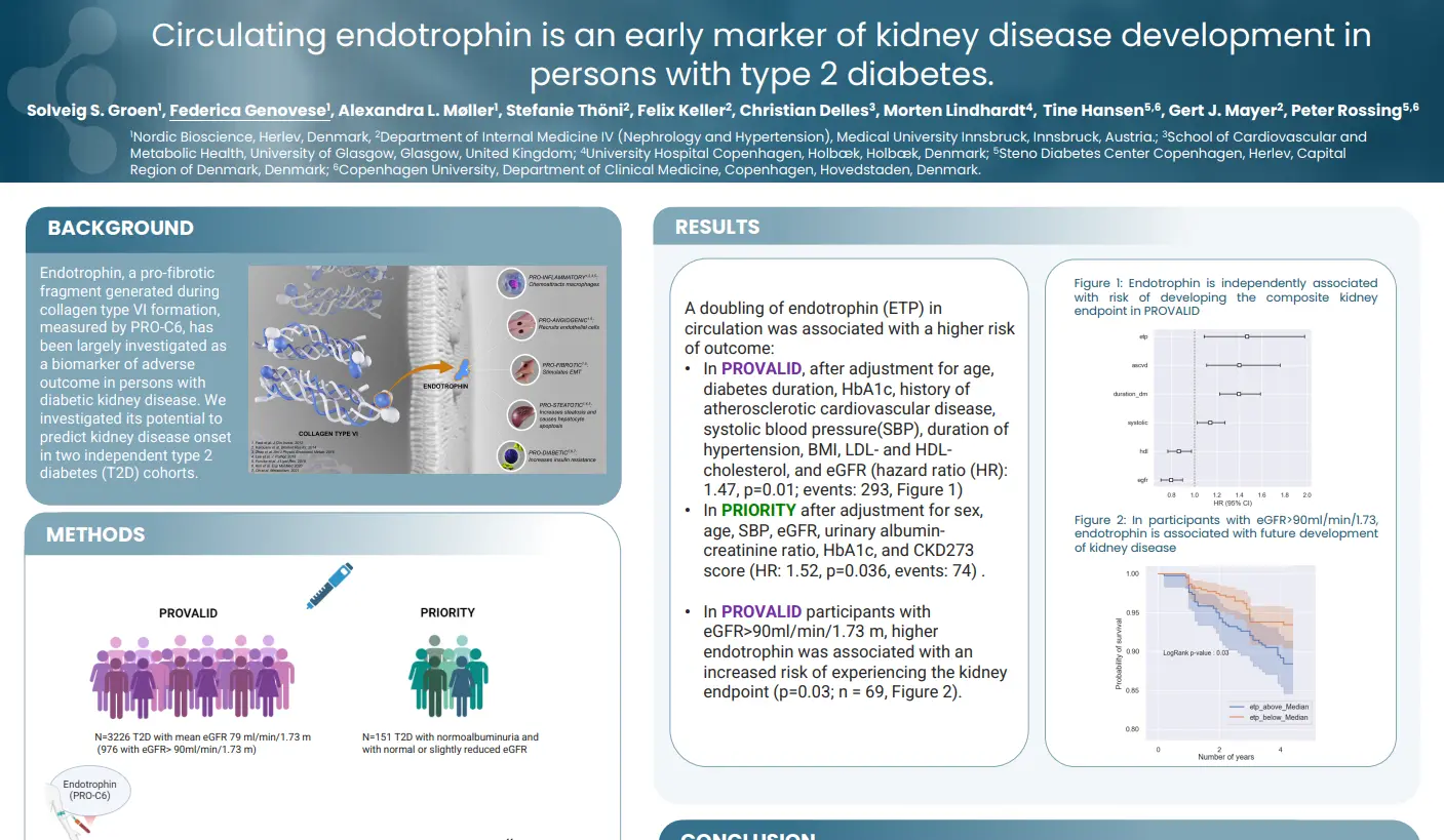
Circulating endotrophin is an early marker of kidney disease development in persons with type 2 diabetes
Introduction
NordicEndotrophin™ (Endotrophin), a pro-fibrotic fragment generated during collagen type VI formation, measured by nordicPRO-C6™, has been largely investigated as a biomarker of adverse outcome in persons with diabetic kidney disease. We investigated its potential to predict kidney disease onset in two independent type 2 diabetes (T2D) cohorts.
Circulating nordicEndotrophin™, measured by the nordicPRO-C6™ assay, was a risk marker for kidney outcomes in people with T2D without or with early kidney disease. This adds to the evidence that nordicEndotrophin™is a relevant biomarker of kidney complications in T2D, even in persons with no or mild kidney disease at baseline.
Get in touch
Are you interested in exploring collaboration possibilities? Enter your information in the form and a representative will contact you shortly.
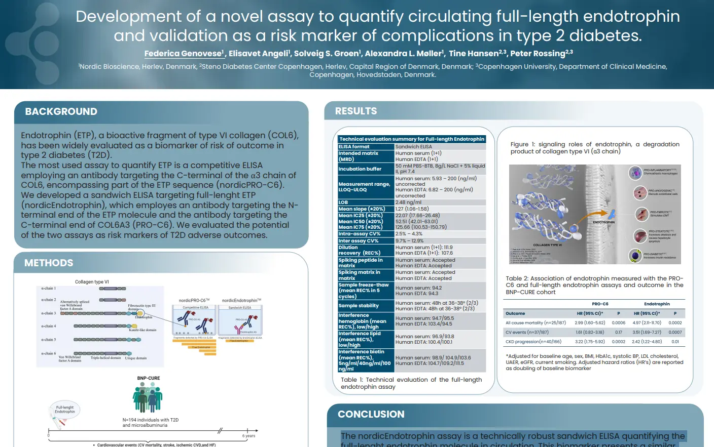
Development of a novel assay to quantify circulating full-length endotrophin and validation as a risk marker of complications in T2D
Introduction
NordicEndotrophin™ (ETP), a bioactive fragment of type VI collagen (COL6), has been widely evaluated as a biomarker of risk of outcome in type 2 diabetes (T2D). The most used assay to quantify ETP is a competitive ELISA employing an antibody targeting the C-terminal of the α3 chain of COL6, encompassing part of the ETP sequence (nordicPRO-C6™).
We developed a sandwich ELISA targeting full-lenght ETP (nordicEndotrophin™), which employes an antibody targeting the N-terminal end of the ETP molecule and the antibody targeting the C-terminal end of COL6A3 (nordicPRO-C6™). We evaluated the potential of the two assays as risk markers of T2D adverse outcomes.
The nordicEndotrophin™ assay is a technically robust sandwich ELISA quantifying the full-lenght endotrophin molecule in circulation. This biomarker presents a similar, or possibly higher prognostic power for complications of T2D than the competitive ELISA nordicPRO-C6™, used so far to quantify nordicEndotrophin™ in circulation.
Get in touch
Are you interested in exploring collaboration possibilities? Enter your information in the form and a representative will contact you shortly.