Activated macrophage-conditioned media induces fibrogenesis in a gastrointestinal scar-in-a-jar model that is quantifiable with serological biomarkers of collagen formation
Introduction
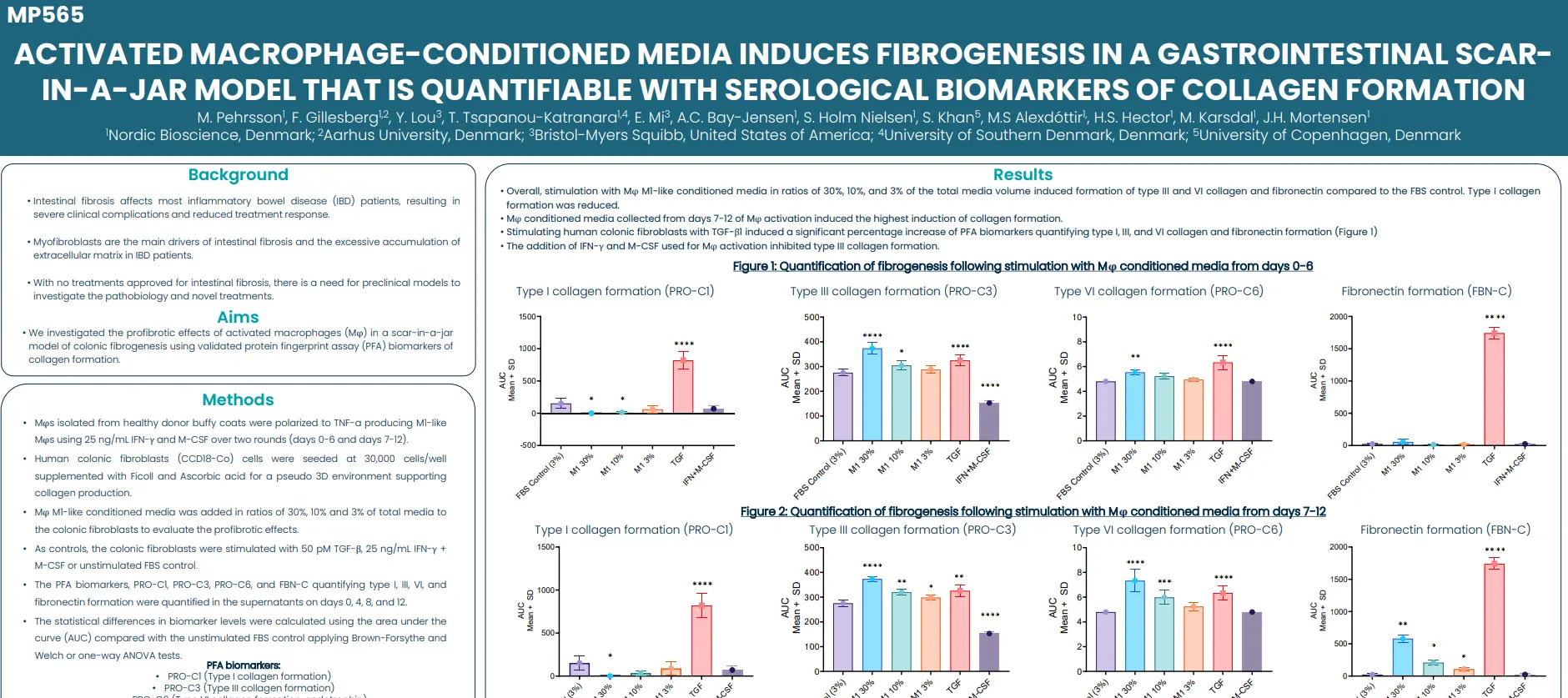
Intestinal fibrosis affects most inflammatory bowel disease (IBD) patients, resulting in severe clinical complications and reduced treatment response. Driven primarily by myofibroblasts, this condition is characterized by the excessive accumulation of extracellular matrix in the intestines. With no treatments approved for intestinal fibrosis, there is a need for preclinical models to investigate the pathobiology and novel treatments.
In this study we investigated the profibrotic effects of activated macrophages (Mφ) in a scar-in-a-jar model of colonic fibrogenesis using validated protein fingerprint assay (PFA) biomarkers of collagen formation.
The noninvasive PFA biomarkers can be used to objectively quantify fibrogenesis in the in vitro Scar-in-a-Jar model, providing a valuable tool for investigating the underlying mechanisms of fibrogenesis.
Get in touch
Are you interested in exploring collaboration possibilities? Enter your information in the form and a representative will contact you shortly.
Degradation of the alveolar basement membrane type IV collagen alpha-3 chain is associated with antifibrotic treatment and pulmonary hypertension in idiopathic pulmonary fibrosis
Introduction
Idiopathic pulmonary fibrosis (IPF) is a rare but devastating disease with inevitable progression and high mortality. Currently, Pirfenidone and Nintedanib are the only approved antifibrotic treatment options to slow disease progression. Additionally, a pulmonary hypertension (PH) complication can further worsen disease outcome and quality of life. Forced vital capacity (FVC) is currently the most employed endpoint in clinical trials to monitor disease progression for antifibrotic treatment (Tx) development.
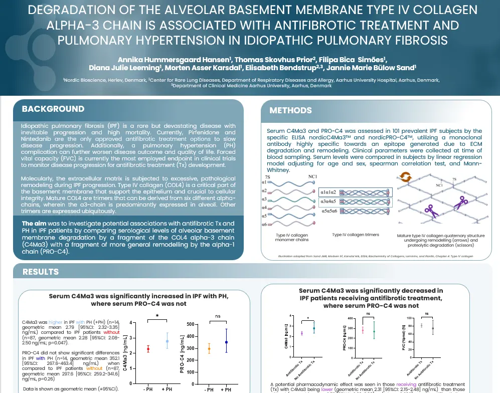
Molecularly, the extracellular matrix is subjected to excessive, pathological remodeling during IPF progression. Type IV collagen (COL4) is a critical part of the basement membrane that support the epithelium and crucial to cellular integrity. Mature COL4 are trimers that can be derived from six different alpha-chains, wherein the a3-chain is predominantly expressed in alveoli. Other trimers are expressed ubiquitously.
The aim was to investigate potential associations with antifibrotic Tx and PH in IPF patients by comparing serological levels of alveolar basement membrane degradation by a fragment of the COL4 alpha-3 chain (C4Ma3) with a fragment of more general remodeling by the alpha-1 chain (PRO-C4).
The COL4 alpha-3 chain has limited tissue distribution and is crucial for alveolar function. In this study, higher levels of COL4 alpha-3 chain degradation (C4Ma3) was:
found in IPF with PH.
associated with no antifibrotic treatment, indicating a pharmacodynamic potential.
In comparison, the COL4 alpha-1 chain marker PRO-C4, indicating ubiquitous basement membrane remodeling, did not show statistically different levels between any of the groups compared in this study. This could highlight the fact that damage done within alveoli are especially relevant when assessing the effect of antifibrotic Tx and PH in IPF.
Get in touch
Are you interested in exploring collaboration possibilities? Enter your information in the form and a representative will contact you shortly.
Reduction of PRO-C3 and PRO-C6 fibrogenesis biomarkers inconnective tissue disease-associated interstitial lung disease:results from the Phase IIb RECITAL trial
Introduction
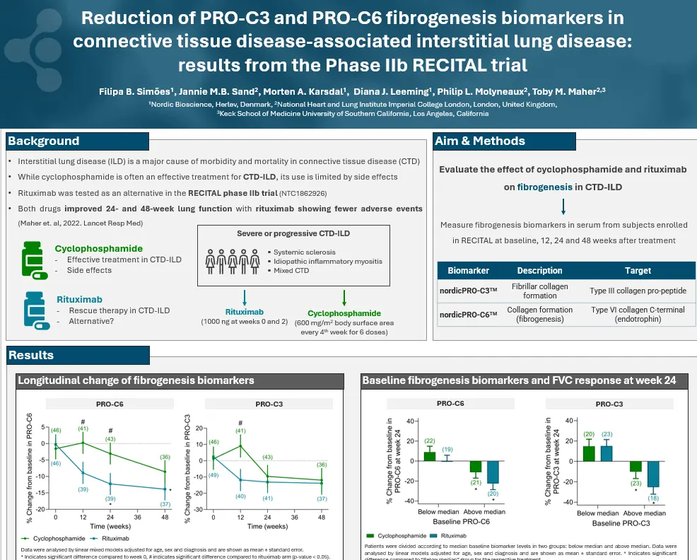
Interstitial lung disease (ILD) is a major cause of morbidity and mortality in connective tissue disease (CTD). While cyclophosphamide is often an effective treatment for CTD-ILD, its use is limited by side effects.
In this study, Rituximab was tested as an alternative in the RECITAL phase IIb trial(NTC1862926). Both drugs improved 24- and 48-week lung function with rituximab showing fewer adverse events.
The decrease in nordicPRO-C6™ and nordicPRO-C3™ suggest that, besides their immunomodulatory effects, these drugs may also reduce fibrogenesis. NordicPRO-C3™ and nordicPRO-C6™, measured at baseline and as % change from baseline, are associated with an FVC response. These findings highlight nordicPRO-C3™ and nordicPRO-C6™ as promising biomarkers for CTD-ILD.
Get in touch
Are you interested in exploring collaboration possibilities? Enter your information in the form and a representative will contact you shortly.
A novel serological biomarker targeting a collagen type-I-derived matricryptin predicts all-cause mortality at admission with ST-elevated myocardial infarction
Introduction
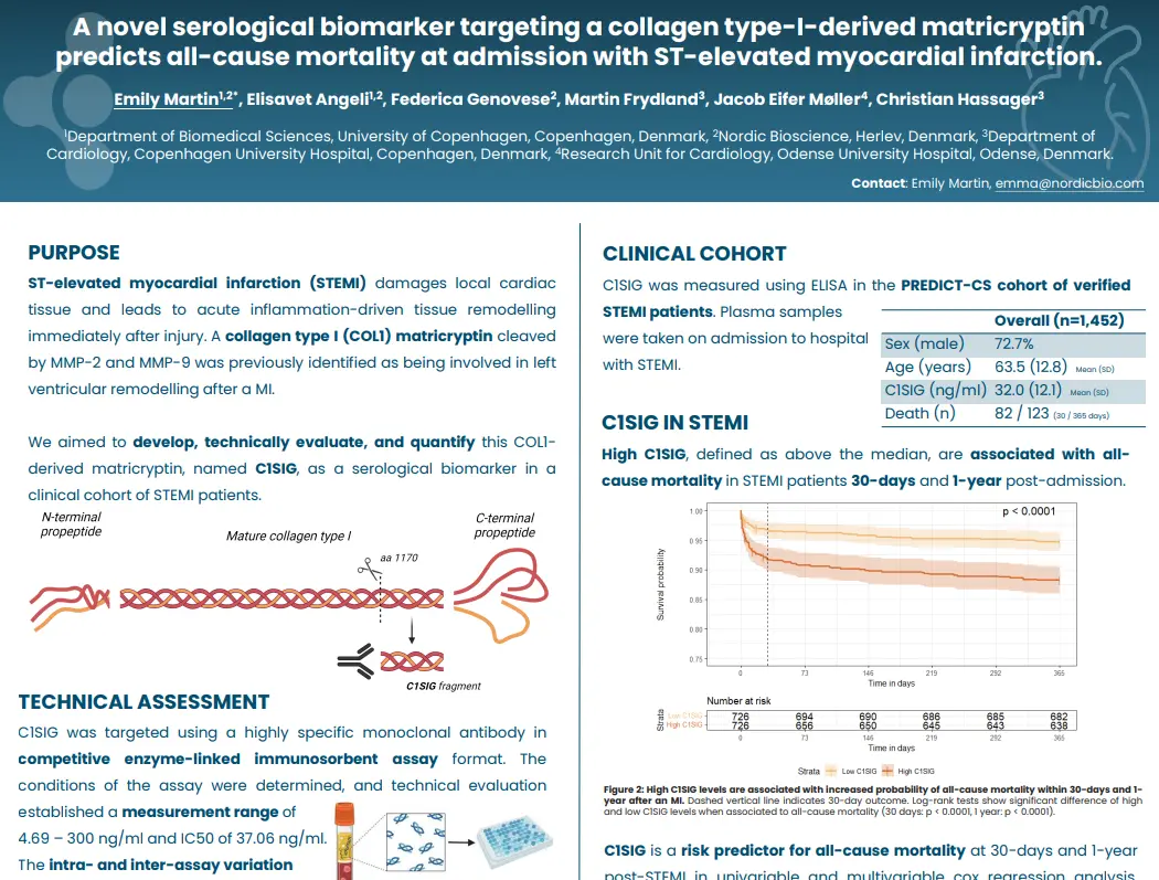
ST-elevated myocardial infarction (STEMI) damages local cardiac tissue and leads to acute inflammation-driven tissue remodeling immediately after injury. A collagen type I (COL1) matricryptin cleaved by MMP-2 and MMP-9 was previously identified as being involved in left ventricular remodeling after a MI.
We aimed to develop, technically evaluate, and quantify this COL1- derived matricryptin, named C1SIG, as a serological biomarker in a clinical cohort of STEMI patients.
C1SIG was developed as a technically robust biomarker and demonstrated as an independent predictor of mortality at 30 days and 1-year after STEMI, even when adjusted for multiple clinically-relevant variables. This COL1 biomarker could be helpful in assessing acute extracellular matrix processing in individuals after suffering a STEMI and could identify a subset of patients at increased risk of long-term outcome.
Get in touch
Are you interested in exploring collaboration possibilities? Enter your information in the form and a representative will contact you shortly.
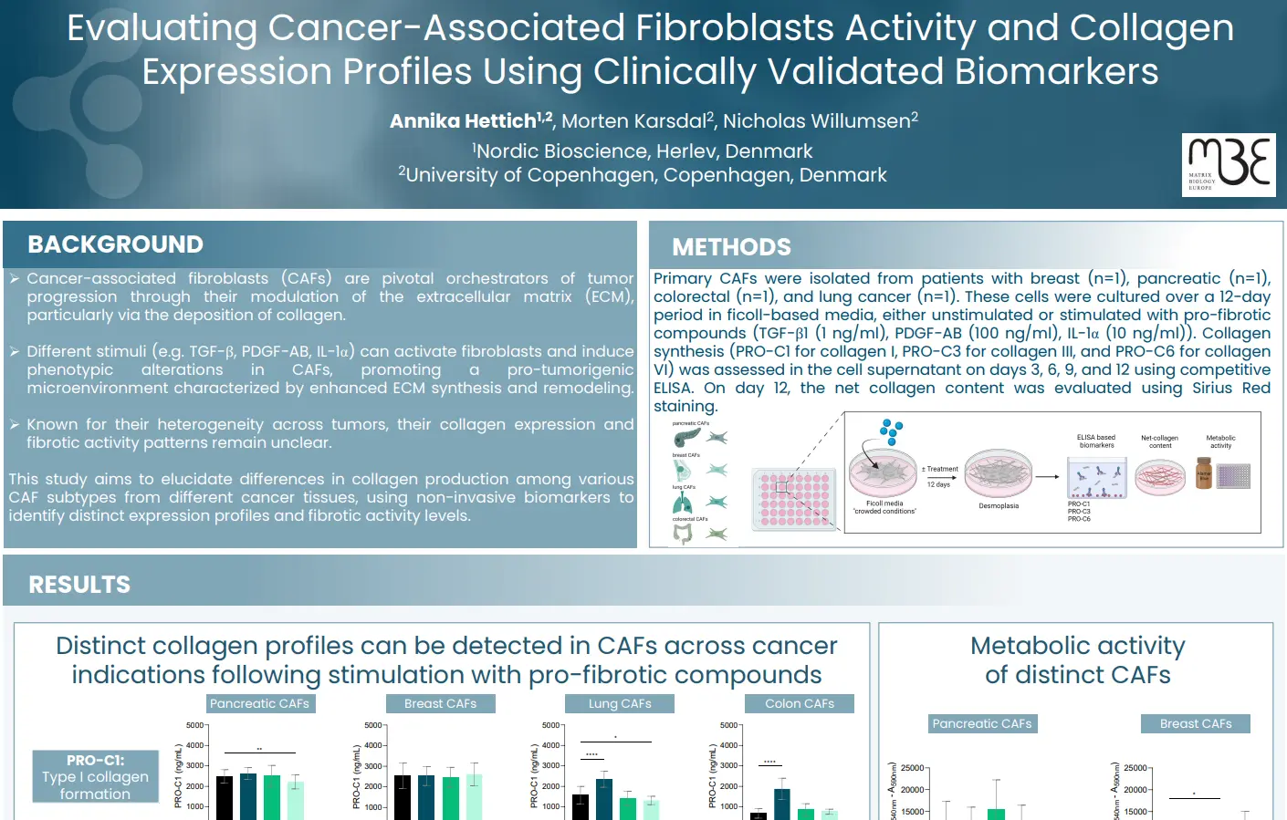
Evaluating cancer-associated fibroblasts activity and collagen expression profiles using clinically validated biomarkers
Introduction
Cancer-associated fibroblasts (CAFs) are pivotal orchestrators of tumor progression through their modulation of the extracellular matrix (ECM), particularly via the deposition of collagen. Different stimuli (e.g. TGF-β, PDGF-AB, IL-1α) can activate fibroblasts and induce phenotypic alterations in CAFs, promoting a pro-tumorigenic microenvironment characterized by enhanced ECM synthesis and remodeling. Known for their heterogeneity across tumors, their collagen expression and fibrotic activity patterns remain unclear.
In this study we aimed to elucidate differences in collagen production among various CAF subtypes from different cancer tissues, using non-invasive biomarkers to identify distinct expression profiles and fibrotic activity levels.
These findings underscore the heterogeneity in collagen production and fibrotic activity among CAFs from different indications, providing valuable insights into the ECM dynamics within distinct TMEs. Collagen-based non-invasive biomarkers demonstrate the capability to differentiate between the fibrotic activity of CAFs isolated from different tissues.
Get in touch
Are you interested in exploring collaboration possibilities? Enter your information in the form and a representative will contact you shortly.
In this webinar leading experts will discuss the critical role fibroblasts play in metabolic disorders. The presentations will highlight recent research findings and explore the therapeutic potential of modulating fibroblast activity to improve patient outcomes.
Agenda
Opening of the session
Metabolic activation of ECM production of fibroblast and the relation to outcomes in heart, liver and kidney diseases – Dr. Morten Karsdal
Metabolic activation and de-activation of fibroblasts in MAFLD by weight loss. The weight-dependent angle – Dr. Jörn M. Schattenberg
Pharmacological modulation of fibroblast activation and the benefit for patients –Dr. Diana J. Leeming
General discussion and questions
Scientific topics
We will explore key aspects of fibroblast activity in metabolic health, starting with the metabolic activation and deactivation of fibroblasts in MAFLD (Metabolic Associated Fatty Liver Disease). It highlights the weight-dependent factors influencing these processes and the impact of weight loss on fibroblast behavior in MAFLD.
The discussion then moves to the metabolic activation of extracellular matrix (ECM) production by fibroblasts and its effects on heart, liver, and kidney diseases. Finally, it addresses the potential benefits of pharmacological interventions in modulating fibroblast activation, offering therapeutic possibilities for patients with these conditions.
Dr. Jörn M. Schattenberg is a Professor of Medicine and Director of the Department of Medicine II at Saarland University Medical Center in Homburg and the University of the Saarland in Germany.
Dr. Schattenberg completed his post-doctoral training at Albert-Einstein College of Medicine in New York, focusing on signaling pathways involved in acute and chronic liver injury, particularly in metabolic dysfunction associated steatotic liver disease (MASLD).
He is board-certified in Internal Medicine, Gastroenterology & Hepatology, and Infectious Disease.
His research specializes in translational sciences and clinical trials in Hepatology, with a focus on new technologies and approaches for the prevention, screening, and treatment of metabolic liver disease.
Dr. Schattenberg is involved in multinational, EU-funded research consortia exploring biomarkers and novel treatment methods for liver diseases.
He is a member of the Policy, Public Health, and Advocacy Committee of the European Association for the Study of the Liver (EASL).
He serves on the Editorial Boards of “Hepatology” and “Alimentary Pharmacology & Therapeutics” and is an Associate Editor for “JHEP Reports.”
Dr. Diana J. Leeming
Dr. Diana Julie Leeming is the Senior Director of Fibrosis, Hepatic, and Pulmonary Research at Nordic Bioscience.
She joined Nordic Bioscience in 2004 and assumed the role of Director of Fibrosis in 2010, later being promoted to Senior Director in 2024.
Dr. Leeming focuses on developing serologically assessed markers to evaluate extracellular matrix remodeling in patients with pulmonary or hepatic fibrosis, aiding in diagnosis and pharmacodynamic evaluation.
She is a principal inventor of the PRO-C3 assay, a fibrogenesis marker utilized in multiple clinical trial studies.
Dr. Leeming has authored over 280 peer-reviewed publications, demonstrating her extensive contributions to the field.
Her H-index is 61, her I10-index is 174, and her research has garnered over 11,825 citations as of March 2024.
Dr. Morten A. Karsdal
Dr. Morten Karsdal joined Nordic Bioscience in 2001 and became CEO in June 2010, leading the company to significant advancements in biomarker development and disease biology.
Dr. Karsdal is an honorary professor of inflammation research at the University of Southern Denmark, where he continues to supervise PhD students, fostering the next generation of researchers.
Dr. Karsdal chairs the Extracellular Matrix Pharmacology Congress, an important forum for advancing drug development by focusing on the extracellular matrix (ECM) as a key factor in most chronic diseases. He is renowned for his deep expertise in fibrosis, rheumatology (including rheumatoid arthritis and osteoarthritis), diabetes, and other chronic conditions, particularly in relation to ECM and biomarker research.
Dr. Karsdal has led the development of FDA-approved and supported molecular diagnostics, as well as more than 100 commercialized biomarker assays, including ELISA assays and high precision automated platforms.
He has extensive experience in clinical trial design and the clinical application of biochemical markers, often serving as a consultant to major pharmaceutical companies for the use of serological biomarkers in clinical trials.
In 2016, he and his research team authored the first edition of “Biochemistry of Collagens, Laminins and Elastin,” published by Elsevier Science. The book, now in its 3rd edition as of 2023, is a key resource on collagens and structural proteins, with a focus on their applications in chronic diseases.
Get in touch
Are you interested in exploring collaboration possibilities? Enter your information in the form and a representative will contact you shortly.
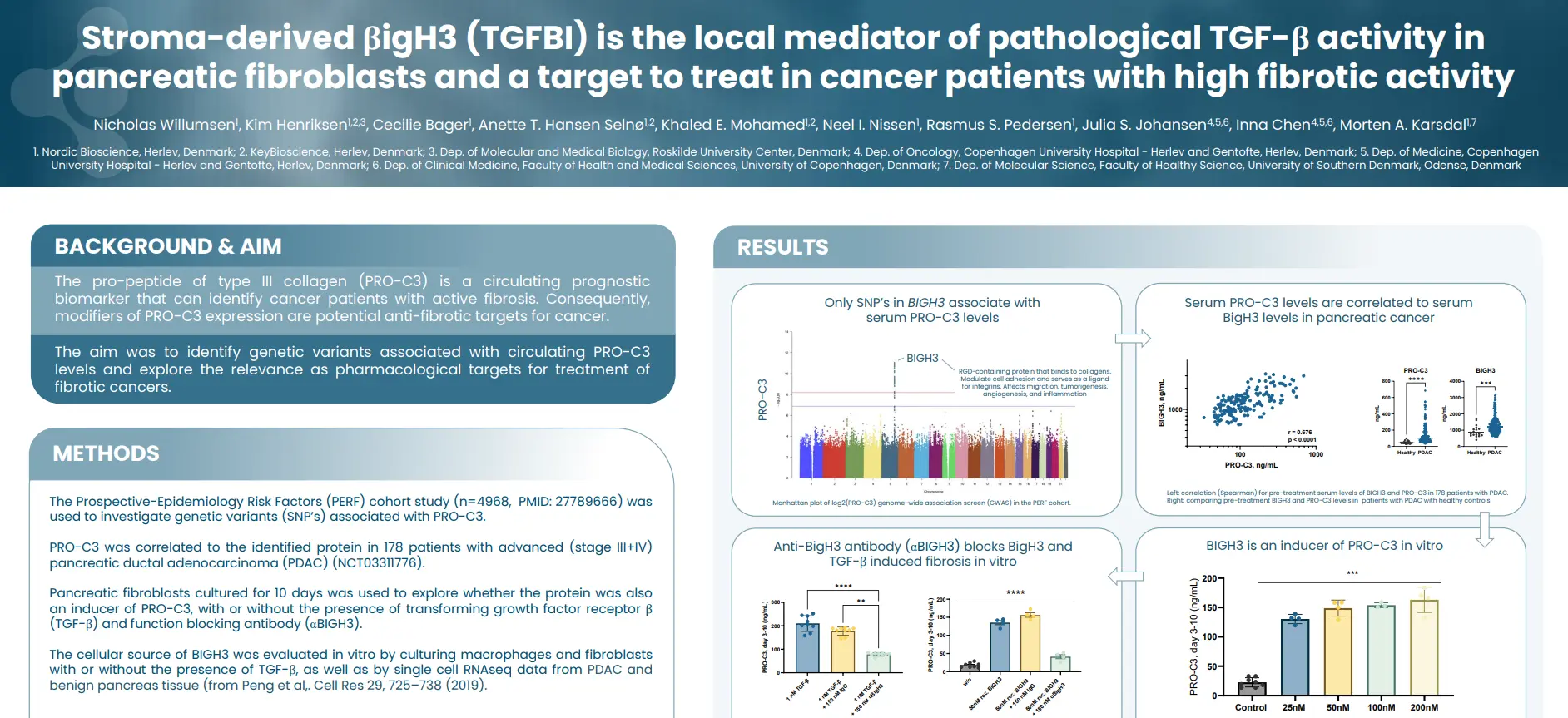
Stroma-derived βigH3 (TGFBI) is the local mediator of pathological TGF-β activity in pancreatic fibroblasts and a target to treat in cancer patients with high fibrotic activity
Introduction
The pro-peptide of type III collagen (nordicPRO-C3™) is a circulating prognostic biomarker that can identify cancer patients with active fibrosis. Consequently, modifiers of nordicPRO-C3™ expression are potential anti-fibrotic targets for cancer. The aim was to identify genetic variants associated with circulating nordicPRO-C3™ levels and explore the relevance as pharmacological targets for treatment of fibrotic cancers.
An association between BigH3 and nordicPRO-C3™ was found by GWAS, in vitro, and in patients with PDAC: TGF-β induce BigH3, which subsequently activate fibroblasts to become fibrotic, resulting in elevated levels of nordicPRO-C3™, which in turn can be modulated by an anti-BigH3 antibody. This highlights the potential for treatment of tumor fibrosis by inhibiting BIGH3 in cancer patients with elevated nordicPRO-C3™ levels.
Get in touch
Are you interested in exploring collaboration possibilities? Enter your information in the form and a representative will contact you shortly.
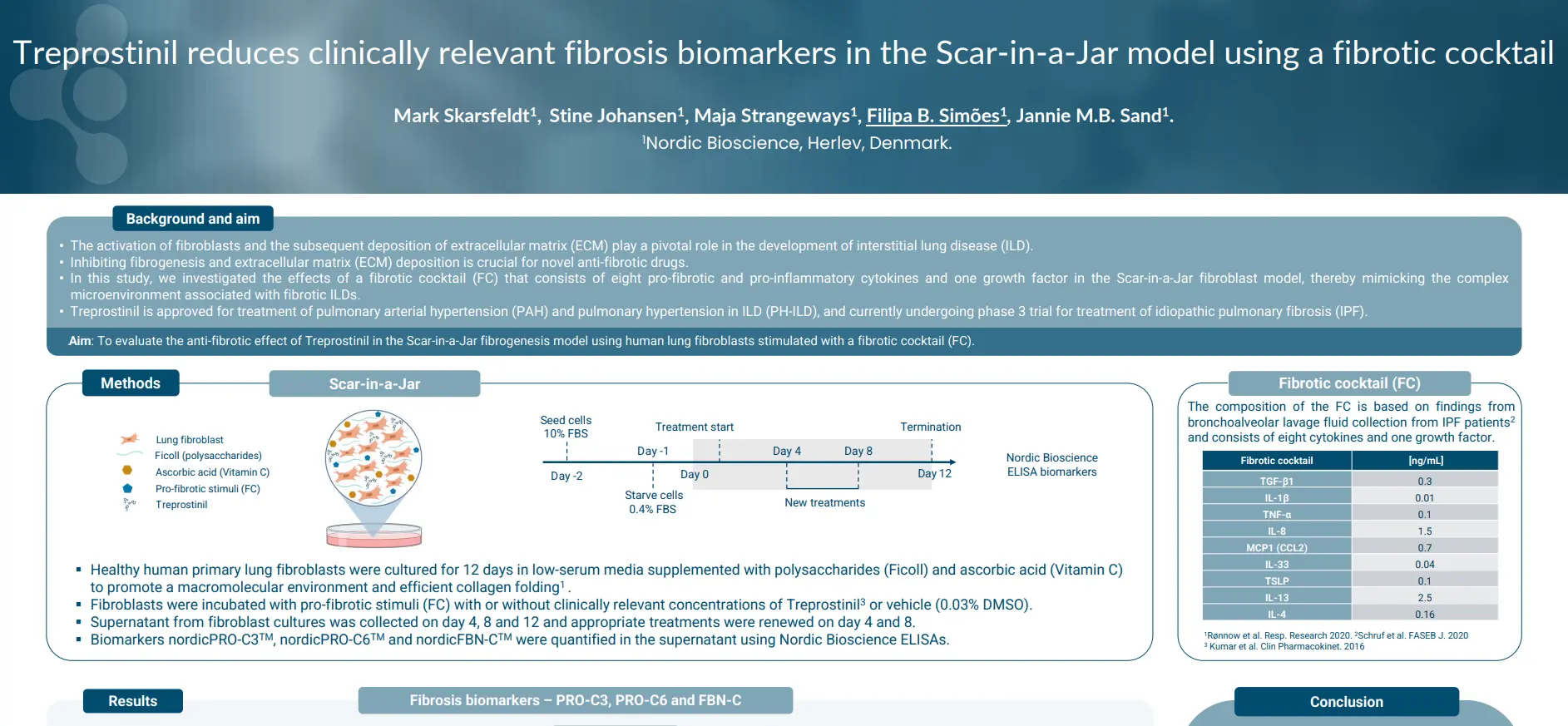
Treprostinil reduces clinically relevant fibrosis biomarkers in the Scar-in-a-Jar model using a fibrotic cocktail
Introduction
The activation of fibroblasts and the subsequent deposition of extracellular matrix (ECM) play a pivotal role in the development of interstitial lung disease (ILD), making it crucial for novel anti-fibrotic drugs.
In this study, we investigated the effects of a fibrotic cocktail (FC) that consists of eight pro-fibrotic and pro-inflammatory cytokines and one growth factor in the Scar-in-a-Jar fibroblast model, thereby mimicking the complex microenvironment associated with fibrotic ILDs.
The FC effectively stimulated fibrogenesis in the Scar-in-a-Jar in-vitro model, leading to elevated levels of the fibrosis biomarkers nordicPRO-C3™, nordicPRO-C6™ and FBN-C™. Treprostinil significantly inhibited fibrogenesis and collagen deposition quantified by ELISA. In conclusion, the Scar-in-a-Jar model is a useful tool for screening novel anti-fibrotic drugs.
Get in touch
Are you interested in exploring collaboration possibilities? Enter your information in the form and a representative will contact you shortly.
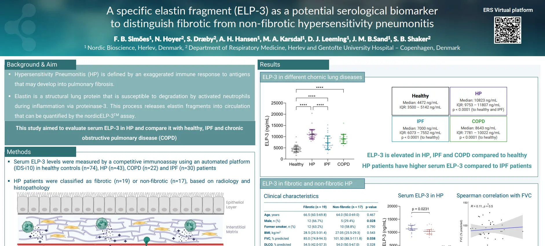
A specific elastin fragment (ELP-3) as a potential serological biomarker to distinguish fibrotic from non-fibrotic hypersensitivity pneumonitis
Introduction
Hypersensitivity Pneumonitis (HP) is defined by an exaggerated immune response to antigens that may develop into pulmonary fibrosis. Elastin, a structural lung protein, is susceptible to degradation by activated neutrophils during inflammation via proteinase-3. This process releases elastin fragments into circulation that can be quantified by the ELP-3 assay.
This study aimed to evaluate serum ELP-3 in HP and compare it with healthy, IPF and chronic obstructive pulmonary disease (COPD).
The serum ELP-3 is elevated in patients with different chronic lung diseases, particularly in HP. Notably, ELP-3 was able to separate non-fibrotic from fibrotic HP patients. These findings highlight the potential value of ELP-3 as a biomarker that provides additional clinical information beyond conventional inflammation markers.
Get in touch
Are you interested in exploring collaboration possibilities? Enter your information in the form and a representative will contact you shortly.
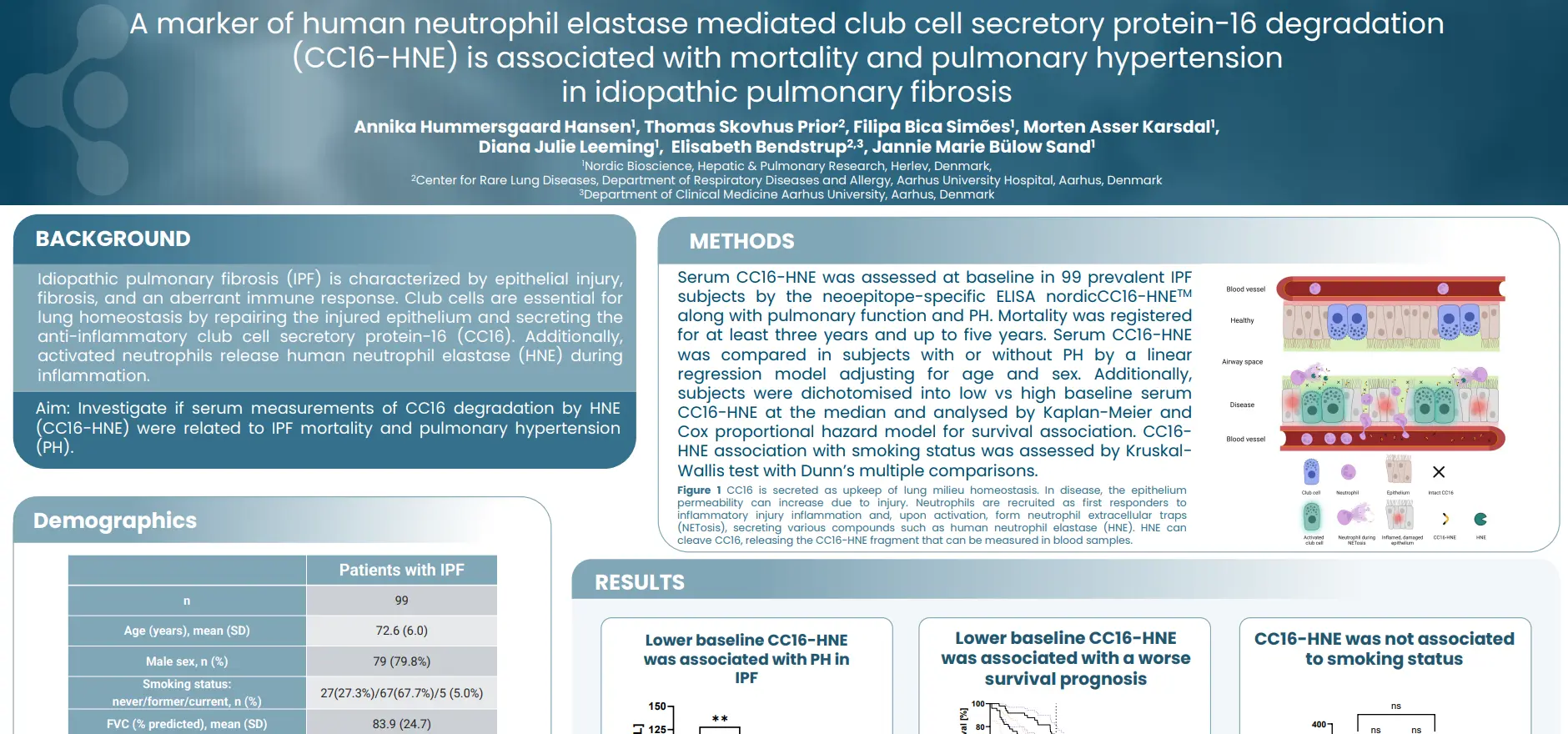
A marker of human neutrophil elastase mediated club cell secretary protein-16 degradation in (CC16-HNE) is associated with mortality and pulmonary hypertension in idiopathic pulmonary fibrosis
Introduction
Idiopathic pulmonary fibrosis (IPF) is characterized by epithelial injury, fibrosis, and an aberrant immune response. Club cells are essential for lung homeostasis by repairing the injured epithelium and secreting the anti-inflammatory club cell secretory protein-16 (CC16). Additionally, activated neutrophils release human neutrophil elastase (HNE) during inflammation. Our aim was to investigate if serum measurements of CC16 degradation by HNE (CC16-HNE) were related to IPF mortality and pulmonary hypertension (PH).
Low serum CC16-HNE at baseline was associated with increased risk of mortality and a PH complication in IPF. These results indicate that a neutrophil immune response and degradation of CC16 is relevant for disease outcome. CC16 degradation by HNE could serve as a prognostic biomarker for IPF and diagnostic for a PH complication.
Get in touch
Are you interested in exploring collaboration possibilities? Enter your information in the form and a representative will contact you shortly.