TNF-α and TGF-β Synergistically Promote Fibrogenesis

TNF-α and TGF-β Synergistically Promote Fibrogenesis in a In Vitro Model of Fibro-inflammation

Introduction

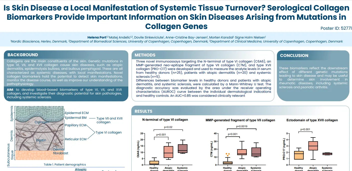
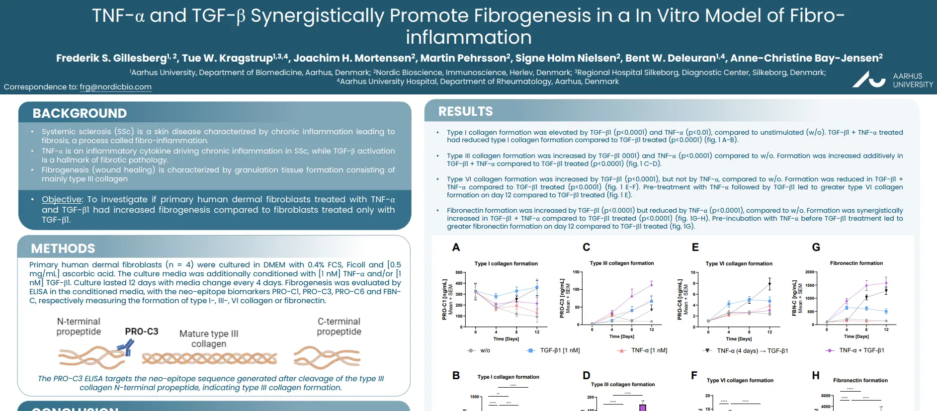
Systemic sclerosis (SSc) is a skin disease characterized by chronic inflammation leading to
fibrosis, a process called fibro-inflammation. TNF-α is an inflammatory cytokine driving chronic inflammation in SSc, while TGF-β activation is a hallmark of fibrotic pathology. Fibrogenesis (wound healing) is characterized by granulation tissue formation consisting of mainly type III collagen.

The aim of this study was to investigate if primary human dermal fibroblasts treated with TNF-α
and TGF-β1 had increased fibrogenesis compared to fibroblasts treated only with TGF-β1.

Poster

Conclusion

Inflammatory TNF-α stimulation increases TGF-β driven fibrogenesis in dermal fibroblasts, by promoting their formation of type III collagen and fibronectin. Consequently, biomarkers of type III collagen formation and fibronectin formation may be markers of early fibrosis in fibro-inflammatory skin disease.

Get in touch

Are you interested in exploring collaboration possibilities?
Enter your information in the form and a representative will contact you shortly.

    Is Skin Disease a Local Manifestation of Systemic Tissue Turnover?

    Is Skin Disease a Local Manifestation of Systemic Tissue Turnover? Serological Collagen Biomarkers Provide Important Information on Skin Diseases Arising from Mutations in Collagen Genes

    Introduction

    Collagens are the main constituents of the skin. Genetic mutations in type VI, VII, and XVII collagen cause skin diseases, such as atopic dermatitis, epidermolysis bullosa, and bullous pemphigoid. These are all
    characterized as systemic diseases, with local manifestations. Novel collagen biomarkers hold the potential to detect skin manifestations, monitor the disease course, as well as improve our understanding of the pathophysiology.

    The aim of this study was to develop blood-based biomarkers of type VI, VII, and XVII collagen, and investigate their diagnostic potential for skin pathologies, including systemic sclerosis.

    Poster

    Conclusion

    These biomarkers reflect the downstream effect of different genetic mutations leading to skin disease and may be useful to determine skin involvement in rheumatic diseases, including systemic sclerosis and psoriatic arthritis.

    Get in touch

    Are you interested in exploring collaboration possibilities?
Enter your information in the form and a representative will contact you shortly.

      C3M, PRO-C7 and VICM Can Identify and Monitor Response to Infliximab (IFX)

      Serum biomarkers of proteolytic tissue destruction, formation and macrophage activity can discern patients with IBD according to infliximab treatment non-response or response

      Introduction

      Characterized by chronic inflammation, patients with Inflammatory Bowel Disease (IBD) experience detrimental remodeling of their intestinal extracellular matrix (ECM). Treatment with anti-inflammatory drugs can reduce inflammation, leading to remission and tissue healing. However, adequate monitoring of patients is critical to ensure and maintain treatment response.

      As potential surrogate markers of ECM remodeling, we investigated blood-based biomarkers of type III and -VII collagen and posttranslational modifications of vimentin in patients with IBD. Our aim was to determine the value of the C3M, PRO-C7, and VICM biomarkers for identifying and monitoring response to infliximab (IFX).

      Poster

      Conclusion

      Quantifying a combination of non-invasive biomarkers of ECM remodeling and macrophage activity provided AUCs of 0.684 to 0.797 identifying responders to IFX treatment. Each biomarker provided value at the three different visits (Visit 1, 2, and 3). Combining all three biomarkers measured at each visit
      resulted in an AUC of 0.797 identifying responders to IFX treatment.

      Get in touch

      Are you interested in exploring collaboration possibilities?
Enter your information in the form and a representative will contact you shortly.

        Neutrophil Elastase Degraded Fragment of Type III Collagen Is Elevated in IBD Patients

        Immune-cell specific biomarker of early intestinal inflammation: Neutrophil elastase degraded fragment of type III collagen is elevated in patients with inflammatory bowel disease

        Introduction

        Inflammatory Bowel Disease (IBD) is characterized by epithelial barrier injury of the gastrointestinal (GI) tract and is driven by abnormal immune responses and excessive secretion of proteases from immune cells. Among these, neutrophils are the first to migrate into the inflamed interstitial matrix, where type III collagen is significantly deposited. Early detection of mucosal inflammation is crucial to prevent cumulative clinical damage, as a delayed diagnosis can hinder effective treatment.

        In this study we aimed to develop a biomarker that reflects early intestinal inflammation prior to it becoming medically evident; allowing us to distinguish patients that would benefit from an anti-inflammatory treatment.

        Poster

        Conclusion

        C3-HNE levels are elevated in patients with IBD compared with HD. This increase is also observed in conditioned media from primary neutrophils activated with lipopolysaccharide (LPS) for six hours. Importantly, C3-HNE reflects the early stages of clinically apparent mucosal damage in experimental models of colitis. As such, this biomarker holds promise for identifying early mucosal injury or acute inflammation in the gastrointestinal tract. Nevertheless, additional studies are needed to evaluate its clinical validity.

        Get in touch

        Are you interested in exploring collaboration possibilities?
Enter your information in the form and a representative will contact you shortly.

          Type III Collagen Biomarkers as Markers of Endoscopic Disease Activity in Ulcerative Colitis

          Blood-based biomarkers of type III collagen remodeling as surrogate markers of endoscopic disease activity in patients with ulcerative colitis

          Introduction

          The chronic inflammation of Ulcerative Colitis (UC) causes excessive extracellular matrix (ECM) remodeling, resulting in clinical complications. Currently, endoscopic evaluation remains the gold standard method for determining disease activity. However, novel methods are wanted due to its
          invasiveness and accompanying patient discomfort.

          As type III collagen is a major component of the intestinal ECM and a target for multiple proteases catalyzing its remodeling in IBD, we sought to investigate two blood-based neoepitope biomarkers of type III collagen degradation, and fibrosis resolution as surrogate markers of disease activity.

          Poster

          Conclusion

          C3M was elevated at Week 0 in UC patients with severe endoscopic disease activity according to the Total Mayo Score. The fibrolysis biomarker, nordicCTX-III™, was significantly elevated in patients with moderate endoscopic disease at week 0 and numerically elevated in mild disease compared to severe disease activity.

          Get in touch

          Are you interested in exploring collaboration possibilities?
Enter your information in the form and a representative will contact you shortly.