Biomarkers aren’t just confined to different omics categories; various classes are referring to the types of serological measurements.
Consider cytokines, growth factors, receptors, kinases, transcription factors, intracellular proteins, extracellular proteins, and a subset within that—Extracellular Matrix (ECM) proteins. The common denominator in most pathologies is a loss of balance between different ECM proteins, especially collagens.
Biomarkers are more than just omics categories
This excessive destruction and deposition of proteins significantly propel the progression of end-stage diseases, culminating in organ dysfunction, failure, and, ultimately, death.
Diverse cytokines and growth factors orchestrating via specific receptors and kinases lead to ECM destruction and deposition, making ECM the converging pathway for multiple stimuli.
By the end of the day, what matters is to reverse ECM deterioration, or even undo, organ damage and function decline. Reversing organ damage can be achieved by stopping ECM deterioration. To truly reverse organ damage and restore organ functionality in patients, we need to repair the ECM to its normal balance.
We at Nordic Bioscience believe that the answer lies in effecting change at the tissue level—the key to reversing organ damage and, in turn, revitalizing organ function.
Have you considered if your treatment or pathway is affecting tissue formation or degradation? If so, feel free to browse our unique ECM biomarker portfolio.
Get in touch
Are you interested in exploring collaboration possibilities? Enter your information in the form and a representative will contact you shortly.
Nordic Bioscience’s extracellular matrix-based dermatology biomarkers offer a unique approach that sets them apart from competitors, providing deeper insights into tissue formation, degradation, immune cell activity, and resolution processes related to skin diseases. Unlike wide-range proteomics arrays, our biomarkers are fundamentally different and more specific, offering a higher level of clarity and precision.
Nordic Bioscience’s ECM-based dermatology biomarkers offer a unique approach
A clear connection
The clear connection between our dermatology biomarkers and skin diseases lies in the fact that immune cell activity, skin tissue remodeling and genetic mutations of collagens are known to cause these conditions. By identifying pathology specific biomarkers, we not only indicate the presence of a disease in a patient but also highlight the local manifestation of an underlying systemic cause. This genetic link and visible manifestation on the skin create a relatable narrative for stakeholders and pharmaceutical companies, potentially paving the way towards a better understanding of the disease.
A key advantage of our skin disease biomarkers is the limited competition in the market, and no approved biomarkers for skin diseases. With no competitors in the tissue-based biomarker space, we have the opportunity to position ourselves as leaders in this area. Supported by compelling data that demonstrates pathology specificity, disease activity, disease progression, and pharmacodynamic response, there is a rare chance for pharmaceutical partners to be among the first movers and pioneers in this field.
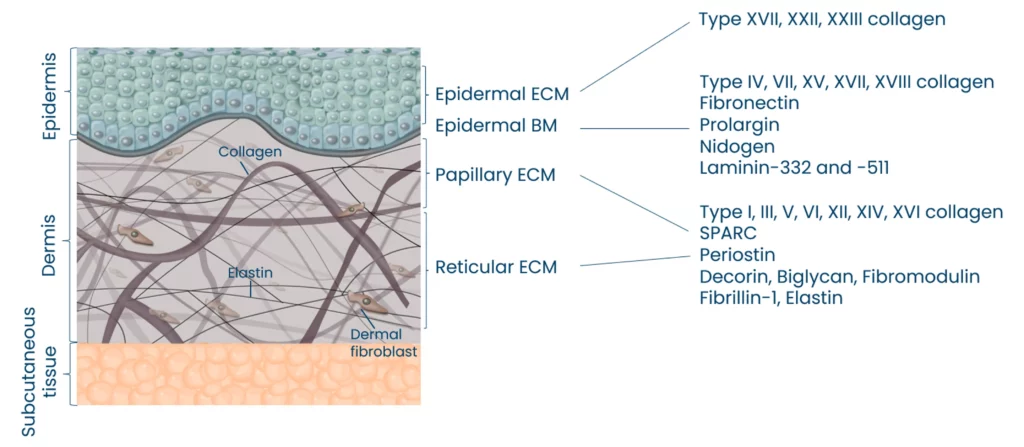
Moreso, we have observed that skin aging and cross-linking collagens are correlated. Our biomarkers can quantify the age of collagens by measuring alpha and beta CTX-III together with the ECM of the epidermis and dermis.
Nordic Bioscience can quantify specific pathological fragments from the different compartments of the skin
Implications for precision medicine
Precision medicine is a crucial aspect empowered by our dermatology biomarkers. By providing information about ongoing pathological processes within the tissue, such as damage and repair, we enable targeted treatments and therapies that enhance patient outcomes. This personalized medicine approach is far more effective than relying on proteomic providers’ hope of hitting the right target by chance. Our markers aid in drug development by specifically identifying target populations and reducing costs, ultimately expediting the development process.
Our skin disease biomarkers play a vital role in elucidating the mechanism of action (MoA) of treatments. Here, we can also shed light on the speed at which a compound modulates tissue and fibroblast derived biomarkers, providing essential information on the efficacy and responsiveness of the treatment.
Furthermore, by analyzing the objective disease activity, we can determine how the compound affects specific factors associated with the patient’s profile, such as neutrophil activity, mast cell activity, macrophage activity, soft tissue destruction, systemic fibroblast activity, and more. This holistic evaluation enables a deeper understanding of how the compound influences different aspects of the disease process, ultimately facilitating the development of more effective and targeted therapies for patients.
Robust technology
Our laboratory technology is robust and meets CLSI validation guidelines, ensuring reliability and accuracy. Moreover, our derma markers can be seamlessly integrated into Roche automated platforms, making them easily accessible worldwide. This scalability and dependability are vital for the development of reliable clinical research tools, further solidifying the value of our biomarkers in the field of dermatology.
A potential case for melanoma studies
While our dermatology markers have broad applicability, there is a particularly intriguing potential use in melanoma. By understanding the tissue formation, degradation, and repair processes specific to this type of skin cancer, we can offer valuable insights for diagnosis and management.
For clients involved in ongoing metastatic melanoma research, we can help establish a connection between extracellular matrix remodeling and melanoma, positioning our biomarkers as a valuable resource for further investigation and improving patient care.
Nordic Bioscience’s ECM-based biomarkers provide a relatable and comprehensive narrative by connecting genetic links, visible skin manifestations, and in-depth insights into ongoing pathological processes. With unparalleled clarity and understanding, our biomarkers empower precision medicine in skin diseases. Supported by robust technology, validated methodologies, and a potential application in melanoma, our biomarkers offer an unprecedented opportunity for stakeholders and pharmaceutical companies.
Get in touch
Are you interested in exploring collaboration possibilities? Enter your information in the form and a representative will contact you shortly.
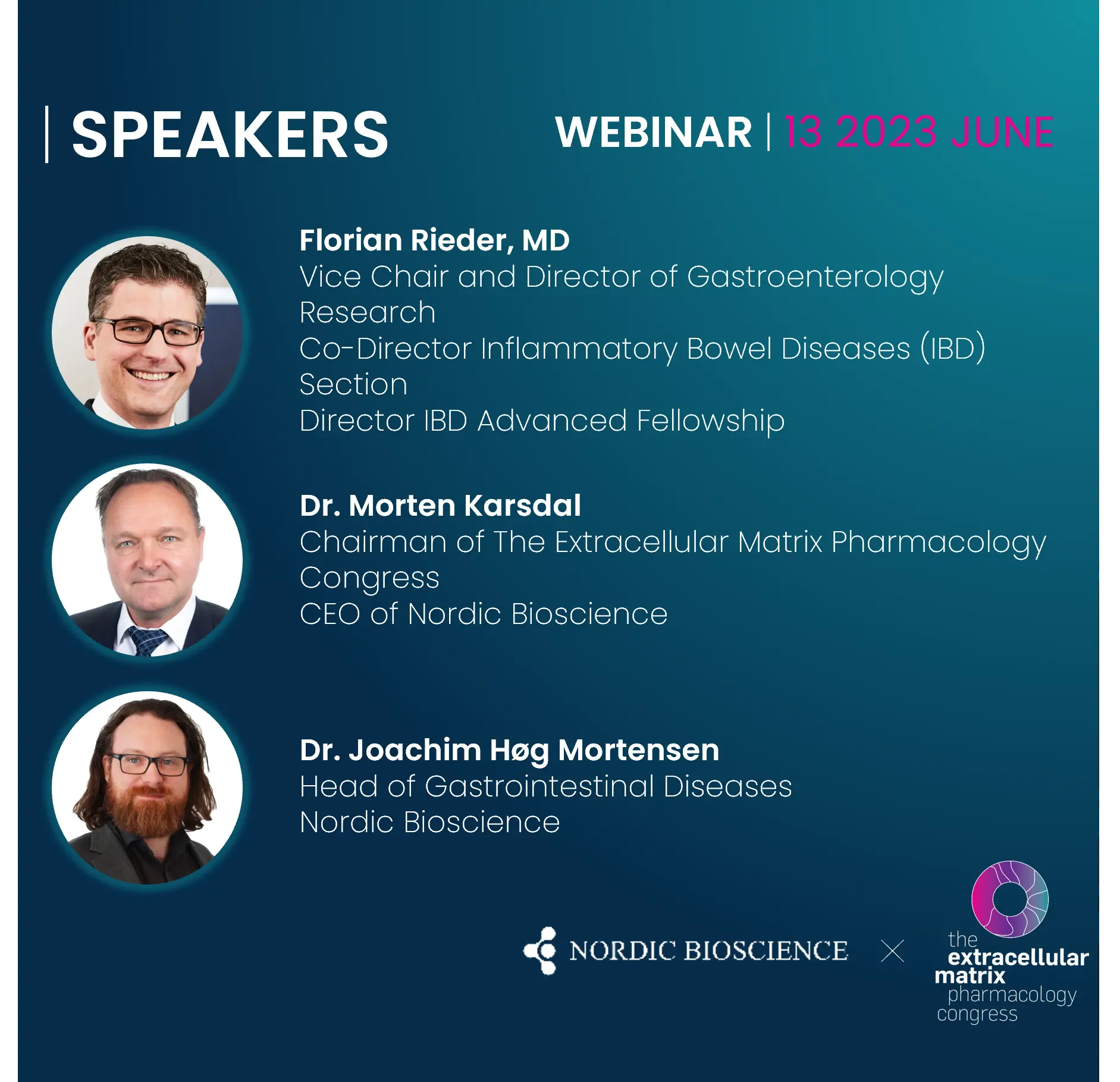
This webinar focuses on the intriguing interplay between inflammatory bowel diseases (IBD) and neutrophil activity, with a special emphasis on the insights provided by the unique CPa9-HNE serum calprotectin.
Our experts will unravel the biological processes of mucosal damage and healing in inflammatory bowel disease (IBD), and how it relates to neutrophil activity and the extracellular matrix (ECM). Specifically, we will delve into the role of mucosal damage, intestinal fibrosis, and fibro-inflammation, and how they impact ECM remodeling and integrity.
The role of neutrophil activity in tissue destruction – Dr. Morten A. Karsdal
Mucosal damage and intestinal fibrosis in IBD – Dr. Florian Rieder
Fibro-inflammation in IBD a new frontier and how to quantify it – Dr. Joachim Høg Mortensen
Questions from the chat
Scientific topics and speakers
In this edition of the ECM Pharmacology Symposium Series, we will address the mechanisms of neutrophil activity in chronic diseases, in particular on fibro-inflammation in inflammatory bowel diseases, focusing in particular on fibro-inflammation and mucosal damage.
We will also present the latest methods for quantifying fibro-inflammation in IBD, such as the novel serum calprotectin biomarker CPa9-HNE, and discuss the implications of these findings for patients. In addition, we will look at current and future treatment options for IBD and the potential impact of these advances on patient outcomes.
This webinar is an excellent opportunity for researchers, clinicians, and healthcare professionals to gain valuable insight into the latest developments in IBD research and treatment, and to gain a deeper understanding of the intricate interplay of neutrophil activity, mucosal damage and fibro-inflammation in IBD.
Dr. Rieder’s clinical focus is patients with IBD with a special emphasis on the field of pathogenesis, prediction, and therapy of intestinal fibrosis.
Dr. Rieder has published over 150 articles (h-index 52) and book chapters and he has been awarded the Sherman Emerging Leaders award.
He is the lead author of the ECCO guidelines on Ulcerative colitis and the first ECCO clinical consensus on ‘Diagnosis and Management of Intestinal Fibrosis’. He is a past associate editor of Clinical and Translational Gastroenterology.
Dr. Rieder serves as an abstract reviewer for all major GI conferences and on several editorial boards of medical journals.
Dr. Rieder has significant ties to the ECCO, having served as the chair of Y-ECCO, a member of the ECCO operational board, a prior Y-ECCO committee member, and a member of the scientific committee.
He was the past chair of REACH-IBD and is currently the Co-Chair of the Professional Education Committee of the Crohn’s and Colitis Foundation.
Dr. Rieder is the leading Principal Investigator (PI) of the international Stenosis Therapy and Research (STAR) Consortium, which aims to test anti-fibrotic medications in stricturing Crohn’s disease.
Dr. Joachim Høg Mortensen
Dr. Mortensen joined Nordic Bioscience in 2013 and currently serves as Head of Department of Gastrointestinal Diseases.
The group’s research activities span from biomarker discovery to preclinical and clinical trial exploration focusing on the development and validation of blood-based biomarkers to quantify fibro-inflammation which covers neutrophil activity, mucosal damage and intestinal fibrosis in serum of patients with gastrointestinal diseases e.g. inflammatory bowel disease.
The group’s primary focus is to evaluate pharmacodynamics changes, efficacy and monitoring of response to various treatments using biomarkers reflecting fibro-inflammation.
Biomarkers are developed to support and understand fibro-inflammation-associated IBD disease progression and response to treatment, where the biomarkers are applied from target selection to clinical trial monitoring.
Dr. Morten A. Karsdal
Dr. Karsdal chairs the Extracellular Matrix Pharmacology Congress, an influential forum dedicated to advancing drug development by identifying changes in the ECM as common denominators in chronic diseases.
Dr. Morten Karsdal joined Nordic Bioscience in 2001 and has been serving as CEO since June 2010.
He is a prolific author, with more than 650 peer-reviewed articles to his name, accumulating over 34,000 citations and an h-index of 95.
He is renowned for his expertise and commitment to biomarker development, extracellular matrix biology, and disease research, particularly in fibrosis, rheumatology, and diabetes. He possesses extensive experience in clinical trial design and the clinical application of biochemical markers, consulting for major pharmaceutical companies.
As an honorary professor of inflammation research at the University of Southern Denmark, Dr. Karsdal supervises PhD students and contributes to advancing scientific knowledge in the field.
In 2016, he authored the first edition of “Biochemistry of Collagens, Laminins and Elastin,” published by Elsevier Science, which is now in its 3rd edition, providing a comprehensive overview of structural proteins and their relevance in chronic diseases.
Dr. Karsdal’s leadership and contributions to the Nordic Life Science community were recognized with the Arthur D. Little Nordic Life Science Award in 2023, highlighting his outstanding management and leadership achievements.
Get in touch
Are you interested in exploring collaboration possibilities? Enter your information in the form and a representative will contact you shortly.
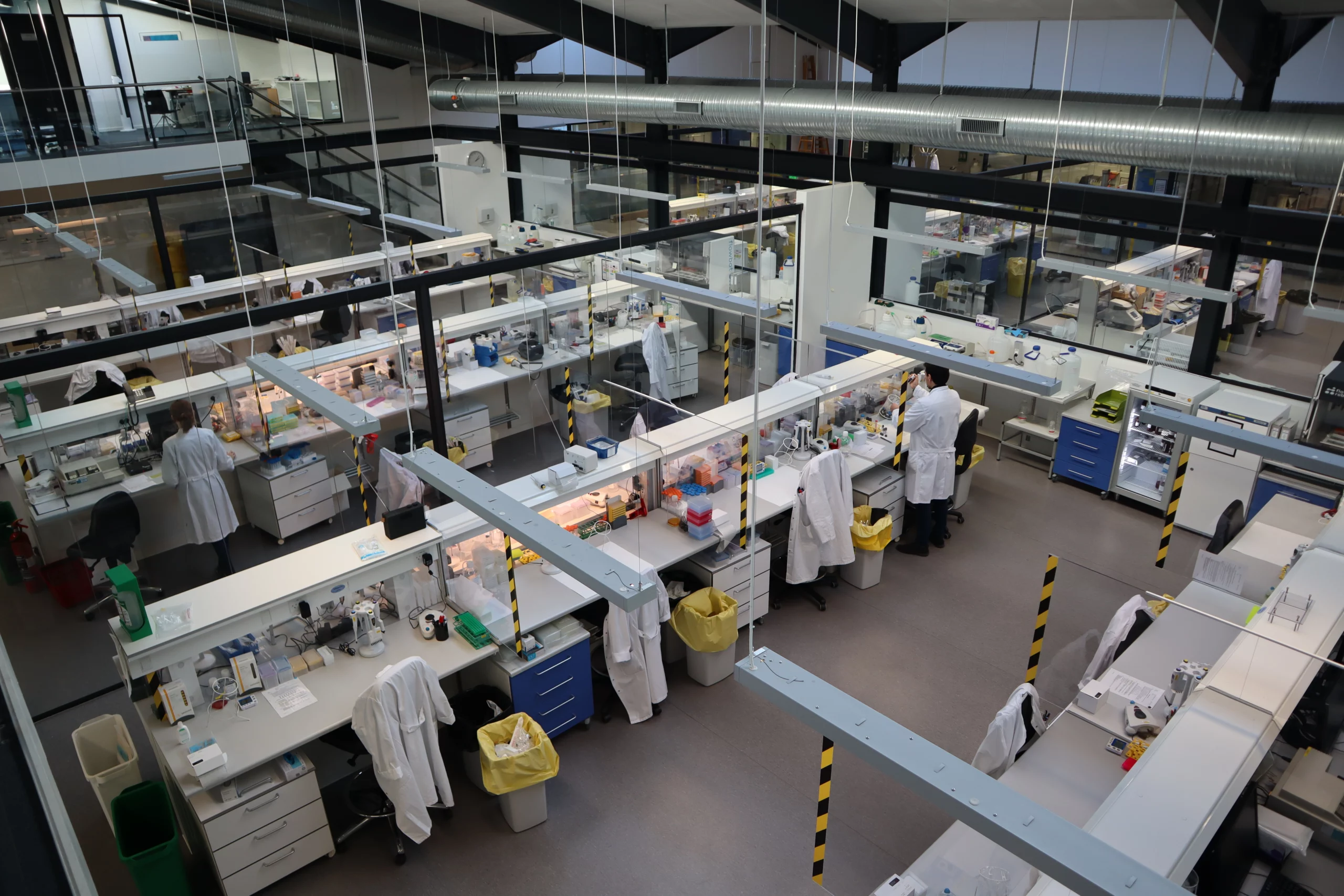
In the field of clinical trials, accuracy and reliability are critical to success. For this reason, Nordic Bioscience Laboratory is dedicated to providing the highest quality laboratory services, with a strong focus on data integrity, customer satisfaction, and scientific excellence.
Our clinical laboratory offers central lab and specialty lab services, including a wide range of diagnostic and exploratory tests that are essential for meeting patients’ care needs and advancing drug development efforts. We use state-of-the-art equipment and technology, as well as a team of highly skilled laboratory professionals, to deliver fast, accurate and reliable results without sacrificing quality.
Nordic Bioscience Clinical Laboratory offers central and specialty lab services
Compliance at every step of the way
We pride ourselves on ensuring that biomarker validation complies with European Medicines Agency (EMA) and Food and Drug Administration (FDA) guidelines. In addition, we provide access to a team of scientific experts who have the experience and expertise to help you navigate the complexities of biomarker research, interpret data, and make informed decisions about your studies.
Our Clinical Laboratory is accredited by the College of American Pathologists (CAP) and all aspects of laboratory services are performed in accordance with international Good Clinical Practice (GCP) and Good Clinical laboratory Practices (GCLP) standards. Our accreditation reflects our unwavering commitment to providing the highest quality laboratory services that adhere to the most rigorous industry standards.
Commitment to improvement
As part of our ongoing commitment to continuous improvement, our processes, and procedures are regularly reviewed and audited by numerous external auditors and accreditation bodies. These audits enable us to continuously improve and innovate our laboratory services to meet the latest regulatory requirements. We also invest in the continuous education and training of our staff to stay abreast of the latest laboratory science. These efforts allow us to maintain our high-quality standards and ensure that our customers receive the best possible laboratory services, as well as scientific excellence that can help them interpret data.
At Nordic Bioscience Laboratory, we hold ourselves to the highest standard of quality and place a high value on providing a personalized experience for clinical testing. We are committed to serving as your trusted partner in delivering accurate and reliable laboratory services that help advance the field of clinical trials.
We hope that you will consider partnering with Nordic Bioscience for your clinical laboratory needs. We look forward to the opportunity to work with you and support your research activities.
Get in touch
Are you interested in exploring collaboration possibilities? Enter your information in the form and a representative will contact you shortly.
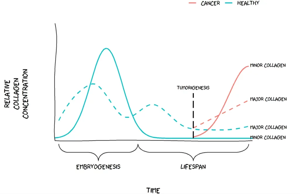
Collagen is a crucial protein that provides structural support and strength to tissues throughout the body. However, there is a group of highly specialized minor collagens that may play a critical role in cancer progression. Most research on the biology of collagens has focused on the abundant collagens, but the highly specialized minor collagens that are expressed only at certain developmental stages may be deregulated in cancer.
The Hypothesis
Minor collagens are widely expressed during embryogenesis, as they organize the extracellular matrix (ECM) to support tissue development. However, in adult tissues, minor collagens become restricted to a few specific organs and are expressed at very low levels.
Recent studies have suggested that minor collagens may become deregulated in cancer, where they are highly upregulated in tumor tissues. This upregulation is thought to occur as the ECM is reorganized during tumor development and progression. We have chosen to call this hypothesis “The Minor Collagen Hypothesis”. We hypothesize that minor collages, which are normally restricted to embryonic tissues, may become active again in adult tissues as cancer progresses. The Minor Collagen Hypothesis proposes that this reactivation may be a key component in in tumor growth and invasion by altering the composition of the ECM.
Visualization of the minor collagen hypothesis. Figure by Jeppe Thorlacius-Ussing.
Applying The Minor Collagen Hypothesis in clinical research
Biomarkers are important for indicating the presence or severity of a disease, and in cancer, they are crucial for early detection, monitoring of cancer progression, and guiding treatment decisions.
In general, several collagen biomarkers are overexpressed in cancer. However, minor collagens may offer a more specific biomarker for certain types of cancer. For example, recent research has shown that certain subtypes of cancer-associated fibroblasts (CAFs) express specific minor collagens. These collagens can be detected in blood, making them potential targets for drug development aimed at CAFs.
This hypothesis offers researchers new possibilities and highlights the need for more specific biomarkers for cancer diagnosis and treatment that could lead to better patient outcomes. Identifying biomarkers that detect specific minor collagens could be a breakthrough in cancer research and might shed light on an overlooked aspect of cancer progression.
Minor collagens are typically expressed during embryogenesis or tissue development and are expressed at low levels afterward. In cancer, however, these specialized collagens are expressed again to form an environment conducive to cancer growth. The abundant major collagens are also expressed during tissue development and growth spurts in childhood and adolescence but tapers off in adulthood. The relative induction as a function of cancer is greater for the minor collagens compared to the major collagens. This could mean better biomarker performance.
Watch this insightful webinar on tumor fibrosis, where we will explore the latest advancements in cancer research with leading experts in the field.
This webinar will provide a unique opportunity to hear from experts in the field and gain valuable insights into the latest advancements in tumor fibrosis research. Join us to learn about cutting-edge approaches to developing new therapeutic targets, and the use of biomarkers for patient stratification and evaluating treatment efficacy.
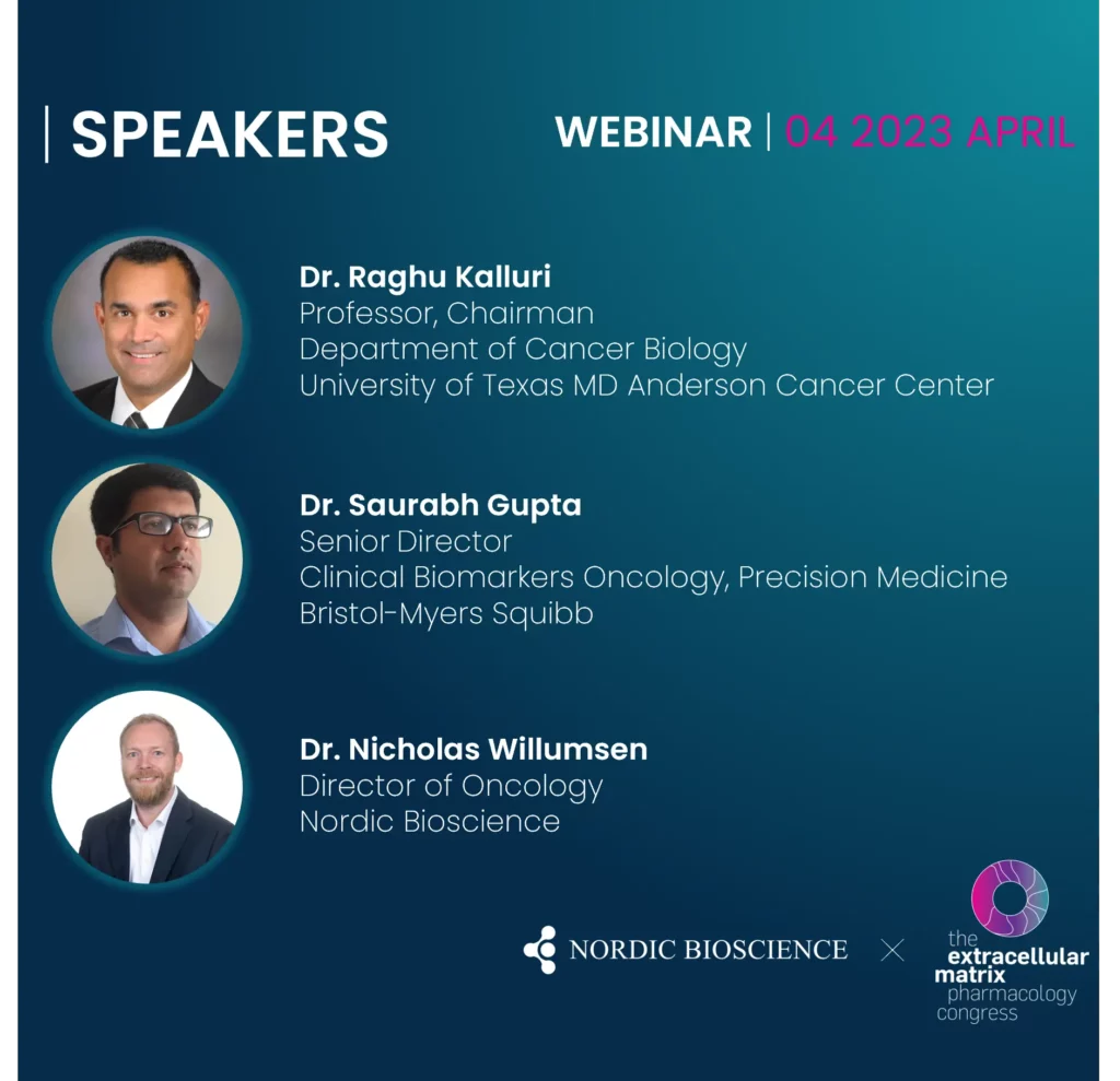
Tumor Fibrosis and Clinical Outcomes: Underlying Mechanisms – Dr. Saurabh Gupta
The Collagen Landscape in Cancer: Fibroblast Activities and New Biomarkers – Dr. Nicholas Willumsen
Unique and unexpected role of collagens in cancer – Dr. Raghu Kalluri
Question from the chat
Expert line-up
Dr. Raghu Kalluri (Professor and Chairman, Department of Cancer Biology, University of Texas MD Anderson Cancer Center), is an expert in the cellular microenvironment and will discuss, among others, the unique and unexpected role of collagens in cancer.
Dr. Saurabh Gupta (Senior Director, Clinical Biomarkers Oncology, Precision Medicine), Bristol-Myers Squibb, is a clinical drug development and biomarker research expert and will provide valuable insights into the underlying mechanisms of tumor fibrosis and the association with clinical outcomes.
Dr. Nicholas Willumsen (Director, Oncology, Nordic Bioscience), will focus on the development and validation of blood-based biomarkers to quantify tumor fibrosis in serum from cancer patients and discuss how this may inform on cancer-associated fibroblast (CAF) activity and subtyping.
Get in touch
Are you interested in exploring collaboration possibilities? Enter your information in the form and a representative will contact you shortly.
Understanding Fibrosis Remodeling in Animal Models
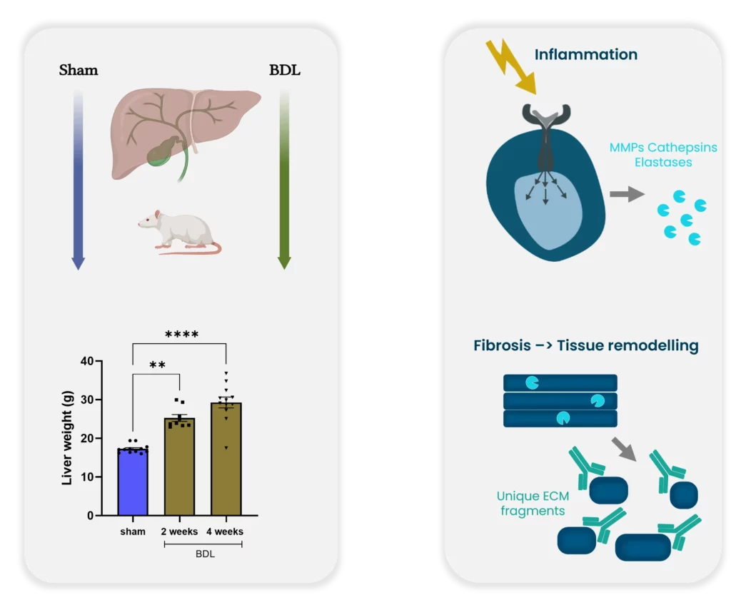
Animal testing is a crucial aspect of drug development, mandated by the FDA for all premature drugs in development. However, a critical question arises: how can we be sure that the pathology observed in rodents is reflective of the same pathology in humans? This is a significant concern for the medical community because of the potential for negative consequences if clinical trials are based on inaccurate preclinical data as we see in humans. Why then is it so important to measure biomarker formations in rodents? Because it matters whether it mimics the right pathology.
Example for bridging preclinical and clinical practice. Bile duct ligation: a preclinical model of liver fibrosis.
The process of transitioning from preclinical to clinical trials is critical to success. Histological stages are built on human tissue sections and used for comparison to create an appropriate animal model. However, none of the existing rodent models currently develop nonalcoholic steatohepatitis (NASH) as seen in humans. This discrepancy raises concerns regarding the usefulness of rodent models for preclinical studies.
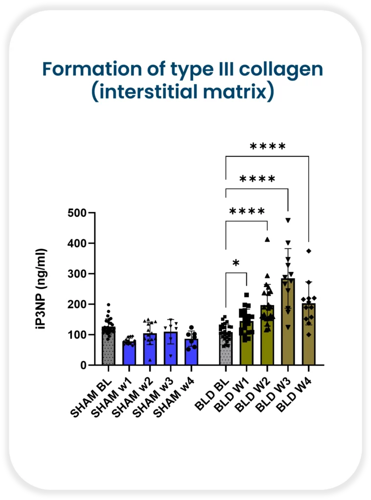
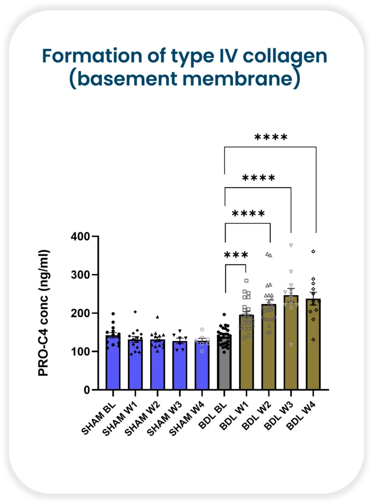
Despite these concerns, it is still essential to measure biomarker formations in rodents. The reason being, it matters whether the biomarker mimics the correct pathology. Nordic Bioscience has demonstrated increasing formation of both type III collagen (interstitial matrix) and type IV collagen (basement membrane) with continued injury in a rat bile duct ligation model. Collagen III has been shown to be upregulated in human liver fibrosis, including NASH, and is associated with increased severity. On the other hand, collagen IV is a measure of early fibrosis remodeling.
Bile duct ligation: a preclinical model of liver fibrosis
It is critical to understand fibrosis remodeling during disease progression in both animals and humans. With Nordic Bioscience’s biomarkers, researchers can improve translation between the preclinical and clinical practice. The use of biomarkers enables researchers to track disease progression and evaluate the effectiveness of potential treatments accurately. Moreover, they can also help identify patients who are likely to respond positively to a particular therapy.
Animal testing remains a critical aspect of drug development, it is essential to understand rodent model limitations. Biomarkers offer an effective way to track the progression of disease accurately. We can identify patients likely to benefit from a specific treatment and improve the transition from preclinical to clinical trials. By leveraging biomarkers in both animal and human models, we can make sure that the right therapy is provided to the right patient—at the right time.
The session will include a stimulating talk by Dr. Morten Karsdal on understanding the mysteries of the different types of collagens. Dr. Alexander Lynge Reese-Petersen will then spotlight collagen type VI and its relationship to various pathologies, as well as the pharmacological and biological properties of the dangerous hormone Endotrophin.
Scientific topics
Extracellular matrix (ECM) dysregulation is the founding cause of tissue fibrosis, a process driving more than 50 pathologies and most chronic diseases. Fibrosis may develop both consequent to either an increase in tissue formation or a decrease in tissue degradation.
Understanding the ECM and the central components of the basement membrane and interstitial ECM will lead to a better understanding of disease mechanisms and to novel treatment strategies. This may be a central part in precision medicine strategies to target the right patients.
ECM remodeling is the common denominator of all fibro-inflammatory disorders
Collagens have signaling molecules that we need to understand to understand how the ECM affects cells in fibrosis and tissue turnover
Endotrophin and other collagen fragments (Vastatin, Tumstatin and Endostatin) are potential signaling molecules derived from the processing of the collagens in the ECM
Understanding ECM turnover may lead to better insight into disease mechanisms and better biological understanding of individual patients
Dr. Alexander Reese’s presentation will cover:
ECM biomarkers are elevated in chronic pathologies and have prognostic significance for outcome. How can we best use this to guide drug development?
Endotrophin, a collagen hormone in multi-organ diseases
Endotrophin in HFpEF, a potentially treatable high-risk endotype
Use of the tissue turnover profile for patient selection
Get in touch
Are you interested in exploring collaboration possibilities? Enter your information in the form and a representative will contact you shortly.
CPa9-HNE ELISA has emerged as a novel serum calprotectin biomarker
Status quo: Crohn’s and Colitis biomarkers
Conventional serum calprotectin biomarkers are often not as clinically useful as the fecal versions, which is related to the half-life of calprotectin in blood (only 5-6 hours), leading to dissociation of calprotectin protein. The short half-life of calprotectin in blood reduces the window in which the current serum calprotectin ELISA assay can detect calprotectin dimer protein, which is composed of monomers S100A8 and S100A9.
By applying the Protein Fingerprint technology, we have identified a neo-epitope of the S100A9 monomer of calprotectin derived from proteolytic cleavage by human neutrophil elastase (HNE). This ELISA assay is referred to as the CPa9-HNE assay and is a new and innovative method for quantification of calprotectin in serum developed by Nordic Bioscience.
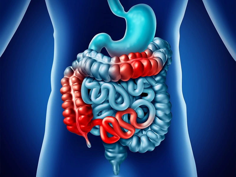
Inflammatory Bowel Disease (IBD) flares in the intestines.
A new tool to measure… calprotectin
Patients with Crohn’s disease and ulcerative colitis have been found to show significantly higher levels of CPa9-HNE in their serum than healthy subjects, with the AUC reaching 0.98 (CI: 0.97-1.00, p<0.0001) for CD and 0.96 (CI: 0.92-1.00, p<0.0001) for UC, proving the assay’s efficacy in distinguishing between disease states.
In comparison, the MRP8/14 serum calprotectin assay from Bühlmann Lab, while also used to identify IBD, showed poorer separation between patients and healthy subjects, with AUCs of 0.72 (CI: 0.59-0.86, p=0.0025) for UC and 0.70 (CI: 0.56-0.84, p=0.0046) for CD, respectively.
The CPa9-HNE biomarker showed a better correlation with the endoscopic score for Crohn’s disease (SES-CD) and ulcerative colitis (MES) than the FCP. At the same time, the MRP8/14 assay and neutrophil count showed no significant correlation with endoscopic scores for IBD patients.
To investigate the accuracy of the CPa9-HNE, we performed a received operator characteristic curve, demonstrating CPa9-HNE with acceptable accuracy to identify patients with IBD in remission vs. active disease.
Finally, we demonstrated that the CPa9-HNE biomarker could detect the calprotectin neo-epitope only in the supernatant of in vitro activated primary human neutrophils, but not in inactive primary human neutrophils. This was in contrast to the MRP8/14 serum CP assay of the Bühlmann lab, as both inactive and activated primary human neutrophils secreted detectable levels of calprotectin.
CPa9-HNE ELISA proved to be a novel serum calprotectin biomarker with significant clinical potential as a biomarker for patients with IBD to monitor disease activity and neutrophil activity.
However, circulating calprotectin metabolites released from locally inflamed mucosa are not be dissociated further. They can be readily quantified using an ELISA-based technique called Protein Fingerprint technology. This refined ELISA technique quantifies protein metabolites or neo-epitopes derived from proteolysis that reflect local tissue inflammation and remodeling.
PRO-C6 Findings In NEJM Evidence to Help Patient Segregation in HFpEF
A persistent problem remains a challenge in HFpEF – patient heterogeneity.
We need the right patients for the right treatment, but how do we get it to them? We believe that one approach to solving this problem is to improve patient segregation and endotyping.
In collaboration with Bristol Myers Squibb and University of Pennsylvania, we recently identified a subset of patients at a very high risk of adverse outcomes, all characterized by increased fibroblast activity. Using our PRO-C6 biomarker assay we can accurately quantify this risk profile and differentiate patients based on how likely they are to be re-hospitalized or have all-cause mortality due to heart failure.
These findings were recently published in NEJM Evidence.
So how do we deal with patient heterogeneity?
We believe it is time to give physicians and clinicians a better tool than what is available now.
This is endotrophin endotyping, heart failure risk stratification redefined.
Get in touch
Are you interested in exploring collaboration possibilities? Enter your information in the form and a representative will contact you shortly.