Tocilizumab Superior to Methotrexate in Collagen Degradation Inhibition

Tocilizumab demonstrates superior inhibition of MMP-mediated basement membrane collagen degradation compared to methotrexate or placebo

Introduction

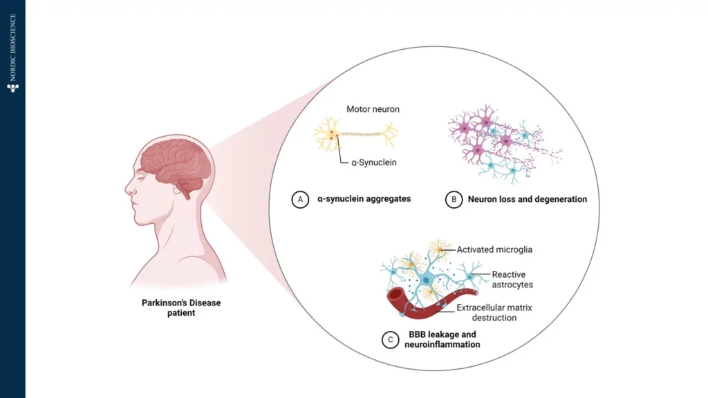
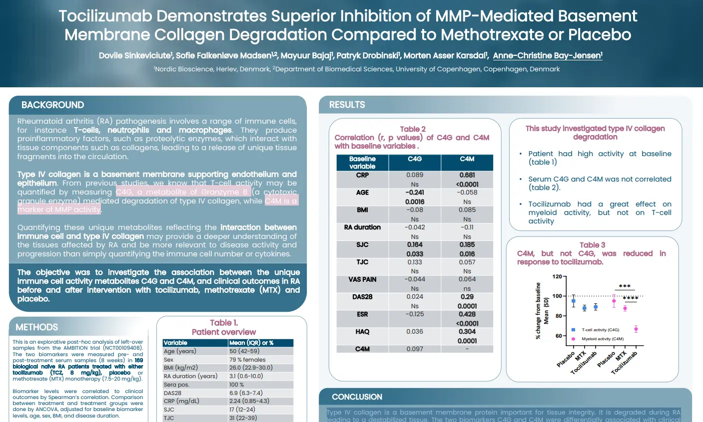
Rheumatoid arthritis (RA) pathogenesis involves a range of immune cells, for instance T-cells, neutrophils and macrophages. They produce proinflammatory factors, such as proteolytic enzymes, which interact with tissue components such as collagens, leading to a release of unique tissue fragments into the circulation. Type IV collagen is a basement membrane supporting endothelium and epithelium. From previous studies, we know that T-cell activity may be quantified by measuring C4G, a metabolite of Granzyme B (a cytotoxic granule enzyme) mediated degradation of type IV collagen, while C4M is a marker of MMP activity. Quantifying these unique metabolites reflecting the interaction between immune cell and type IV collagen may provide a deeper understanding of the tissues affected by RA and be more relevant to disease activity and progression than simply quantifying the immune cell number or cytokines.

The aim of this study was to investigate the association between the unique immune cell activity metabolites C4G and C4M, and clinical outcomes in RA before and after intervention with tocilizumab, methotrexate (MTX) and placebo.

Poster

Conclusion

Type IV collagen is a basement membrane protein important for tissue integrity. It is degraded during RA
leading to a destabilized tissue. The two biomarkers studied, C4G and C4M, were differentially associated with clinical outcome measures. Importantly, only C4M, a marker of MMP-derived tissue destruction, could be inhibited by tocilizumab. None of the markers were modulated by MTX.

Get in touch

Are you interested in exploring collaboration possibilities?
Enter your information in the form and a representative will contact you shortly.

    α-synuclein—a critical protein in Parkinson’s disease

    Understanding Parkinson’s Disease Progression Through Protein Biomarkers

    Parkinson’s Disease (PD) is a complex neurodegenerative disorder, primarily affecting the brain’s control over movement, thought, memory, and emotion. Early symptoms often manifest as tremors due to impaired motor skills. Underlying these visible symptoms is a cascade of molecular changes, beginning with alterations in specific proteins—one of which, α-synuclein, plays a critical role in PD pathology. In Figure 1, we have illustrated how α-Synuclein aggregates, which impairs the motor neuron.

    Figure 1. Patients diagnosed with Parkinson’s Disease is affected by A) α-Synuclein aggregates affecting the motor neurons, B) Neuron loss and degeneration, and C) Blood-Brain-Barrier (BBB) leakage and neuroinflammation, by activated microglia, reactive astrocytes and extracellular matrix destruction.

    In a healthy brain, α-synuclein supports neuronal communication. However, in Parkinson’s, this protein undergoes abnormal processing, driven partly by the enzyme Calpain-1, which cleaves α-synuclein into smaller, altered fragments. This early cleavage disrupts cellular function and promotes the formation of toxic aggregates, which accumulate, kill neurons, and drive disease progression. Intriguingly, these fragmented proteins can cross the blood-brain barrier and enter the bloodstream, providing a potential “window into the brain” for tracking disease activity from a simple blood sample.

    At Nordic Bioscience, we have developed an innovative approach to harness this biomarker potential. Using our ProteinFingerPrint Biomarker Technology™, we can detect Calpain-1-cleaved α-synuclein fragments in blood serum with high precision. Our specific assay, α-SYN-C, captures the unique “fragment fingerprint” of PD by quantifying these cleaved fragments, which are significantly elevated in the blood of PD patients compared to healthy individuals, as illustrated in Figure 2.

    Figure 2. Patients with Parkinson’s Disease has significant higher levels of α-SYN-C in serum, compared to healthy donors. The α-SYN-C biomarker detects levels of α-Synuclein cleaved by Calpain-1 in serum. The assay is technically validated for measurements in human blood samples.

    This biomarker offers a non-invasive, accessible tool for monitoring Parkinson’s Disease progression and evaluating therapeutic responses. By examining α-SYN-C levels in blood samples, our technology not only provides insights into PD mechanisms but also opens doors for developing targeted therapies that address the disease’s underlying pathology. Through this work, we aim to support more accurate PD diagnostics and more effective, individualized treatments in the fight against neurodegeneration.

    Get in touch

    Are you interested in exploring collaboration possibilities?
Enter your information in the form and a representative will contact you shortly.

      FAP-Activity Is Associated with Survival Outcomes in Patients with PDAC

      Fibroblast Activation Protein (FAP) generates a specific type III collagen fragment detectable in serum, which is associated with survival outcomes in patients with PDAC

      Introduction

      FAP expression is very low in healthy tissues, and highly upregulated in tumors Fibroblast activation protein (FAP) has unique proteolytic activity. The disease specific expression and unique proteolytic activity have made FAP an interesting protein to be utilized for drug targeting purposes. Therefore, it is important to identify the patients with FAP activity.

      In this study we aimed to measure FAP activity indirectly through its proteolytic degradation of type III collagen in serum from patients with PDAC and evaluate its prognostic value.

      Poster

      Conclusion

      FAP-activity can be assessed non-invasively through quantification of FAP-cleaved type III collagen and is
      associated with survival outcome in patients with PDAC.

      Get in touch

      Are you interested in exploring collaboration possibilities?
Enter your information in the form and a representative will contact you shortly.