Novel ECM Biomarkers Are Associated with Prolonged QTc and PAH in Patients with SSc

Novel tissue turnover biomarkers are associated with prolonged QTc and pulmonary arterial hypertension in patients with systemic sclerosis

Introduction

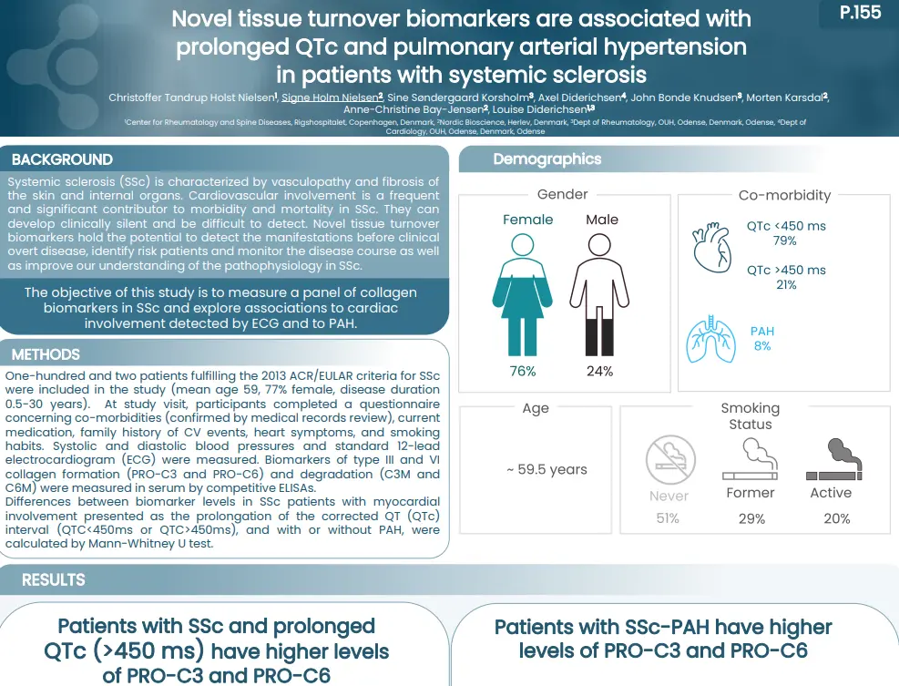
Systemic sclerosis (SSc) is characterized by vasculopathy and fibrosis of the skin and internal organs. Cardiovascular involvement is a frequent and significant contributor to morbidity and mortality in SSc. They can develop clinically silent and be difficult to detect. Novel tissue turnover biomarkers hold the potential to detect the manifestations before clinical overt disease, identify risk patients and monitor the disease course as well as improve our understanding of the pathophysiology in SSc.

The objective of this study is to measure a panel of collagen biomarkers in SSc and explore associations to cardiac involvement detected by ECG and to PAH.

Poster

Conclusion

Patients with SSc and prolonged ECG presented an altered tissue turnover, by an increased level of nordicPRO-C3™ and nordicPRO-C6™. In addition, SSc patients with presence of PAH had increased levels of nordicPRO-C3™ and nordicPRO-C6™ as well. presented an altered tissue turnover in presence of PAH, and with a QTc>450 ms. Our study indicates that they could serve as biomarkers of these manifestations and warrant further studies in cardiac disease in SSc.

Get in touch

Are you interested in exploring collaboration possibilities?
Enter your information in the form and a representative will contact you shortly.

    TNF-α and TGF-β Synergistically Promote Fibrogenesis

    TNF-α and TGF-β Synergistically Promote Fibrogenesis in a In Vitro Model of Fibro-inflammation

    Introduction

    Systemic sclerosis (SSc) is a skin disease characterized by chronic inflammation leading to
    fibrosis, a process called fibro-inflammation. TNF-α is an inflammatory cytokine driving chronic inflammation in SSc, while TGF-β activation is a hallmark of fibrotic pathology. Fibrogenesis (wound healing) is characterized by granulation tissue formation consisting of mainly type III collagen.

    The aim of this study was to investigate if primary human dermal fibroblasts treated with TNF-α
    and TGF-β1 had increased fibrogenesis compared to fibroblasts treated only with TGF-β1.

    Poster

    Conclusion

    Inflammatory TNF-α stimulation increases TGF-β driven fibrogenesis in dermal fibroblasts, by promoting their formation of type III collagen and fibronectin. Consequently, biomarkers of type III collagen formation and fibronectin formation may be markers of early fibrosis in fibro-inflammatory skin disease.

    Get in touch

    Are you interested in exploring collaboration possibilities?
Enter your information in the form and a representative will contact you shortly.

      Is Skin Disease a Local Manifestation of Systemic Tissue Turnover?

      Is Skin Disease a Local Manifestation of Systemic Tissue Turnover? Serological Collagen Biomarkers Provide Important Information on Skin Diseases Arising from Mutations in Collagen Genes

      Introduction

      Collagens are the main constituents of the skin. Genetic mutations in type VI, VII, and XVII collagen cause skin diseases, such as atopic dermatitis, epidermolysis bullosa, and bullous pemphigoid. These are all
      characterized as systemic diseases, with local manifestations. Novel collagen biomarkers hold the potential to detect skin manifestations, monitor the disease course, as well as improve our understanding of the pathophysiology.

      The aim of this study was to develop blood-based biomarkers of type VI, VII, and XVII collagen, and investigate their diagnostic potential for skin pathologies, including systemic sclerosis.

      Poster

      Conclusion

      These biomarkers reflect the downstream effect of different genetic mutations leading to skin disease and may be useful to determine skin involvement in rheumatic diseases, including systemic sclerosis and psoriatic arthritis.

      Get in touch

      Are you interested in exploring collaboration possibilities?
Enter your information in the form and a representative will contact you shortly.

        Hidradenitis Suppurutiva (HS) is a sytemic disease with local manifestations

        Hi-drad-uh-NIE-tis sup-yoo-ruh-TIE-vuh – also known as hidradenitis suppurutiva (HS) – is a pathologically complicated skin condition, where chronic skin inflammation leads to abscesses and scarring. It is a systemic disease with local manifestations, meaning that the chronic insult to the skin is systemic, but it is physically located where skin rubs against skin, such as the armpits, groins and under the breasts. It is well known that immune cells, such as neutrophils and mast cells are involved, but what do we know about tissue remodeling?

        When the beautiful collagens of the skin become a part of disease pathogenesis

        Patients with HS not only experience pain from the neutrophil-rich tunnels but also from excessive tissue remodeling that causes scarring of the skin. These patients have an imbalance in tissue formation and tissue repair, partly due to the excessive activity of immune cells, which release enzymes that degrade the skin.

        One group of tissue-degrading enzymes are matrix metalloproteinases, abbreviated as MMPs. These are released by macrophages, the most numerous inflammatory cells found in HS patients. MMPs infiltrate and contribute to HS pathology, signaling that increased activity of MMPs degrades the proteins of the skin tissue, such as collagens. This process can be quantified by specific blood-based biomarker assays targeting this pathological process.

        Pathological fragments in HS may be used to identify disease types

        In HS, biomarkers of tissue remodeling such as type III collagen degraded by MMPs (C3M), are associated with disease severity (Hurley Staging).

        Figure 1. Biomarkers of tissue remodeling associate with Hurley Stages

        C3M is released upon MMP activation and measures dermal tissue remodeling. This raises the question – can we use C3M to identify subtypes of patients based on their disease activity, and potentially molecular endotypes to help select the right treatment for the patients?

        To address this, the levels of C3M are different depending on how active the disease is when divided into the Sartorious Score (HSS).

        Finding the patients with high C3M levels reflects high disease activity, and may indicate a different subtype of patients needing a different treatment type.

        Figure 2. Type II collagen degradation biomarker C3M as a patient stratification tool

        Get in touch

        Are you interested in exploring collaboration possibilities?
Enter your information in the form and a representative will contact you shortly.