Biomarkers in diabetes

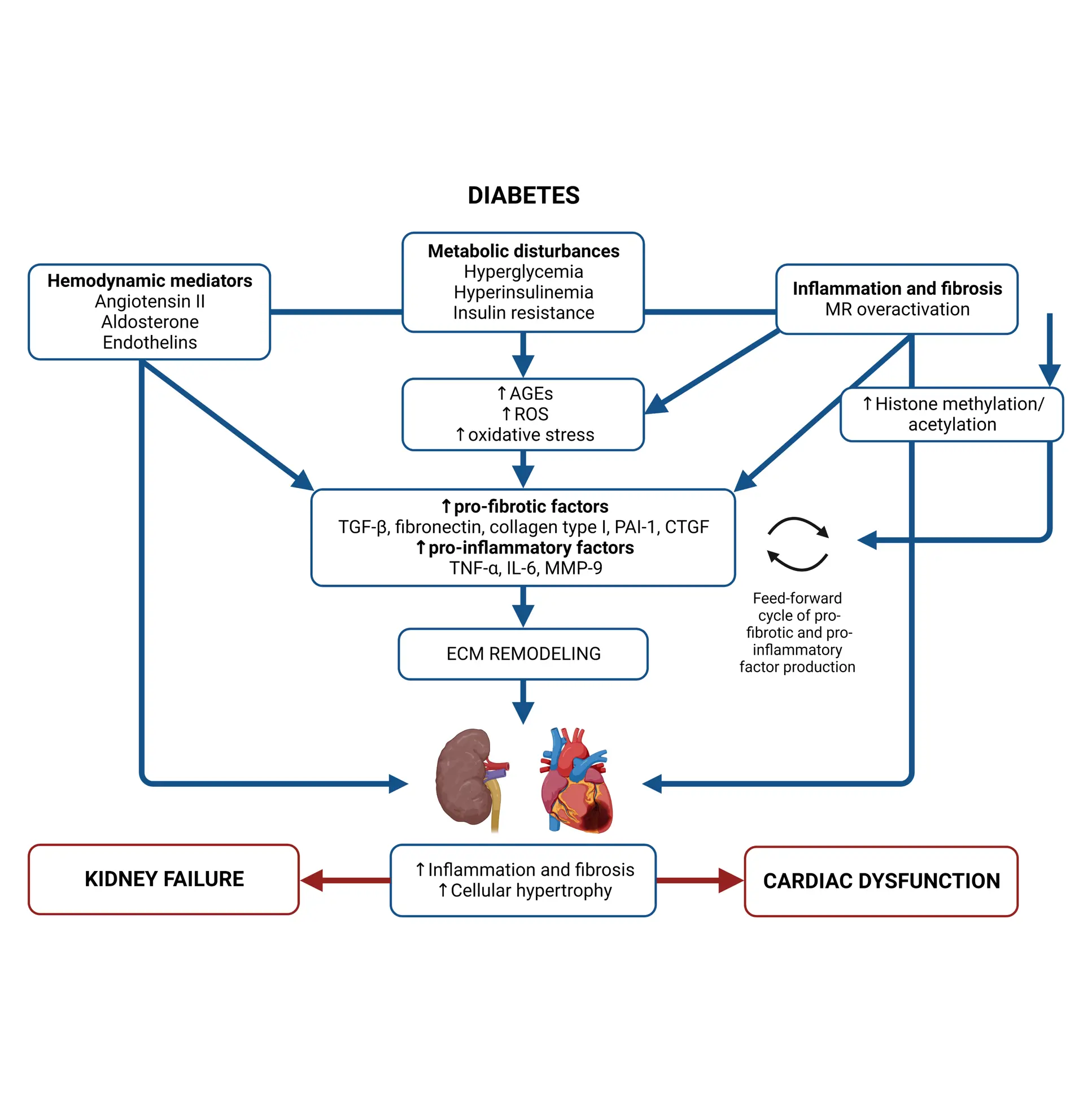
Diabetes significantly accelerates fibrotic and inflammatory processes in vital organs, yet current treatments leave a high residual risk of complications. The right biomarkers may offer a way to detect and quantify these underlying changes, particularly in kidney and cardiovascular tissues. By capturing early shifts in tissue dynamics, these biomarkers enable better risk stratification, selection, and evaluation of treatment efficacy—supporting more targeted and effective management of diabetic complications.

Despite the success of novel therapies, the residual risk of outcomes in persons with both type 1 diabetes (T1D) and type 2 diabetes (T2D) remains high. Therefore, novel biomarkers are needed to predict complications before clinical manifestations. There are currently no approved antifibrotic drugs capable of halting and/or reversing progression of kidney and/or cardiac fibrosis.

As novel antifibrotic therapies enter clinical development in early phases of chronic kidney disease (CKD), diabetic kidney disease (DKD), and cardiovascular disease (CVD), specific markers for selecting individuals and evaluating efficacy of treatment are needed.

Nordic ProteinFingerPrint Technology™ biomarkers in diabetes

The Nordic ProteinFingerPrint Technology™ allows the quantification of protein fragments generated by extracellular (ECM) turnover in both circulation and urine, thereby monitoring the processes of fibrogenesis and fibrolysis.

Non-invasive biomarkers of formation and/or degradation of different ECM proteins, such as collagens, can be used to study and advance our understanding of the fibrotic processes in the kidney and heart in people with diabetes.

Biomarkers of extracellular matrix (ECM) remodeling are valuable tools for assessing disease activity, identifying individuals at increased risk of experiencing adverse outcomes, selecting individuals who could benefit from preventive treatment, and enriching the clinical trial populations based on the risk of outcome.

Separation between healthy and type 2 diabetes

There is a significant separation between healthy people and individuals with T2D based on serum levels of extracellular matrix (ECM) remodeling biomarkers. Across all eight biomarkers, individuals with T2D show consistently elevated median levels compared to age-and gender matched healthy controls, indicating upregulated fibrotic activity and matrix turnover characteristic of diabetic tissue pathology. Data are median (IQR); T2D (n = 267); healthy subjects (n = 79).

NordicPRO-C3™ and nordicPRO-C6™, markers of collagen type III and VI formation respectively, are markedly higher in T2D, pointing to increased fibrogenesis and pro-fibrotic signaling—particularly through Endotrophin in the case of nordicPRO-C6™. The nordicPRO-C3™/C3M ratio, which captures the imbalance between collagen III synthesis and degradation, is also significantly elevated, suggesting a shift toward net matrix accumulation.

Markers of degradation—C1M (collagen I), C3M (collagen III), and C4M (collagen IV)—are elevated in T2D as well, though the increase in formation markers is generally more pronounced. TUM (tumstatin, a fragment of collagen IV cleaved by MMP-2 and MMP-9) and TIM (titin fragment from MMP-12-mediated breakdown) further reinforce a systemic increase in MMP-driven proteolysis within diabetic individuals.

This biomarker profile reflects dysregulated ECM turnover in T2D, integrating increased collagen deposition with enhanced matrix degradation, a fibrotic phenotype consistent with known vascular and organ complications. The clear separation from healthy subjects across all markers supports the use of these ECM metrics for stratifying disease burden and evaluating therapeutic interventions targeting tissue remodeling in metabolic disease.

Disease progression in diabetes

The survival curves demonstrate that elevated baseline levels of serum nordicPRO-C6™ are predictive of adverse clinical outcomes in individuals with T2D and microalbuminuria. Stratification by nordicPRO-C6™ tertiles shows progressively poorer outcomes with increasing biomarker concentration.

Cumulative survival decreases most rapidly in the highest nordicPRO-C6™ tertile group, reflecting elevated all-cause mortality risk. A similar pattern can be observed for major adverse cardiovascular events (CV death, stroke, ischemic cardiovascular disease, and heart failure), with the highest tertile experiencing significantly more events over time. Kidney disease progression—defined as a ≥30% decline in eGFR—is again most prominent in the highest tertile, indicating predictive validity of nordicPRO-C6™ for kidney deterioration.

Hazard ratios per doubling of nordicPRO-C6™ levels are statistically significant after adjustment for conventional risk factors including age, sex, systolic blood pressure, LDL cholesterol, smoking status, HbA1c, plasma creatinine, and urinary albumin excretion. This confirms the independent prognostic value of nordicPRO-C6™ across multiple organ systems. Its incorporation into risk stratification algorithms can enhance early identification of individuals at highest risk for mortality, cardiovascular complications, and kidney decline, thereby informing therapeutic prioritization and long-term disease management.

Pharmacodynamic modulation in type 2 diabetes

In the AWARD-7 trial, we showed pharmacodynamic modulation of ECM remodeling biomarkers in participants with T2D. Changes in urinary C3M (a marker of collagen type III degradation), normalized to urinary creatinine, can be observed in Dulaglutide treatment results in a marked increase over 52 weeks, suggesting enhanced matrix turnover. In contrast, insulin glargine leads to a progressive decline, indicating reduced ECM degradation. These divergent trends suggest that dulaglutide promotes remodeling dynamics, potentially reversing fibrotic activity.

At the same time, serum nordicPRO-C6™ levels showed that dulaglutide significantly reduced nordicPRO-C6™ from baseline, whereas insulin glargine drives a steady increase over time. The data support the potential of GLP-1 receptor agonists like dulaglutide to not only manage glycemic parameters but also favorably modulate fibrotic processes, positioning these ECM markers as valuable tools for pharmacodynamic profiling in T2D drug development.

NordicPRO-C6™ signals treatment efficacy

Longitudinal plasma nordicPRO-C6™ levels in individuals with T2D over a 5-year period (from the DC-ren study), compared the effects of renin-angiotensin system inhibitor (RASi) monotherapy versus combination therapy with RASi and a sodium-glucose cotransporter 2 inhibitor (SGLT2i).

Participants on RASi monotherapy exhibited an upward trajectory in nordicPRO-C6™ concentrations, indicating persistent or increasing pro-fibrotic signaling over time. In contrast, those receiving combined RASi + SGLT2i therapy show a downward trend in plasma nordicPRO-C6™ levels, with a statistically significant difference in slope (p = 0.002), suggesting that combination therapy actively suppresses fibrogenic processes.

Given that nordicPRO-C6™ reflects collagen type VI formation and endotrophin-mediated fibrosis, this decline aligns with the observed kidney benefit—specifically a slower decline in eGFR—among individuals on dual therapy.

Prediction of treatment response in type 2 diabetes

We demonstrated the predictive utility of baseline serum nordicPRO-C6™ levels for treatment response to pioglitazone, an insulin sensitizer and PPARγ agonist, in individuals with T2D from the BALLET trial. Participants were stratified into tertiles based on their initial nordicPRO-C6™ concentrations, and their glycemic outcomes were tracked over 26 weeks.

Only participants in the middle and high nordicPRO-C6™ tertiles exhibit substantial reductions in HbA1c, with the greatest decline observed in the upper tertile. In contrast, individuals in the lowest tertile show minimal to no glycemic improvement.

Incorporating nordicPRO-C6™ profiling into clinical decision-making can enable more targeted use of insulin sensitizers, avoiding unnecessary exposure in predicted non-responders and enhancing precision in T2D therapy.

Mechanisms of DKD and diabetic cardiomyopathy

Diabetic kidney disease (DKD) and diabetic cardiomyopathy are initiated by systemic changes, triggering metabolic, hemodynamic, inflammatory, and fibrotic processes contributing to disease progression.

Metabolic disturbances lead to the accumulation of advanced glycation end products, generation of reactive oxygen species, and increased oxidative stress, which induce the production of pro-fibrotic and pro-inflammatory factors and lead to altered metabolism, inflammation, and ECM remodeling.

An imbalance in the formation and/or degradation of ECM components is highly linked with the development of fibrosis. Together these changes mediate cellular hypertrophy and proliferation, inflammation, and fibrosis, ultimately resulting in organ failure (MR, mineralcorticoid receptor; TGF-β, transforming growth factor-β; PAI-1, plasminogen activator inhibitor-1; CTGF, connective tissue growth factor; TNF-α, tumor necrosis factor-α; IL-6, interleukin-6; MMP-9, matrix metalloproteinase-9).

About diabetes

  • Diabetes, a cardiometabolic disease, is one of the largest global health emergencies of the 21st century. Today, diabetes is considered a cardiometabolic disease, defined as “a cluster of interrelated risk factors”1. Vascular and metabolic factors, such as hyperglycemia, dyslipidemia, hypertension, obesity, elevated inflammatory and fibrotic markers, and complications of diabetes, fall within cardiometabolic disease. The major complications of diabetes are generally separated into macrovascular, affecting the heart and vasculature, and microvascular, affecting the kidneys, eyes, and nerves.


    CVD is the most prevalent cause of mortality and morbidity among people with diabetes. Several factors increase the risk of a CV event, such as increasing age, diabetes, CKD, current smoking, dyslipidemia, hypertension, unhealthy diet, family history, obesity, and physical inactivity. Diabetic cardiomyopathy is characterized by abnormal cardiac structure and function in the absence of other CV risk factors. The diagnosis is based on left ventricular (LV) diastolic dysfunction and/or reduced LV ejection fraction, pathological LV hypertrophy, and interstitial fibrosis.


    CKD among people with diabetes is common. It has been estimated that up to 40% of persons with diabetes will develop CKD during their lifetime2. CKD can progress to end-stage kidney disease (ESKD) or kidney failure, requiring dialysis or kidney transplantation. DKD typically develops after a diabetes duration of 10 years in T1D but may already be present at diagnosis of T2D.

  • Biomarkers are measurable indicators of biological processes, such as tissue remodeling or inflammation. In diabetes, they help track disease progression, predict complications like kidney or heart disease, and assess treatment response—enabling earlier intervention and more personalized care.

  • Extracellular matrix (ECM) biomarkers reflect fibrotic activity in organs like the kidneys and heart. Measuring collagen formation (e.g., nordicPRO-C3™, nordicPRO-C6™) and degradation (e.g., C3M, C4M) helps identify people at higher risk for adverse outcomes and monitor how therapies influence tissue remodeling in real time.

  • Yes. Elevated levels of nordicPRO-C6™, a marker of type VI collagen formation and endotrophin activity, are associated with higher risks of mortality, cardiovascular events, and kidney disease progression. This biomarker has independent prognostic value beyond conventional risk factors.

  • The Nordic ProteinFingerPrint Technology™ captures dynamic changes in ECM turnover—offering a more precise view of fibrotic processes than traditional clinical markers. These serum and urine-based tests support risk stratification, clinical trial enrichment, and precision medicine strategies in diabetes and its complications.

Get in touch

Are you interested in exploring collaboration possibilities?
Enter your information in the form and a representative will contact you shortly.

    Nordic Bioscience’s assays, products, and services are for research use only and are not intended for medical or diagnostic purposes.