Scleroderma (Systemic Sclerosis)

Biomarkers for investigating scleroderma (systemic sclerosis)

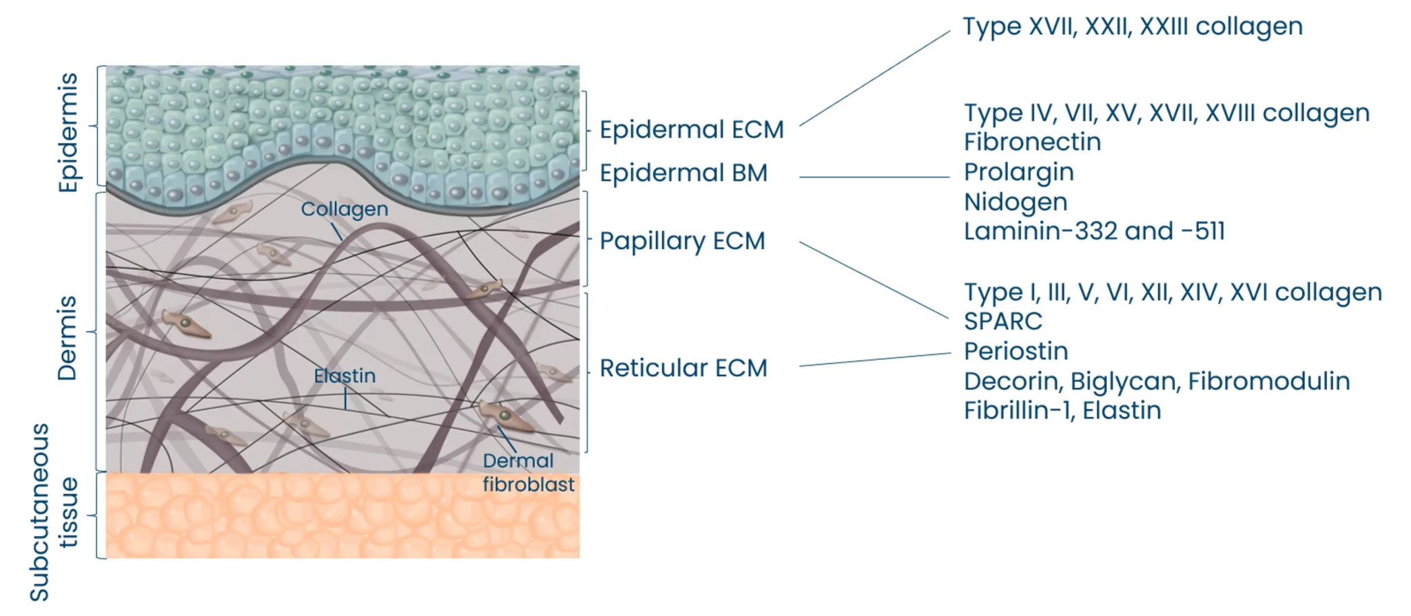
Autoimmune diseases, such as scleroderma—systemic sclerosis (SSc)—affect several tissues, leading to fibrotic tissue buildup of the skin and internal organs. Fibrosis is caused by an increased synthesis of extracellular matrix (ECM), which plays a key role in SSc pathogenesis.

Did you know?

Extracellular matrix-specific biomarkers that help us quantify and monitor fibrosis of the skin internal organs are key when we want to understand the disease mechanisms and prediction of scleroderma/SSc progression. Utilizing biomarkers can help address several aspects of unmet needs in SSc. A well-designed biomarker program enables precise diagnosis, prediction of clinical course and organ involvement, evaluation of therapeutic responsiveness, and identification of novel therapeutic targets.

Biomarkers associated with early diffuse SSc

The distribution of skin involvement is a key factor in classifying SSc patients into limited or diffuse cutaneous subtypes. Diffuse cutaneous SSc is characterized by widespread skin involvement, affecting the torso, face, and both the upper and lower limbs. Compared to limited cutaneous SSc, diffuse SSc is more aggressive, with greater organ involvement and higher mortality rates. In a study published in PLoS One, in 2018 , biomarkers of type III and VI collagen formation PRO-C3 and PRO-C6, as well as type III and VI collagen degradation C3M and C6M, were generally elevated in both early (<2Y) and late stages (>10Y) of diffuse SSc compared to asymptomatic controls (Figure 1). Additionally, early diffuse SSc patients tend to present upregulated ECM biomarker levels compared to late diffuse SSc patients. This study suggests that ECM biomarkers are valuable tools both for investigating disease pathogenesis and serving as diagnostic tools.

Figure 1. Type III and VI collagen biomarker levels of fibrosis and fibrolysis are higher in early diffuse patients compared to asymptomatic controls.

Nordic Bioscience can quantify the different layers of the skin

The skin is the largest organ of the human body, and consists of three different layers; epidermis, dermis and subcutis. These three layers have distinct tissue architectures and functions.

Skin functions are dependent on a complex composition of extracellular matrix (ECM) proteins. The ECM of the skin can be divided into the epidermal ECM, epidermal basement membrane and the papillary/reticular ECM. The major ECM components of the skin are collagens, which are located in the different tissue compartments to maintain tissue architecture.

The Nordic ProteinFingerPrint Technology™ allows for the quantification of the different layers of the skin, and also the quantification of immune cell activity involved in different pathologies (Mast Cells, Neutrophils, and macrophages).


The different layers of the skin

Pharmacodynamics in SSC

Understanding the relationship between SSc and circulating biomarkers is key for drug development, as it facilitates patient stratification and the assessment of therapeutic responses, e.g. pharmacodynamic effect of ECM biomarkers in tocilizumab treatment.

Efforts are underway to identify circulating molecular biomarkers in SSc to predict disease progression and assess drug mechanisms. A recent study from the fouSSced trial, published in Clinical Immunology, showed that 48 weeks of tocilizumab treatment led to treatment-related reductions in C3M and C4M levels (Figure 3 A and B). This occurs as the inhibition of IL-6 leads to reduced MMP-9 activity, which in turn reduces the cleavage of type III and IV collagen.

The TFG-β pathway biomarker PRO-C3 correlated with mRSS (ρ = 0.3). However, treatment with tocilizumab did not impact the fibrosis component of the disease, hence the levels of PRO-C3, which reflect fibrosis, were not modulated. These findings suggest that our biomarkers can be used to evaluate the effect of treatment in the fibrolysis and fibrosis component of the disease.

Figure 3. Degradation ECM biomarkers show a pharmacodynamic effect with Tocilizumab (anti IL-6 receptor).

Biomarkers associated with SSc disease progression

A balanced ECM is necessary to preserve organ integrity and tissue homeostasis. Quantification of ECM remodelling offers valuable insight into the structural and functional changes associated with disease progression. A study published in The Lancet, in 2021, showed a dysregulation of neo-epitopes of type III and IV collagens in patients with systemic sclerosis with progressive fibrosis compared to non-progressive patients.

The fibrosis biomarkers PRO-C3 and the type IV collagen turnover PRO-C4 were associated with disease progression (10% decline in FVC%, or increased mRSS >25% during a one-year clinical follow-up). PRO-C3 and PRO-C4, together with tissue type III and IV degradation biomarkers C3M and C4M, were upregulated in progressive disease compared to stable disease (Figure 2).

These findings are explained by feedback loops in extracellular matrix remodelling, in which an increase in collagen synthesis leads to an increase in ECM degradation processes and vice versa. This study suggests that ECM biomarkers are valuable tools for identifying patients in risk of disease progression.

The Nordic ProteinFingerPrint Technology™ in rheumatic disorders

Unsure about how our technology can benefit your clinical trial in rheumatic disorder? Watch this short video and get an understanding of the benefit you gain from our biomarkers .

About systemic sclerosis (SSc)

  • Systemic sclerosis (SSc) is a chronic rheumatic disease with one of the highest mortalities within rheumatology.


    It is an autoimmune, fibro-inflammatory skin disease characterized by a distinctive triad of microvascular damage (vasculopathy), immune activation with autoimmunity, and generalized fibrosis of multiple organs, including the skin and lungs.


    The great heterogeneity of disease cause and presentation creates considerable challenges in developing treatment and treating patients.


    The prevalence of SSc varies considerably around the world. Northern Europe and parts of Asia have a prevalence of >150 per million, whereas southern Europe, North America, and Australia have a higher prevalence of up to 443 per million.


    As with other autoimmune diseases, women are about six times more likely to get SSc than men.

  • Skin fibrosis is assessed by the modified Rodnan Skin Score (mRSS) and the presence of vascular damage, including Raynaud’s Phenomenon, is included in the diagnostic assessment.


    Furthermore, the presence of auto-antibodies and pulmonary involvement (either presented as interstitial lung disease or pulmonary arterial hypertension) is included in the diagnosis.


    There is no cure or disease-modifying drug for SSc, but symptoms and inflammation can be treated using NSAIDs, steroids, or biologics.


    The type of treatment depends on the symptoms and organs involved. Several novel drugs are in development targeting either the inflammatory and/or the fibrotic axis.

  • Systemic Sclerosis is characterized by microvascular damage (vasculopathy), immune activation with autoimmunity, and generalized fibrosis of multiple organs, including the skin and lungs.


    The different organs that may be affected by SSc have tissues consisting mainly of collagens.


    The collagens are remodeled as part of normal homeostasis of the organ– that is, collagens are broken down and rebuilt as part of normal repair and maintenance of the tissue.


    In SSc, the balance of tissue remodeling, i.e. formation and degradation of ECM proteins, is interrupted resulting in fibrosis of the organs.


    Similarly, vascular remodeling leads to increased or disrupted vascularization, which may be detected by increased turnover of the ECM proteins of the vasculature.


    Nordic Bioscience’s biomarkers can directly quantify this fibrosis and microvascular damage in a serum sample.

  • Yes, studies have shown that biomarkers such as PRO-C3 and PRO-C4 are associated with clinical deterioration, such as FVC decline or skin score increase. These markers provide a molecular readout of fibrotic activity and can identify patients with progressive disease.

    ECM biomarkers respond to treatments like tocilizumab, making them useful for evaluating pharmacodynamic effects. Reductions in degradation markers (e.g., C3M and C4M) indicate therapeutic impact on tissue breakdown processes, helping guide treatment decisions and clinical trial design.

Get in touch

Are you interested in exploring collaboration possibilities?
Enter your information in the form and a representative will contact you shortly.

    Nordic Bioscience’s assays and services are research use only products and services and do not qualify for medical or diagnostic purposes.