Navigating the drug development process successfully is essential and not without challenges and uncertainties. Employing strategies that enhance the probability of success is crucial. The FDA and EMA acknowledge that biomarker-based drug development strategies, paired with translational research, could provide a viable solution. This approach offers a biology-centric approach that reduces both time and costs while improving the overall likelihood of success.
A major cornerstone of our approach is the combination of translational models with clinically validated biomarkers to improve drug development. To this end, we have evaluated our biomarkers' capability to bridge the gap between hepatic fibrosis patients and various model systems. These findings emphasize the pharmacodynamic and translational application of Nordic ProteinFingerPrint Technology™.
Browse our biomarker portfolio of hepatic biomarkers to guide your translational research!
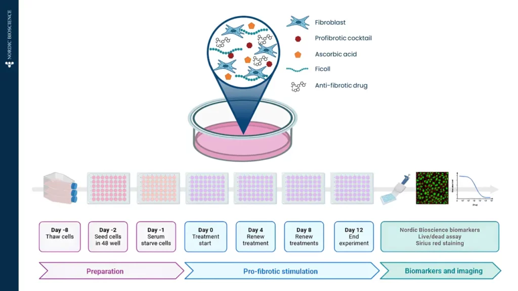
The prolonged Scar-in-a-Jar is an innovative model that utilizes macromolecular crowding to enhance the formation, maturation, and deposition of the extracellular matrix in vitro. The extracellular matrix is essential for providing structural support to cells and regulating tissue repair and regeneration by serving as a reservoir for growth factors and cytokines. Understanding the dynamics of the extracellular matrix is crucial for developing therapeutic strategies for fibrosis. By employing macromolecular crowding, the model gains a three-dimensional like structure and increased complexity, making it a suitable translational model. This modulation demonstrates the model’s effectiveness for screening antifibrotic effects directly on hepatic stellate cells. | Liver Scar-in-a-Jar (SIAJ) |
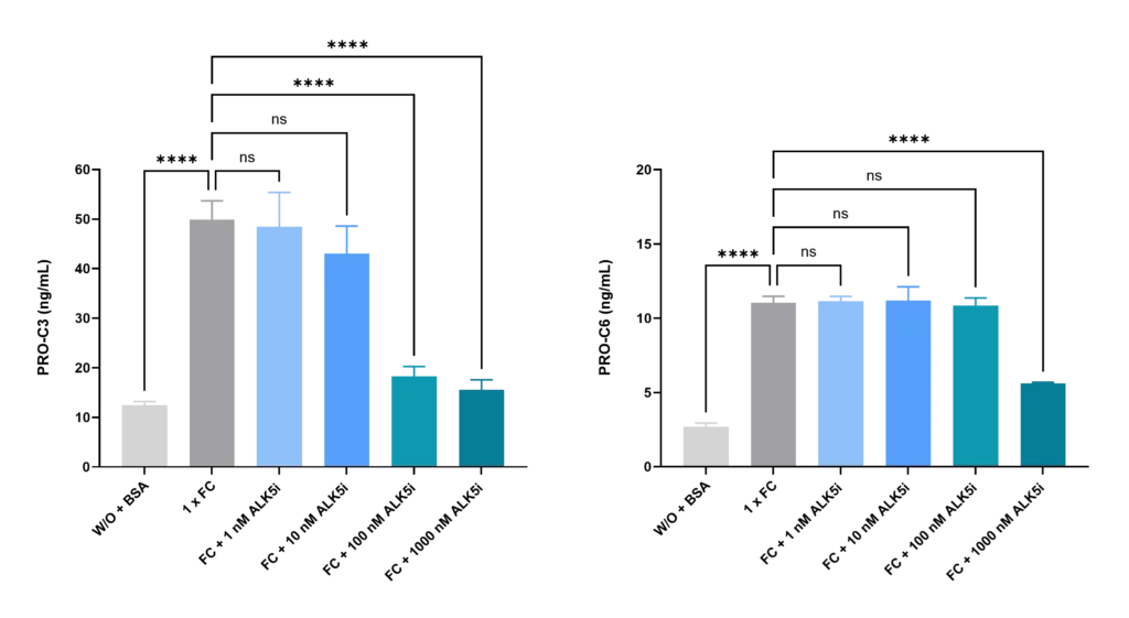
Figure 1. Effect of Fibrotic Cocktail and ALK5 Inhibitor on PRO-C3 and PRO-C6 Levels in Hepatic Stellate Cells: A Model for Antifibrotic Screening
The figures illustrate the increase in NordicPRO-C3 (formation of type III collagen; PRO-C3) and NordicPRO-C6™ (formation of type VI collagen) levels when hepatic stellate cells are stimulated with a fibrotic cocktail (FC), which is a mix of cytokines that induces fibrosis. In addition, the levels of PRO-C3 and PRO-C6 can be decreased by treating the stimulated fibroblasts with ALK5 inhibitor. This modulation demonstrates the model’s suitability for screening of antifibrotic effects directly on the hepatic stellate cells.
The GAN-DIO model has become an important translational model for hepatic fibrosis. The model allows us to study hepatic fibrosis in vivo. To assess efficacy, Nordic ProteinFingerPrint™ biomarkers that are associated with prognosis and pharmacodynamic effects in clinical practice can be measured in the blood of the animals.
In the Intercept study, using the AMLN ob/ob-MASH mouse model, the serum marker PRO-C4, which reflects type IV procollagen processing, closely followed tissue levels of fibrillar and basement membrane collagens over time. The reduction in PRO-C4 observed in INT-767-treated animals correlated with tissue staining for both type I and type IV collagens, while C4M, a degradation product of type IV collagen, showed a trend toward decreased circulating levels.
PRO-C4 and C4M in-vivo: Serum PRO-C4 concentration in vehicle (n=13) and INT-767 (n=14) treated animals. Levels of significance: ** p=0.0094 between groups (2-way ANOVA). (C) Serum C4M concentration in vehicle (n=13) and INT-767 (n=14) treated animals. Levels of significance: n.s. p=0.117 between groups (2-way ANOVA mixed effects analysis)
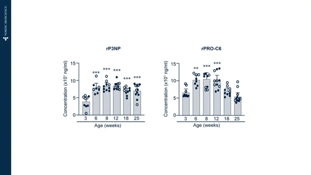
The rodent pro-peptide of type III collagen (rP3NP) and the Endotrophin-associated marker rPRO-C6, correlated significantly with liver fibrosis progression. rP3NP, a known tissue repair and fibrosis biomarker, showed sustained elevation from 6 to 25 weeks (Left figure). Similarly, rPRO-C6, associated with fibrosis, steatosis, and inflammation, was significantly elevated in NIF mice between 6 and 12 weeks, normalizing thereafter (Figure 2). As rPRO-C6 is elevated in advanced fibrosis in MASH patients, these findings suggest the potential utility of rP3NP and rPRO-C6 as markers for assessing anti-fibrotic therapies in NIF mice.
Figure 2. Non-fasting serum levels of rP3NP (left) and rPRO-C6 biomarkers (right) in male (o) and female (•) NIF mice at different ages (n = 8–12 mice/age group). One-way ANOVA was used to compare the mean of the 3-week age group with the mean of the other age groups. Dunnetts post-tests yielded ***p < 0.001 and **p < 0.01, vs. 3-week-old NIF mice.
Search and find publications that we have published.
Please don't hesitate to contact us if you have any questions or other inquiries.