Translational Models in Rheumatology

Strategic drug development in rheumatology

A drug development route for success is key but often challenging and full of uncertainties. Strategies that can increase the likelihood of success are vital. The FDA and EMA recognize that biomarker-based drug development strategies could be the solution in combination with translation research. This approach offers a biology-focused pathway that reduces time and cost and enhances the overall likelihood of success.

Nordic Bioscience combines innovative translational models with the ProteinFingerPrint Technology™ to translate findings effectively. This is facilitated by utilizing the same biomarkers across different clinical stages of development, creating a link between the preclinical model and the clinic. This strategically selected development route greatly enhances the efficiency of drug development pipelines.

The integration of biomarker-based drug development strategies in translational research accelerates the progression of therapeutics and increases the likelihood of success. Through advanced translational models combined with the ProteinFingerPrint™ biomarkers, we contribute to optimizing decision-making processes in clinical development, ensuring better patient treatments.

Translational biomarkers in rheumatology research

Our major cornerstone is combining translational models with clinically validated biomarkers to enhance drug development. For this purpose, we have assessed the ability of our biomarkers to bridge the gap between patients with a rheumatological disease and our various model systems. Our investigations have revealed that ProteinFingerPrint™ biomarkers, such as the C1MC2M, and C3M, reflecting cartilage and soft tissue destruction can be modulated by anti-catabolic treatments. We have assessed all biomarkers in the blood of patients with rheumatoid- and osteoarthritis and the supernatants of our ex-vivo models (Figure 1).

These findings underline the use of the Nordic ProteinFingerPrint TechnologyTM pharmacodynamically and translationally.

Ex Vivo model—human cartilage and synovial membrane explants

By excising cartilage or synovial membranes from human or bovine knees, an explant model system can be set up to mimic rheumatological diseases (Figure 2).

Depending on the specific experimental setup and stimulations, catabolic and anabolic treatment effects on cartilage and synovium can be investigated by measuring the translational Nordic ProteinFingerPrint™ biomarkers.

Figure 2. Nordic Bioscience explant model vivo system model mimics rheumatic diseases

Figure 3. showcases how compounds with different MoA’s affect joint remodeling differently using the ARGC2M, and PRO-C2 (nordicPRO-C2™) biomarkers, quantifying aggrecan degradation and type II collagen degradation and formation. Stimulating human cartilage with proinflammatory cytokines (Figure 3A+B) induces increased aggrecan and type II collagen degradation. Increasing concentrations of an ADAMTS5 inhibitor (Figure 3A) or Fostamatinib, a SYK inhibitor, (Figure 3B) significantly reduces cartilage destruction. Additionally, the biomarker PRO-C2 (nordicPRO-C2™) allowed the assessment of cartilage formation in explants stimulated with Sprifermin, a recombinant human FGF18, (Figure 3C).

Figure 3. Nordic Bioscience tissue destruction and formation biomarkers

Figure 4. Extracellular matrix remodeling in-vitro Scar-in-a-Jar model


In Vitro Model— Scar-in-a-Jar with Fibroblast-Like-Synoviocytes

The prolonged Scar-in-a-Jar is a novel model that employs macro-molecular crowding to promote extracellular matrix formation, maturation, and deposition in vitro (Figure 4).

The extracellular matrix plays a crucial role in providing structural support to cells and regulating tissue repair and regeneration by acting as a reservoir for growth factors and cytokines.

Understanding the extracellular matrix dynamics is vital for developing therapeutic strategies, even in rheumatoid diseases where patients can present with a fibrotic endotype. Using macromolecular crowding, the model becomes more 3D-like and increases complexity, making it a suitable translational model.


By stimulating fibroblast-like-synoviocytes with pro-fibrogenic cytokines, we can model joint fibrosis in vitro and evaluate the efficacy of treatment using the Nordic ProteinFingerPrint™ biomarkers. Thus, we can determine efficacy by measuring collagen formation in the supernatants, allowing direct translation to clinical settings.

As shown in Figure 5., we can induce fibrogenesis with profibrotic stimuli and subsequently inhibit fibrogenesis by increasing anti-fibrotic or anti-inflammatory treatment concentrations. Measuring the nordicPRO-C3™ (PRO-C3) and nordicPRO-C6™ (PRO-C6) biomarkers, reflecting type III and VI collagen formation, allowed for the quantification of the anti-fibrotic effects of an ALK5 inhibitor (Figure 5A), nintedanib (Figure 5B), and tofacitinib (Figure 5C).

Figure 5. Fibrogenesis profibrotic stimuli on antifibrotic and anti-inflammatory treatment

Figure 6. Nordic Bioscience in-vivo arthritic models with the biomarkers C1M and C3M

In Vivo—arthritic models

Various in vivo models mimicking RA or OA, such as the collagen-induced-arthritis model, represent important translational tools to study arthritis in vivo. By collecting blood from these animals, joint remodeling can be evaluated using the Nordic ProteinFingerPrint™ biomarkers to assess drug efficacy and pharmacodynamic effects in preparation for clinical trials.

The graph (Figure 6.) showcases increased synovial tissue destruction quantified with the C1M and C3M biomarkers in a rodent collagen-induced arthritis model.

The Nordic ProteinFingerPrint Technology™ in rheumatic disorders

Unsure about how our technology can benefit your clinical trial in rheumatic disorder? Watch this short video and get an understanding of the benefit you gain from our biomarkers .

  • 1. Blair, J. P. M., Bager, C., Platt, A., Karsdal, M., & Bay-Jensen, A.-C. (2019). Identification of pathological RA endotypes using blood-based biomarkers reflecting tissue metabolism. A retrospective and explorative analysis of two phase III RA studies. PloS One, 14(7), e0219980. https://doi.org/10.1371/journal.pone.0219980


    2. Hannani, M. T., Thudium, C. S., Karsdal, M. A., Ladel, C., Mobasheri, A., Uebelhoer, M., Larkin, J., Bacardit, J., Struglics, A., & Bay-Jensen, A.-C. (2024). From biochemical markers to molecular endotypes of osteoarthritis: a review on validated biomarkers. Expert Review of Molecular Diagnostics, 24(1–2), 23–38. https://doi.org/10.1080/14737159.2024.2315282


    3. Kjelgaard-Petersen, C. F., Platt, A., Braddock, M., Jenkins, M. A., Musa, K., Graham, E., Gantzel, T., Slynn, G., Weinblatt, M. E., Karsdal, M. A., Thudium, C. S., & Bay-Jensen, A.-C. (2018). Translational Biomarkers and Ex Vivo Models of Joint Tissues as a Tool for Drug Development in Rheumatoid Arthritis. Arthritis & Rheumatology (Hoboken, N.J.), 70(9), 1419–1428. https://doi.org/10.1002/art.40527


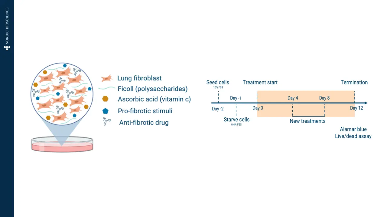
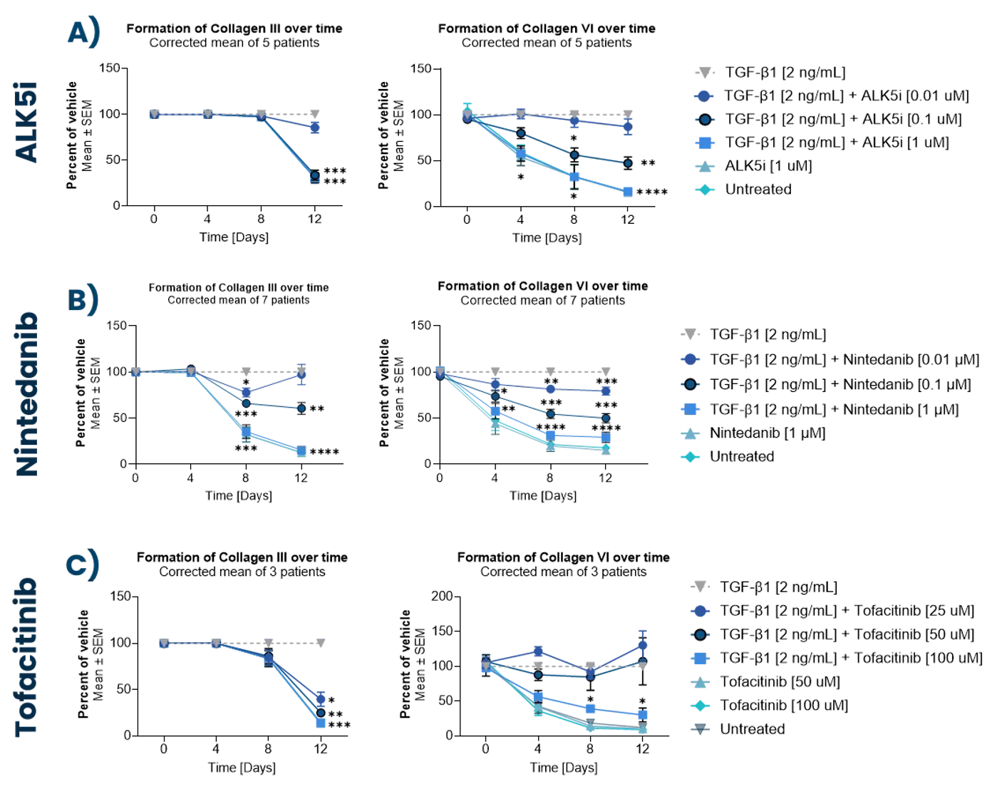
    4. Madsen, S. F., Madsen, S. S., Madrid, A. S., Andersen, M. R., Bay-Jensen, A.-C., & Thudium, C. S. (2024). Fibrotic remodeling in joint diseases: induction and inhibition of fibrosis in fibroblast-like synoviocytes. Translational Medicine Communications, 9(1), 18. https://doi.org/10.1186/s41231-024-00180-0


    5. Reker, D., Siebuhr, A. S., Thudium, C. S., Gantzel, T., Ladel, C., Michaelis, M., Aspberg, A., Berchtold, M. W., Karsdal, M. A., Gigout, A., & Bay-Jensen, A. C. (2020). Sprifermin (rhFGF18) versus vehicle induces a biphasic process of extracellular matrix remodeling in human knee OA articular cartilage ex vivo. Scientific Reports, 10(1), 6011. https://doi.org/10.1038/s41598-020-63216-z


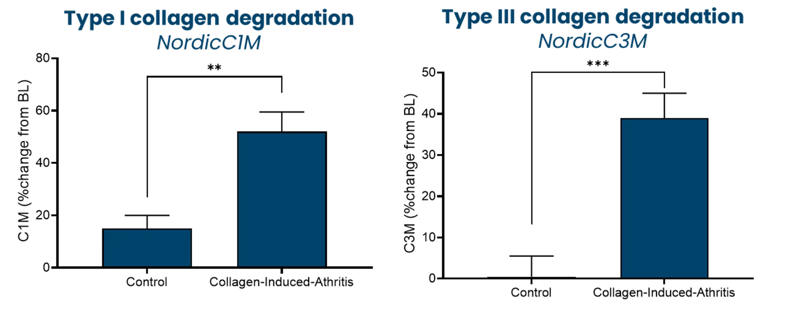
    6. Siebuhr, A. S., Wang, J., Karsdal, M., Bay-Jensen, A.-C., Y, J., & Q, Z. (2012). Matrix Metalloproteinase-dependent turnover of cartilage, synovial membrane, and connective tissue is elevated in rats with collagen induced arthritis. Journal of Translational Medicine, 10(1), 195. https://doi.org/10.1186/1479-5876-10-195


    7. Siebuhr, A. S., Werkmann, D., Bay-Jensen, A.-C., Thudium, C. S., Karsdal, M. A., Serruys, B., Ladel, C., Michaelis, M., & Lindemann, S. (2020). The Anti-ADAMTS-5 Nanobody® M6495 Protects Cartilage Degradation Ex Vivo. International Journal of Molecular Sciences, 21(17). https://doi.org/10.3390/ijms21175992


    8. Thudium, C. S., Bay-Jensen, A. C., Cahya, S., Dow, E. R., Karsdal, M. A., Koch, A. E., Zhang, W., & Benschop, R. J. (2020). The Janus kinase 1/2 inhibitor baricitinib reduces biomarkers of joint destruction in moderate to severe rheumatoid arthritis. Arthritis Research & Therapy, 22(1), 235. https://doi.org/10.1186/s13075-020-02340-7

Get in touch

Are you interested in exploring collaboration possibilities?
Enter your information in the form and a representative will contact you shortly.

    Nordic Bioscience’s assays and services are research use only products and services and do not qualify for medical or diagnostic purposes.