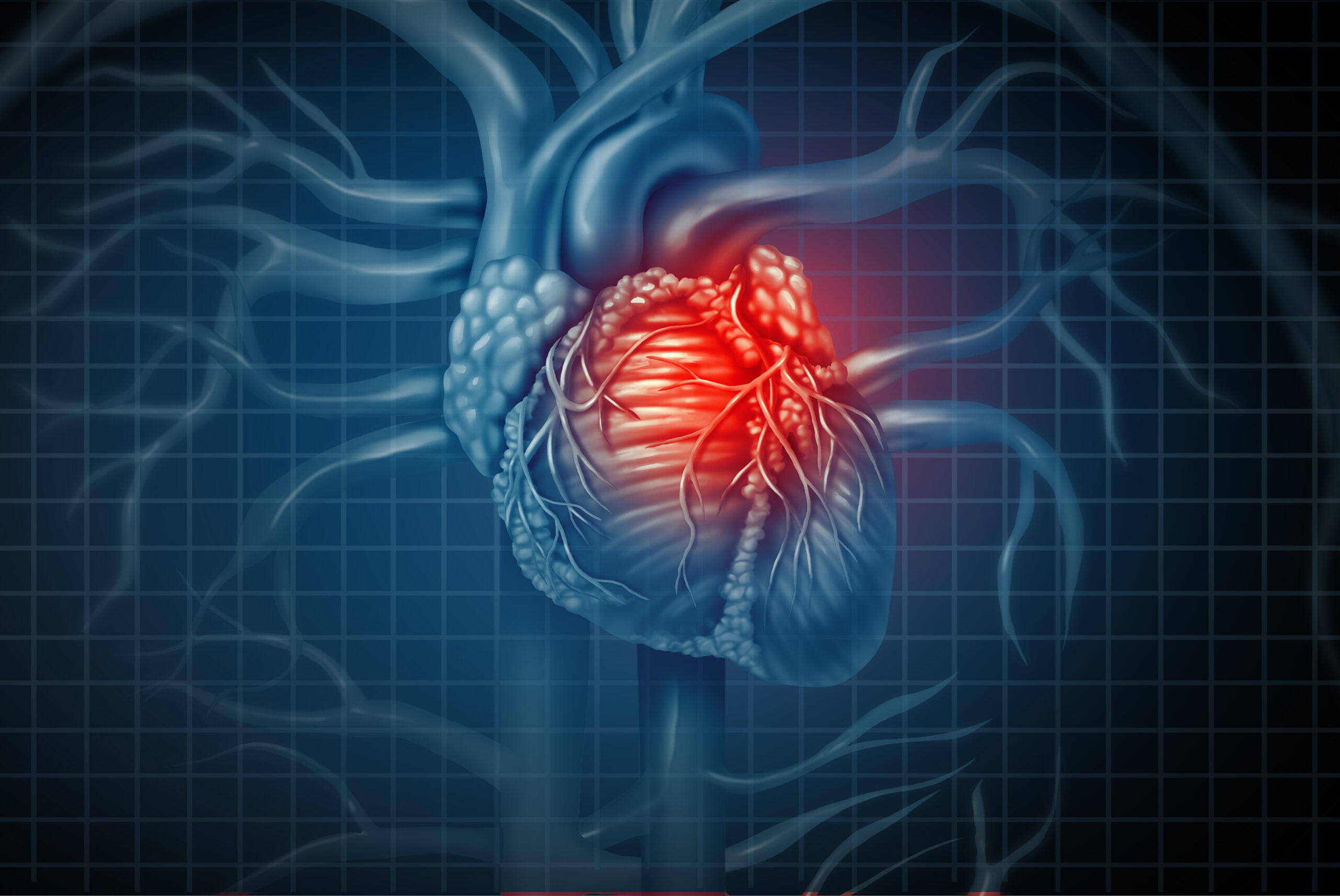
Cardiomyopathies are clinically diagnosed and managed with the use of genetic testing, assessment of clinical symptoms, electrocardiogram (ECG) measurements, and cardiac imaging to detect functional, structural, and morphological alterations.
In hypertrophic cardiomyopathy, diagnosis is performed with ECG as a first screening, followed by echocardiography or magnetic resonance imaging (MRI). The main diagnostic characteristic is ECG abnormalities caused by a characteristic left ventricular hypertrophy ≥1.5 cm (LVH).
In dilated cardiomyopathy, diagnosis is performed based on echocardiographic, computer tomography (CT), or MRI evidence of LV enlargement and impaired systolic function. In arrhythmogenic cardiomyopathy, diagnosis is performed by assessing the size of the right ventricle normalized to body surface area and less frequently with right ventricular endomyocardial biopsy.
Other criteria include the presence of epsilon waves, left bundle morphology ventricular tachycardia, and premature ventricular contractions >500 in 24 h. In restrictive cardiomyopathy, diagnosis is performed with hemodynamic or echocardiographic Doppler, and the main diagnostic characteristic is restrictive ventricular filling, with normal systolic ventricular function, normal or reduced end-diastolic ventricular dimensions, and absence of LVH.
Although detailed imaging is helpful endomyocardial biopsy is necessary to create causality in RCM, especially for non-genetic forms.
In left ventricular non-compaction cardiomyopathy, diagnosis is performed with higher-resolution echocardiography, CT, and MRI, and the main diagnostic characteristic is hyper trabeculation of the LV apex.
According to the latest guidelines, LVNC is considered a common cardiac trait that sometimes occurs with various forms of cardiomyopathy, and which also occurs in people without cardiomyopathy.