Lupus Nephritis

Biomarkers for investigating lupus nephritis

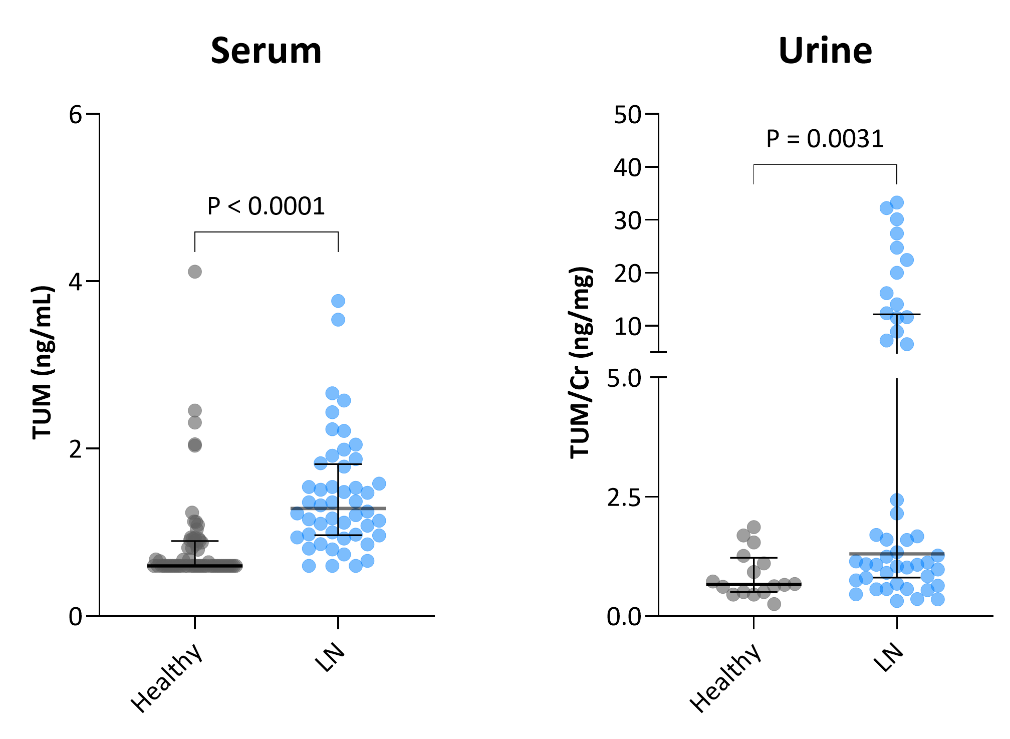
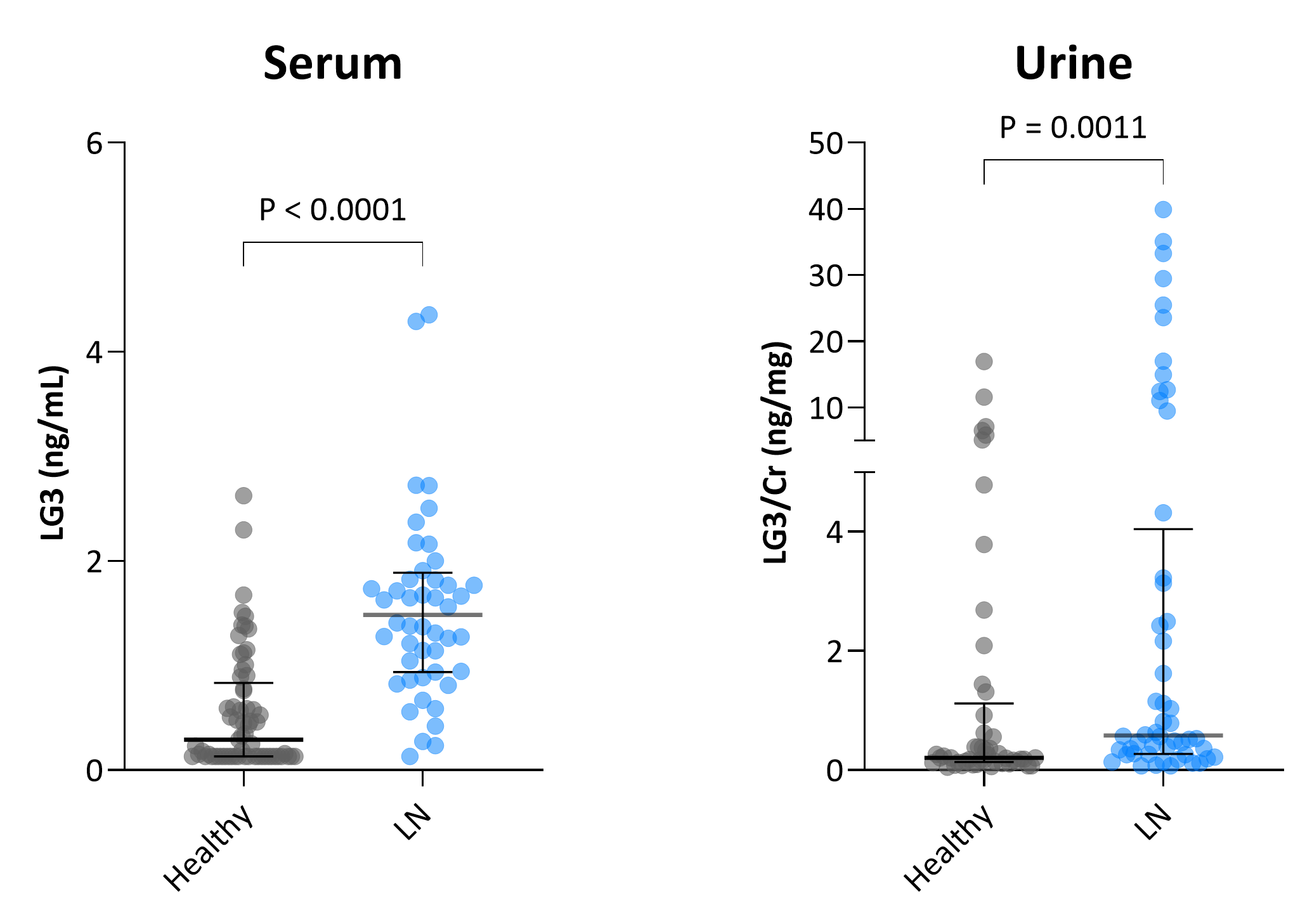
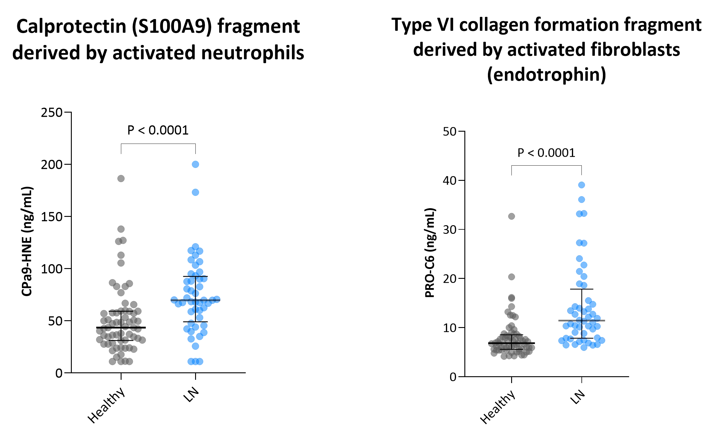
Lupus nephritis (LN) is a serious manifestation of systemic lupus erythematosus and a leading cause of kidney damage in affected patients. Despite immunosuppressive therapies, many patients experience ongoing renal injury that leads to fibrosis and progressive loss of kidney function.

Traditional clinical markers such as proteinuria and serum creatinine often fail to detect early fibrotic changes or capture dynamic treatment responses. This highlights the need for more sensitive and mechanistic biomarkers that can provide deeper insight into underlying disease processes, particularly tissue remodeling. In LN, extracellular matrix (ECM) accumulation and turnover are central to the development of chronic kidney scarring. ECM-derived biomarkers can offer a direct, non-invasive, and biologically relevant assessment of disease activity by capturing ongoing fibrogenesis and fibrolysis.

Accelerating drug development in lupus nephritis with Nordic ProteinFingerPrint™ biomarkers

Tissue remodeling and immune cell activity are key events in LN. Quantifying disease activity and remodeling provides unique insight into the structural and functional changes in pathogenesis.

ECM-derived biomarkers enable earlier detection of fibrotic progression, more precise pharmacodynamic monitoring, and improved patient stratification—critical factors in optimizing drug development in LN. By integrating these biomarkers into clinical trials, pharmaceutical companies can better evaluate therapeutic response, shorten trial timelines, and reduce development risk.

Compared to broad proteomic approaches, our targeted biomarker strategy identifies relevant biological signals with greater specificity and efficiency—while helping reduce costs and increasing the chances of clinical success.

Elevated glomerular basement membrane degradation in lupus nephritis

The glomerular basement membrane (GBM) of the kidney is an essential component of the blood filtration barrier and mainly consists of laminin, type IV collagen, nidogen, and heparan sulfate proteoglycans. The type IV collagen heterotrimer found in the glomerular GBM is composed of α3, α4, and α5 chains.

Our biomarker TUM quantifies circulating and urinary levels of tumstatin, a matrikine fragment released from the α3 chain of type IV collagen, TUM is elevated in serum and urine samples from patients with LN compared to healthy controls (Figure 1A-B).

Figure 1a-b. Tumstatin biomarker TUM is elevated in serum and urine samples in lupus nephritis patients

Perlecan is a major proteoglycan of the glomerular basement membrane. LG3, a matrikine fragment of perlecan, is elevated in serum and urine samples from LN samples compared to healthy controls (Figure 2A-B).

The elevated glomerular basement membrane fragments assessed by TUM and LG3 reflect the active degradation of the GBM in this highly inflammatory disease.

Figure 2a-b. LG3 glomerular basement membrane biomarker is elevated in serum and urine samples in lupus nephritis patients

Immune-mediated Inflammation and fibroblast activity in lupus nephritis

Neutrophils play a key role in the pathogenesis of LN, contributing to inflammation and tissue injury through the release of reactive oxygen species, proteases, and neutrophil extracellular traps (NETs). Persistent neutrophil activation is associated with ongoing immune-mediated damage and correlates with disease severity.

In parallel, fibroblasts are central to the development of kidney fibrosis, as they become activated in response to injury and produce excess extracellular matrix components. Chronic fibroblast activation leads to irreversible scarring and progressive loss of renal function, a major driver of long-term morbidity in LN.

Our biomarkers, nordicCPa9-HNE reflecting activated neutrophils, and nordicPRO-C6 reflecting type VI collagen formation derived by activated fibroblasts, are elevated in serum samples from patients with LN compared to healthy controls (Figure 3A-B). Hence, these biomarkers can potentially identify the patients with active disease progression involving inflammation and tissue injury.

Figure 3a-b. Biomarkers of immune-mediated inflammation and fibroblast activity in lupus nephritis

About lupus nephritis

  • Lupus nephritis (LN) is a serious kidney complication of systemic lupus erythematosus that can lead to chronic damage and kidney failure. Early detection of fibrosis and inflammation is essential to prevent irreversible kidney scarring and preserve long-term function.

  • ECM-derived biomarkers, such as nordicPRO-C6™ and nordicCPa9-HNE™, provide insights into tissue remodeling and immune cell activity. These biomarkers allow for earlier and more accurate monitoring of disease progression and treatment response, even when traditional clinical markers like proteinuria remain unchanged.

  • The glomerular basement membrane (GBM) is a key structure in kidney filtration, and its degradation is a hallmark of LN. Biomarkers such as TUM (tumstatin) and LG3 reflect GBM breakdown and are elevated in the blood and urine of patients with active lupus nephritis.

  • By providing specific, mechanistic insight into tissue damage and repair, ECM biomarkers enhance patient stratification, monitor pharmacodynamic effects, and reduce trial timelines—making them valuable tools in the development of new lupus nephritis treatments.

Would you like to learn more? Contact us!

    Nordic Bioscience’s assays and services are research use only products and services and do not qualify for medical or diagnostic purposes.