IgA Nephropathy (Berger’s Disease)

Biomarkers for IgA nephropathy

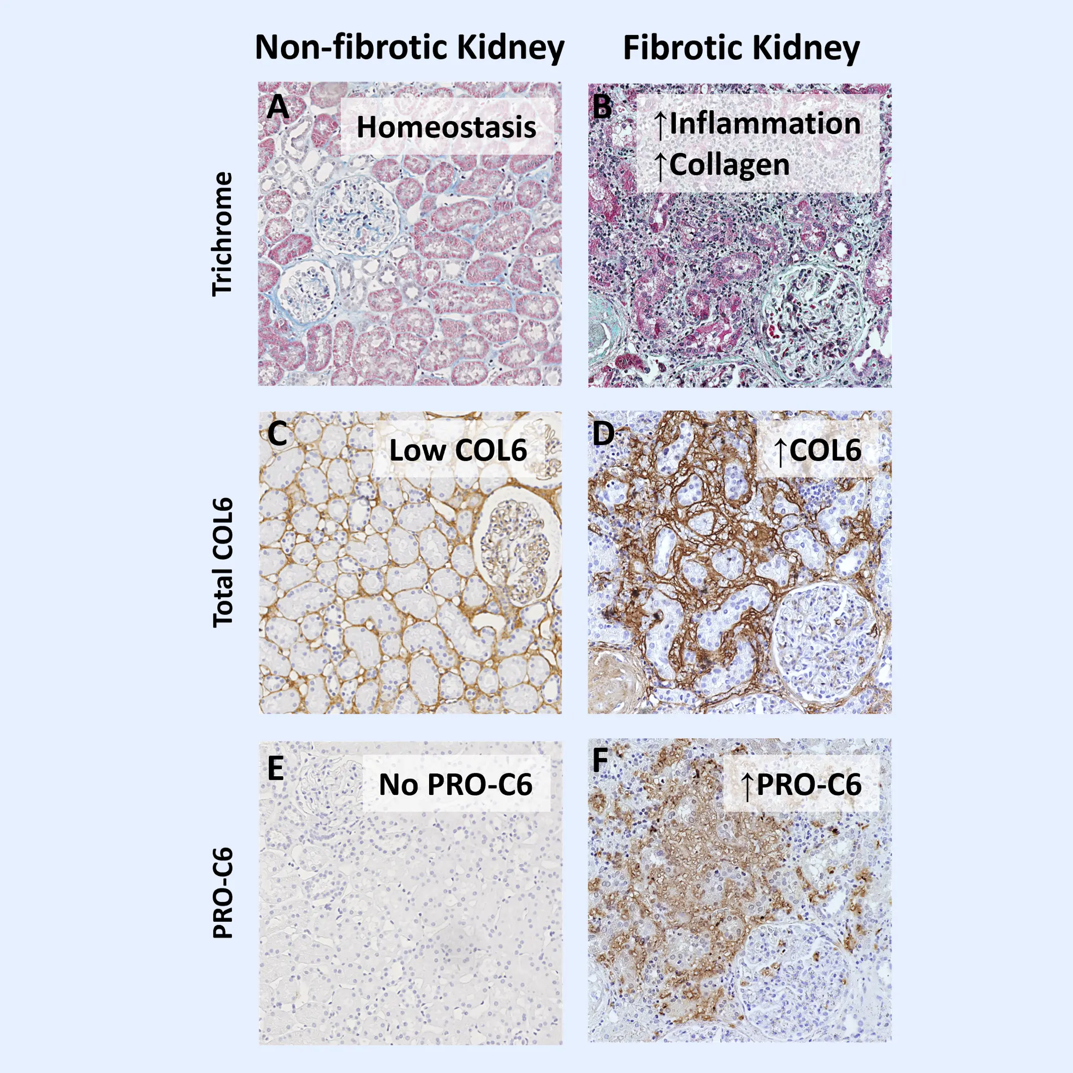
IgA nephropathy (IgAN) is the most common form of glomerulonephritis worldwide and a leading cause of end-stage kidney disease (ESKD). IgAN is characterized by the deposition of IgA in the glomerular basement membrane. While the clinical trajectory of IgAN varies among individuals, its progression is frequently driven by subclinical and persistent kidney fibrosis. Excessive accumulation of extracellular matrix (ECM) proteins occurs during fibrosis, which disrupts cellular organization and function within the kidney.

Current clinical markers are limited in their ability to detect early fibrotic changes or to monitor dynamic treatment responses. Biomarkers offer a powerful tool to uncover dynamic disease processes and enable earlier, more informed decision-making. Biomarkers of the ECM can provide direct insight into active fibrogenesis—the key mechanism underlying irreversible kidney damage.

Advancing drug development in IgA nephropathy with Nordic ProteinFingerPrint™ Biomarkers

Our biomarker assays quantify ECM fragments released into the blood or urine during active fibrogenesis and fibrolysis —processes central to IgAN progression. These markers provide disease-relevant insights into kidney tissue remodeling, including matrix formation, degradation, and fibrosis resolution. Unlike broad omics approaches, our biomarkers are highly specific to ECM dynamics, offering precise detection of fibrotic activity that directly reflects pathological changes in the kidney.

In IgAN, progressive fibrosis is a key driver of long-term kidney function decline and a critical determinant of patient prognosis. By measuring ECM-derived biomarkers, we can non-invasively assess the extent and activity of fibrosis, even before conventional markers show decline. This enables earlier identification of high-risk patients and more accurate tracking of pharmacodynamic effects. Integrating ECM biomarkers into IgAN clinical trials accelerates development timelines and enhances decision-making across all trial phases.

Increased type VI collagen deposition and nordicPRO-C6™ in fibrotic kidneys

Type VI collagen is located near the basement membrane and has several bindings sites for basement membrane proteins. Upon maturation of new type VI collagen molecules produced by highly activated fibroblasts, a bioactive fragment called Endotrophin is released. Endotrophin has pro-fibrotic, pro-tumorigenic and pro-inflammatory signalling roles. We measure nordicEndotrophin™ and formation of type VI collagen with our biomarker nordicPRO-C6™ (PRO-C6).

Type VI collagen is highly upregulated in fibrotic kidney sections (Fig. 1B & 1D) compared to the non-fibrotic (Fig 1A &1C). The fragment measured by PRO-C6 is virtually absent in the non-fibrotic kidneys (Fig. 1E), while it shows a significant presence in fibrotic kidneys (Fig. 1F).

Markers of tissue turnover are modulated by anti-inflammatory treatment

During fibrosis, elevated circulating levels of nordicPRO-C6™ and reduced urinary levels of C3M, reflecting type III collagen degradation, are observed.

Patients with IgAN treated with budenoside in the NEFIGAN trial showed a reversal of this process, by reducing nordicPRO-C6™ and increasing C3M (Figure 2).

Figure 2. Anti-inflammatory treatment modulates tissue turnover in IgAN

About IgA nephropathy (Berger’s Disease)

  • IgA nephropathy (IgAN) is a kidney disease caused by the buildup of IgA antibodies in the glomeruli, leading to inflammation and scarring. Fibrosis—the accumulation of extracellular matrix (ECM) proteins—is a key driver of irreversible kidney damage in IgAN and strongly predicts progression to end-stage kidney disease (ESKD).

  • Biomarkers offer a non-invasive way to detect early fibrotic changes and monitor disease progression. ECM-based biomarkers can track active tissue remodeling in real time, providing critical insights into disease activity, risk stratification, and treatment response in IgAN patients.

  • Yes, clinical data show that biomarkers like PRO-C6 and uC3M are modulated by anti-inflammatory treatments. For example, budesonide therapy in IgAN patients has been associated with decreased PRO-C6 levels and increased uC3M, indicating reduced fibrosis and improved ECM turnover.

  • These biomarkers provide highly specific and mechanistic insights into fibrogenesis and fibrolysis. By using them in clinical trials, researchers can monitor drug effects more precisely, select appropriate patient subgroups, and make faster, more informed decisions throughout the development pipeline.

Would you like to learn more? Contact us!

    Nordic Bioscience’s assays and services are research use only products and services and do not qualify for medical or diagnostic purposes.