Translational Models in Kidney Diseases

De-risking kidney drug development with translational models and ECM Biomarkers

Successful drug development is essential but often challenging and uncertain. To improve outcomes, it’s important to use strategies that increase predictability and lower risk. Regulatory agencies such as the FDA and EMA recognize the value of biomarker-driven approaches, especially when combined with robust translational research, to guide decision-making and accelerate progress through the pipeline.

Preclinical translational models play a vital role in understanding kidney disease mechanisms and evaluating the efficacy of novel therapeutics before entering clinical trials. In models of kidney fibrosis, fibroblast activation and extracellular matrix (ECM) remodelling are key processes that closely reflect human disease progression. ECM-derived biomarkers offer a non-invasive and quantitative way to assess fibrogenesis and fibrolysis.

By integrating these biomarkers into preclinical studies, pharmaceutical developers and researchers can better monitor therapeutic effects, gain mechanistic insights, and improve the translational relevance of animal models. This integrated approach supports earlier and more confident go/no-go decisions, ultimately reducing development time and increasing the likelihood of clinical success.
Animal free precision cut kidney slices


The precision cut kidney slice model

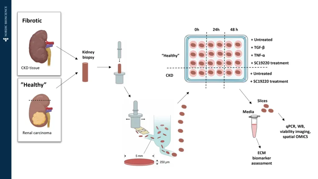
The precision cut kidney slice model (PCKS) is an animal-free drug screening platform based on human kidney slices. A key feature of PCKS is that the slices contain all cell types and acellular components of the whole organ in the original configuration while preserving cell-cell and cell-matrix interactions.

Understanding the dynamics of the ECM is crucial for developing therapeutic strategies for fibrosis.

The PCKS model has been shown to be an extremely valuable translational model for renal (patho)physiology, bridging the gap between preclinical and clinical research.

ECM biomarkers in kidney translational models

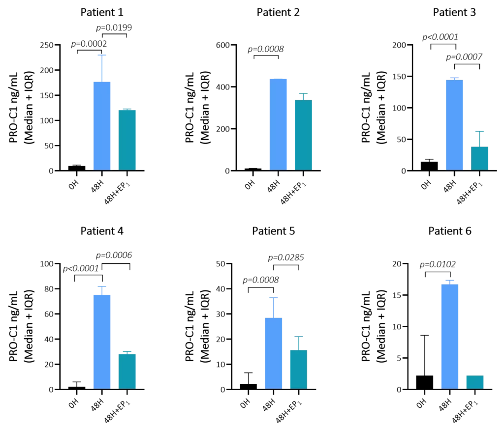
Biomarkers of ECM remodeling can be measured in the tissue culture supernatant, giving insights into the dynamics of tissue remodeling following treatment. Collagen type I formation, measured by our PRO-C1 marker increase after 48 hours of culture of kidney slices from patients with chronic kidney disease (CKD).

In addition, the levels of PRO-C1 could be decreased by an EP1 receptor inhibitor (Figure 1). This modulation demonstrates the model’s suitability for screening of antifibrotic effects on the fibrotic kidney tissue and cells.

Figure 1. Type I collagen formation (PRO-C1) increases after 48 hours and the addition of EP1 receptor inhibitor reduces PRO-C1 levels in the fibrotic kidney slices. 

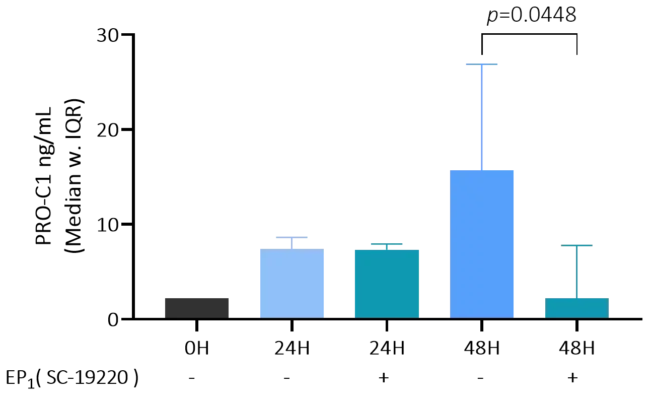
Lastly, PRO-C1 levels also increased after 48 hours in the PCKS model using healthy kidney slices (taken from healthy regions of renal carcinoma-affected kidneys) and showed a similar pharmacodynamic reduction as observed in the fibrotic kidney slices. (Figure 2).

Figure 2. Type I collagen formation (PRO-C1) increases after 48 hours and the addition of EP1 receptor inhibitor reduces PRO-C1 levels in the “healthy” kidney tissue.

Translational application of ECM biomarkers in rodent models

The Nordic ProteinFingerPrint™ biomarkers can be measured in blood and urine of numerous in vivo kidney disease models, where some of these are listed below.

5/6 nephrectomy Anti-Thy 1.1 nephropathy In vivo models Unilateral nephrectomy with  ischemia reperfusion (IRI)
Adenine nephropathy Bilateral ischemia reperfusion (IRI)  Munich Wistar Fromter Unilateral Ureteral Obstruction (UUO)
Adriamycin-induced nephropathy Cisplatin induced injury Nephrotoxic nephritis model Zucker diabetic fatty (ZDF)
Anti-Glomerular Basement Membrane (GBM) Dahl salt-sensitive rats The Streptozotocin (STZ) ZSF-1

The Adriamycin-induced nephropathy in vivo model

The Adriamycin-induced nephropathy rat model is a well-established in vivo system for studying progressive kidney disease, characterized by the onset of proteinuria followed by renal fibrosis. This model enables prognostic evaluation of fibrotic biomarkers in relation to the development and potential prevention of fibrosis. It provides a robust platform for investigating the mechanisms of kidney fibrosis and therapeutic intervention.

To evaluate the impact of Adriamycin, Nordic ProteinFingerPrint™ biomarkers—associated with both prognosis and pharmacodynamic response in clinical settings—can be quantified in blood and urine samples from the animals.

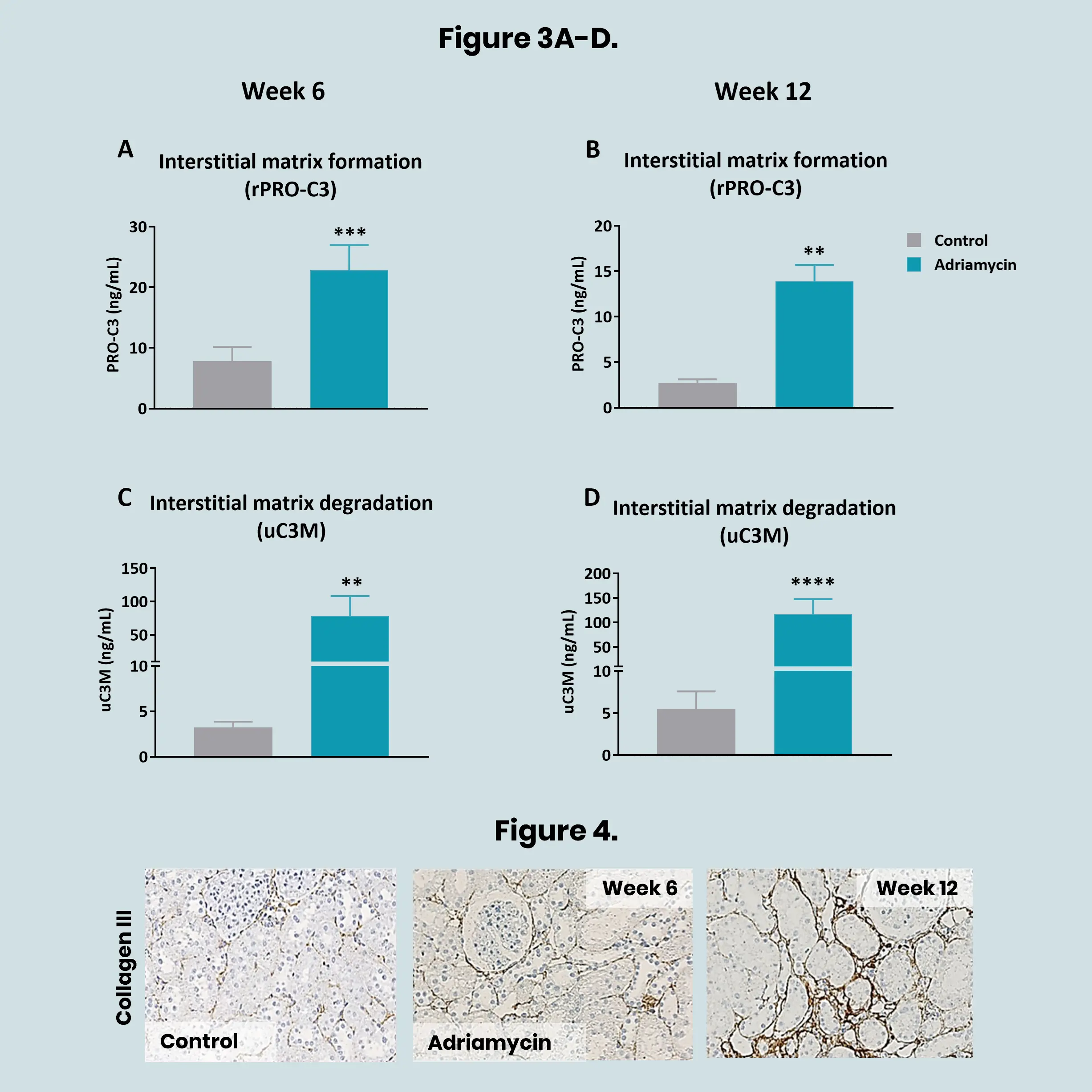
From week 6, urinary levels of type III collagen formation (rPRO-C3) (Figure 3A-B) and degradation (uC3M) (Figure 3C-D) were elevated in the Adriamycin-induced rats compared to controls prior to histological detection of accumulated type III collagen in the kidneys (Figure 4). The findings suggest that these biomarkers could be an early non-invasive markers of kidney fibrosis.

Figure 3A-D. Effects of Adriamycin on collagen formation (rPRO-C3) and degradation (uC3M) markers.

Figure 4. Expression of interstitial type III collagen in controls and in Adriamycin-induced nephropathy rat model at week 6 and 12.

The Zucker diabetic fatty rat model

The Zucker Diabetic Fatty (ZDF) rat is a widely used preclinical model of type 2 diabetes that spontaneously develops obesity, insulin resistance, and hyperglycemia—hallmarks of diabetic kidney disease (DKD). As the disease progresses, ZDF rats exhibit key features of human DKD, including albuminuria, glomerulosclerosis, and tubulointerstitial fibrosis. This makes the model particularly relevant for studying ECM remodeling.

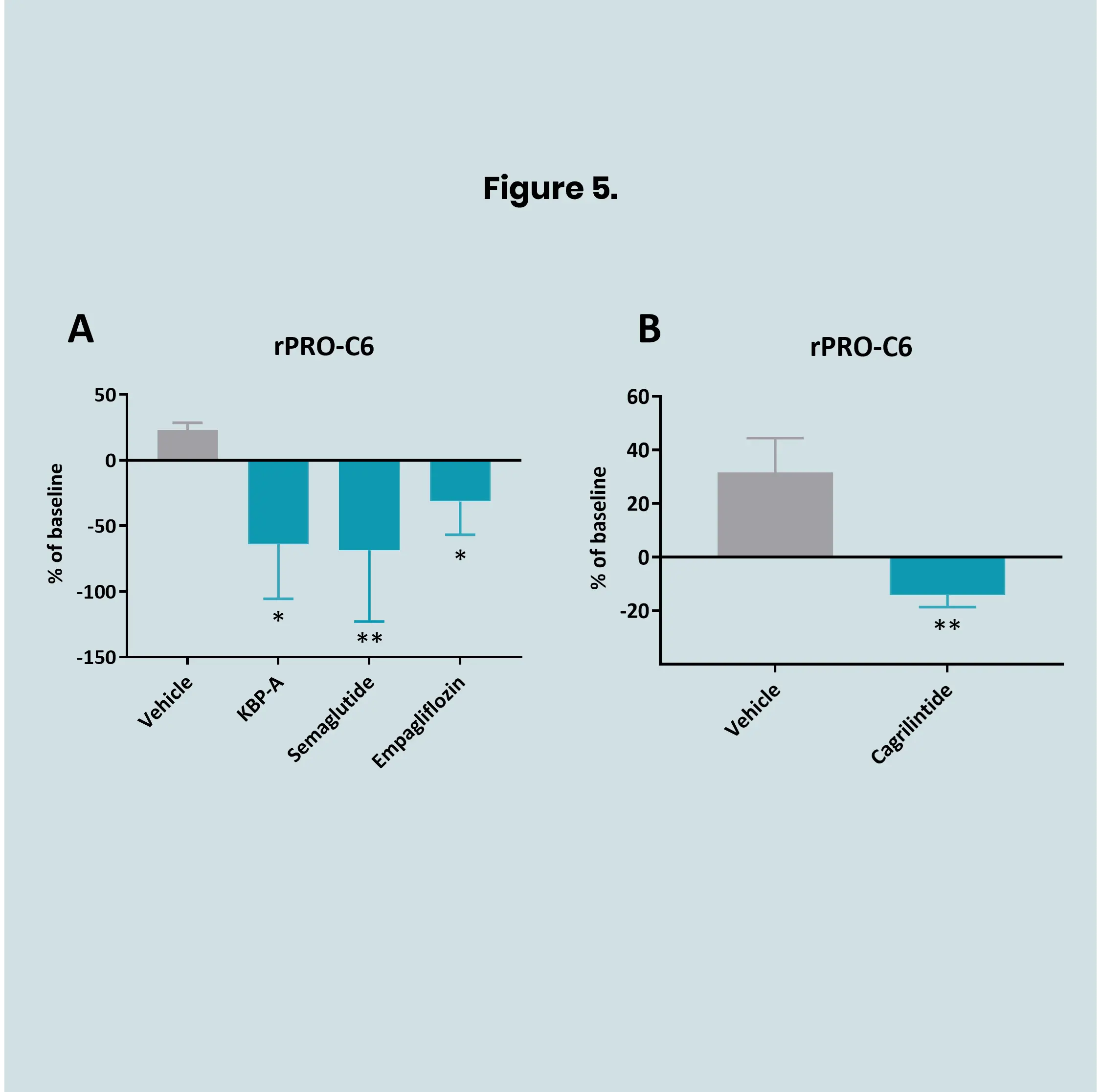
In ZDF rats, anti-diabetic drugs including KBP-A, Semaglutide, Empagliflozin, and Cagrilintide reduced serum levels of rPRO-C6 compared to the control group (Figure 5). This modulation of rPRO-C6 levels by anti-diabetic treatment demonstrates the model’s suitability for examining anti-diabetic effects on T2D-related fibrosis.

Figure 5. Serum levels of rPRO-C6 in ZDF rats. Rats received treatment with KBP-A, Semaglutide, Empagliflozin (A), or Cagrilintide (B).

Would you like to learn more? Contact us!

    Nordic Bioscience’s assays and services are research use only products and services and do not qualify for medical or diagnostic purposes.